Cardiac Conduction Autorhythmic cardiac muscle cells depolarize at


- Slides: 8

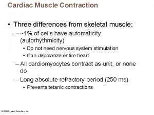
Cardiac Conduction Autorhythmic: cardiac muscle cells depolarize at regular intervals p Cardiac Conduction system: cardiac cells that are specialized to generate AP’s to control contractions of the heart p Pacemaker: the sinoatrial (SA) node, found in the R atrium starts HB and controls rate of contraction. AP’s from node travel down walls of atrium p

Cardiac Conduction Atrioventricular (AV) node: found by the AV valve, acts as a gateway carrying the AP’s from the SA node to the ventricles p AV bundle: a pathway for the AP’s from the AV node to travel through the septum as it splits into R and L branches toward apex of heart p Purkinje fibers: branches of the AV bundle that travel from the apex upward p

Cardiac Rhythm Sinus rhythm: normal heartbeat 7080/min p Nodal rhythm: caused by SA node damage 40 -50/min, AV node still working but ectopic focus working for SA node p If both SA and AV nodes are damaged then ectopic foci keep heart beating at 2040/min body lives but brain dies due to lack of oxygen p

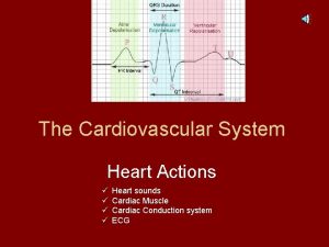
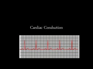
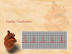
Electrocardiogram (ECG or EKG): a visual record of the heart’s electrical activity p Systole: contraction of a heart chamber p Diastole: heart chamber relaxes and fills with blood p P wave: caused by the signal from the SA node as it spreads through the atria depolarizing the walls causing systole p

Electrocardiogram PQ segment: time it takes the signal to travel from the SA to AV node, p QRS complex: caused by the signal traveling through the two ventricles, p QRS interval: atria repolarization and diastole p ST segment: ventricular systole p T wave: the repolarization of the ventricles before diastole p

Cardiac Output (CO): CO= HR x SV normal CO =5250 ml/min so all blood in the body will pass through and return to the heart in about a min p Cardiac reserve: the difference of CO at rest and max CO p

Heart Rate Pulse: the heart rate measured at an artery p Tachycardia: persistent heart rate at rest above 100 bpm p Bradycardia: persistent heart rate at rest below 60 bpm p

Stroke Volume Preload: the stretch on the myocardium before systole p Contractility: how hard the myocardium can contract for a certain preload p Afterload: the blood pressure in the vessels that resists the blood flow out of heart p
