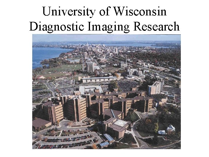
University of Wisconsin Diagnostic Imaging Research Lecture 1


- Slides: 22
University of Wisconsin Diagnostic Imaging Research
Lecture 1: Introduction (1/2) – History, basic principles, modalities Class consists of: 1) Deterministic Studies - distortion - impulse response - transfer functions All modalities are non-linear and space variant to some degree. Approximations are made to yield a linear, space-invariant system. 2) Stochastic Studies SNR (signal to noise ratio) of the resultant image - mean and variance
Course Objectives • Learn basics of 2 D to n-dimensional system theory and signal processing – Emphasis on duals between space and frequency domain – Emphasis on intuitive understanding • Understand underlying physics of medical imaging modalities • Study the deterministic and stochastic descriptions of medical imaging systems – Theory is applicable beyond medical imaging
Prerequisites and Postrequisites • System Theory – ECE 330, BME/MP 573 • Statistics Helpful but Not Required – Mean and variance of stochastic processes – ECE 331, BME/MP 574, ECE 730 • Other Courses • Microscopy of Life • BME 568/ MP 568 MRI ( less math)
Wilhelm Röntgen, Wurtzburg Nov. 1895 – Announces X-ray discovery Jan. 13, 1896 – Images needle in patient’s hand – X-ray used presurgically 1901 – Receives first Nobel Prize in Physics – Given for discovery and use of X-rays. Radiograph of the hand of Röntgen’s wife, 1895.
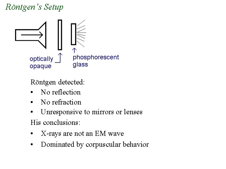
Röntgen’s Setup Röntgen detected: • No reflection • No refraction • Unresponsive to mirrors or lenses His conclusions: • X-rays are not an EM wave • Dominated by corpuscular behavior
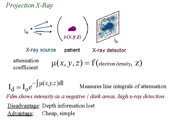
Projection X-Ray attenuation coefficient Measures line integrals of attenuation Film shows intensity as a negative ( dark areas, high x-ray detection Disadvantage: Depth information lost Advantage: Cheap, simple
Sagittal Coronal
Early Developments • Intensifying agents, contrast agents all developed within several years. • Creativity of physicians resulted in significant improvements to imaging. - found ways to selectively opacify regions of interest - agents administered orally, intraveneously, or via catheter
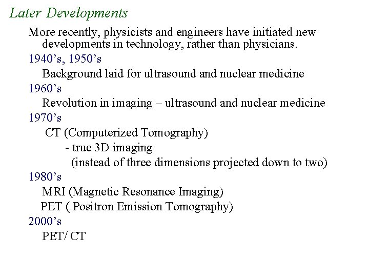
Later Developments More recently, physicists and engineers have initiated new developments in technology, rather than physicians. 1940’s, 1950’s Background laid for ultrasound and nuclear medicine 1960’s Revolution in imaging – ultrasound and nuclear medicine 1970’s CT (Computerized Tomography) - true 3 D imaging (instead of three dimensions projected down to two) 1980’s MRI (Magnetic Resonance Imaging) PET ( Positron Emission Tomography) 2000’s PET/ CT
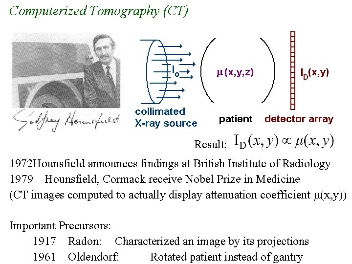
Computerized Tomography (CT) Result: 1972 Hounsfield announces findings at British Institute of Radiology 1979 Hounsfield, Cormack receive Nobel Prize in Medicine (CT images computed to actually display attenuation coefficient m(x, y)) Important Precursors: 1917 Radon: Characterized an image by its projections 1961 Oldendorf: Rotated patient instead of gantry
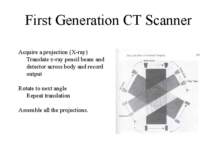
First Generation CT Scanner Acquire a projection (X-ray) Translate x-ray pencil beam and detector across body and record output Rotate to next angle Repeat translation Assemble all the projections.
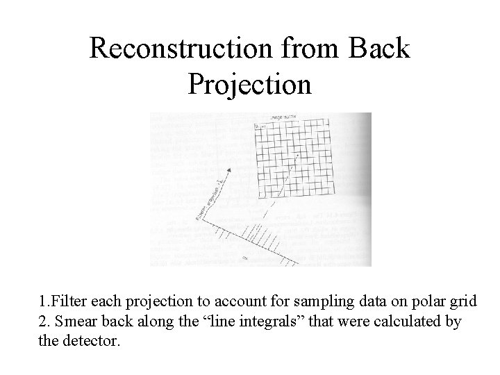
Reconstruction from Back Projection 1. Filter each projection to account for sampling data on polar grid 2. Smear back along the “line integrals” that were calculated by the detector.
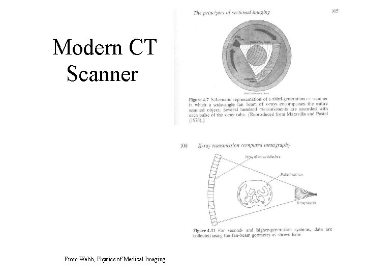
Modern CT Scanner From Webb, Physics of Medical Imaging
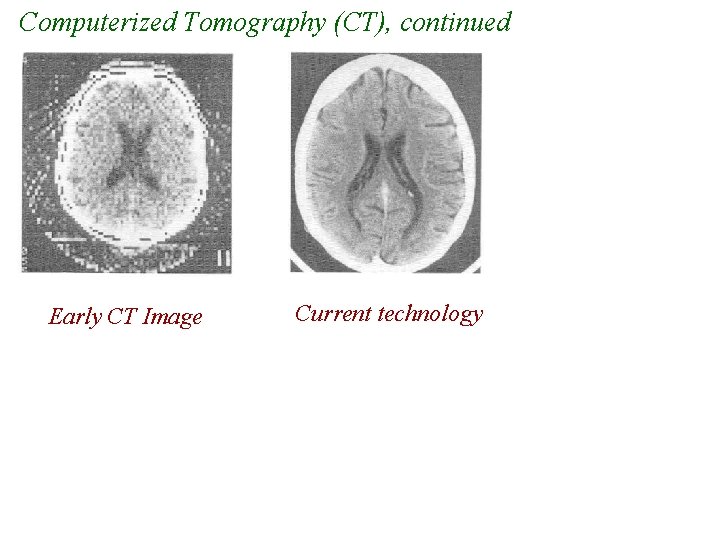
Computerized Tomography (CT), continued Early CT Image Current technology
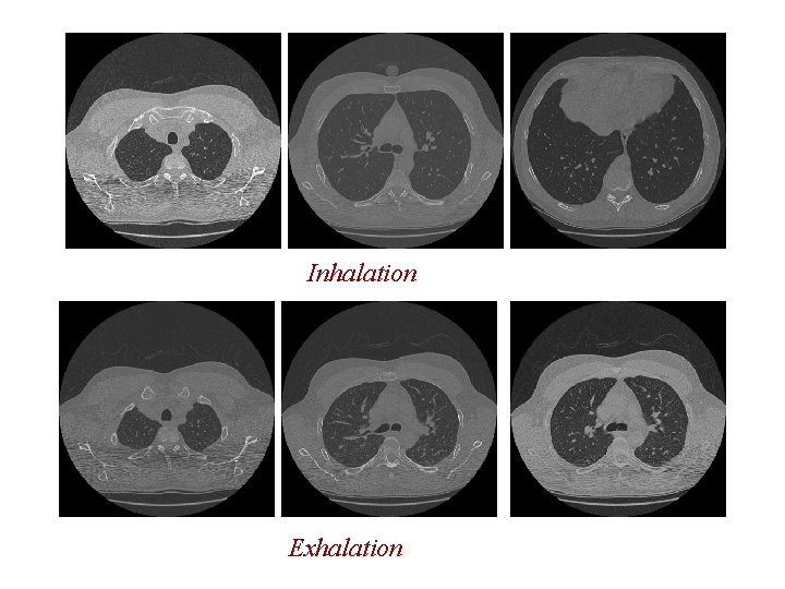
Inhalation Exhalation
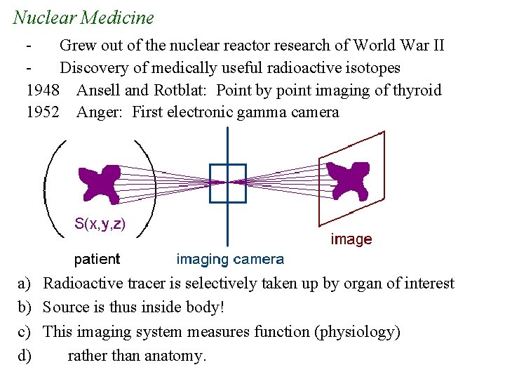
Nuclear Medicine Grew out of the nuclear reactor research of World War II Discovery of medically useful radioactive isotopes 1948 Ansell and Rotblat: Point by point imaging of thyroid 1952 Anger: First electronic gamma camera a) Radioactive tracer is selectively taken up by organ of interest b) Source is thus inside body! c) This imaging system measures function (physiology) d) rather than anatomy.
Nuclear Medicine, continued Very specific in imaging physiological function - metabolism - thyroid function - lung ventilation: inhale agent Advantage: Direct display of disease process. Disadvantage: Poor image quality (~ 1 cm resolution) Why is resolution so poor? Very small concentrations of agent used for safety. - source within body Quantum limited: CT 109 photons/pixel Nuclear ~100 photons/pixel Tomographic systems: SPECT: single photon emission computerized tomography PET: positron emission tomography
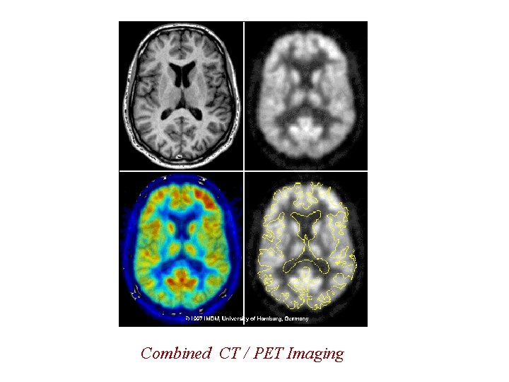
Combined CT / PET Imaging
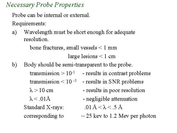
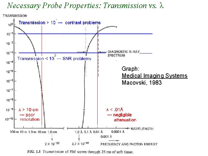
Necessary Probe Properties Probe can be internal or external. Requirements: a) Wavelength must be short enough for adequate resolution. bone fractures, small vessels < 1 mm large lesions < 1 cm b) Body should be semi-transparent to the probe. transmission > 10 -1 - results in contrast problems transmission < 10 -3 - results in SNR problems λ > 10 cm - results in poor resolution λ <. 01Å - negligible attenuation Standard X-rays: . 01 Å < λ <. 5 Å corresponding to ~ 25 kev to 1. 2 Mev per photon
Necessary Probe Properties: Transmission vs. λ Graph: Medical Imaging Systems Macovski, 1983
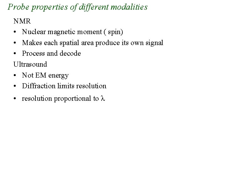
Probe properties of different modalities NMR • Nuclear magnetic moment ( spin) • Makes each spatial area produce its own signal • Process and decode Ultrasound • Not EM energy • Diffraction limits resolution • resolution proportional to λ