Presenting author Serena Girardelli OP 12 02 Mapping
- Slides: 1
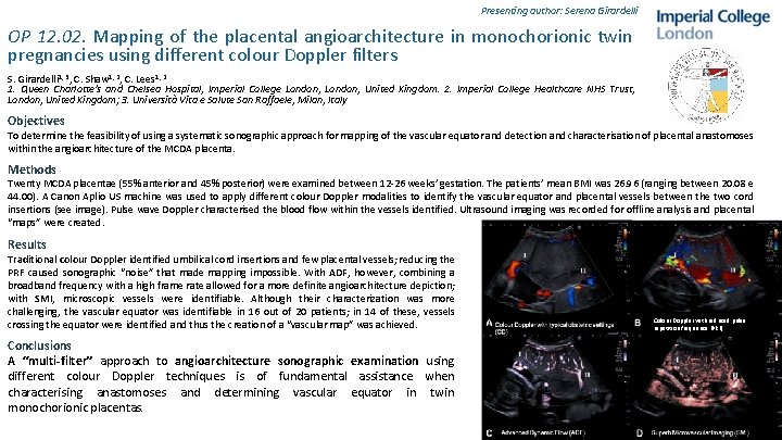
Presenting author: Serena Girardelli OP 12. 02. Mapping of the placental angioarchitecture in monochorionic twin pregnancies using different colour Doppler filters S. Girardelli 1, 3, C. Shaw 1, 2, C. Lees 1, 2 1. Queen Charlotte's and Chelsea Hospital, Imperial College London, United Kingdom. 2. Imperial College Healthcare NHS Trust, London, United Kingdom; 3. Universita Vita e Salute San Raffaele, Milan, Italy Objectives To determine the feasibility of using a systematic sonographic approach for mapping of the vascular equator and detection and characterisation of placental anastomoses within the angioarchitecture of the MCDA placenta. Methods Twenty MCDA placentae (55% anterior and 45% posterior) were examined between 12 -26 weeks’ gestation. The patients’ mean BMI was 26. 96 (ranging between 20. 08 e 44. 00). A Canon Aplio US machine was used to apply different colour Doppler modalities to identify the vascular equator and placental vessels between the two cord insertions (see image). Pulse wave Doppler characterised the blood flow within the vessels identified. Ultrasound imaging was recorded for offline analysis and placental “maps” were created. Results Traditional colour Doppler identified umbilical cord insertions and few placental vessels; reducing the PRF caused sonographic “noise” that made mapping impossible. With ADF, however, combining a broadband frequency with a high frame rate allowed for a more definite angioarchitecture depiction; with SMI, microscopic vessels were identifiable. Although their characterization was more challenging, the vascular equator was identifiable in 16 out of 20 patients; in 14 of these, vessels crossing the equator were identified and thus the creation of a “vascular map” was achieved. Conclusions A “multi-filter” approach to angioarchitecture sonographic examination using different colour Doppler techniques is of fundamental assistance when characterising anastomoses and determining vascular equator in twin monochorionic placentas. Colour Doppler with reduced pulse repetition frequence (PRF)