Metathalamus and epithalamus DIENCEPHALON The diencephalon is a


- Slides: 11

Metathalamus and epithalamus

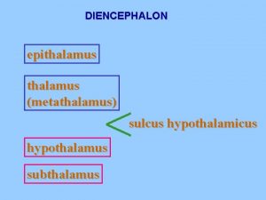
DIENCEPHALON The diencephalon is a middle structure which is largely embedded in the cerebrum its cavity forms the greater part of the third ventricle. The hypothalamic sulcus, extending from the interventricular foramen to the cerebral aqueduct, divides each half of the diencephalon into dorsal and ventral parts. Dorsal Part of Diencephalon 1. Thalamus 2. Metathalamus 3. Epithalamus,

Ventral Part of Diencephalon 1. Hypothalamus, and 2. Subthalamus (ventral thalamus).

Metathalamus Medial Geniculate Body It is an oval elevation situated just below the pulvinar of the thalamus and lateral to the superior colliculus. The inferior brachium connects the medial geniculate body to the inferior colliculus. Afferents: Lateral lemniscus, Efferents: It gives rise to the acoustic (auditory) radiation going to the auditory area of the cortex Function Medial geniculate body is the last relay station on the pathway of auditory impulses to the cerebral cortex.

Lateral Geniculate Body It is a small oval elevation situated anterolateral to the medial geniculate body, and is connected to the superior colliculus by the superior brachium. Structure It is six-layered structure. Layers 1, 4 and 6 (pink) receive contralateral optic fibres, and layers 2, 3 and 5 (light blue) receive ipsilateral optic fibres. Connections Afferents: Optic tract (lateral root).

Efferents: It gives rise to optic radiations going to the visual area of cortex Function Lateral geniculate body is the last relay station on the visual pathway to the occipital cortex.

Six layers of lateral geniculate body

Epithalamus The epithalamus consists of: 1. The right and left habenular nuclei, 2. The pineal body 3. The habenular commissure. 4. The posterior commissure. Pineal Body/Pineal Gland The pineal body is a small, conical organ, projecting backwards and downwards between the two superior colliculi. It consists of a conical body about 8 mm long, and a stalk or peduncle

Structure The pineal gland is composed of two types of cells, pinealocytes and neuroglial cells, with a rich network of blood vessels and sympathetic fibres. Calcareous concretions are constantly present in the pineal after the 17 th year of life and may form aggregations (brain sand). Functions It is an endocrine gland of great importance. The best known hormone is melatonin which causes changes in skin colour in some species. The synthesis and discharge of melatonin is remarkably influenced by exposure of the animal to light and is more during dark period.

Components of the epithalamus

THANK YOU
