FRET Fluorescence Frster Resonance Energy Transfer Jelaina Holroyd


- Slides: 8

FRET Fluorescence (Förster) Resonance Energy Transfer Jelaina Holroyd, Kate Berger, Nicole Liang, Jeff Chen, Jonas Richter Oct. 3 2016

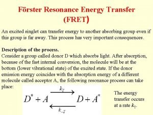
What is FRET? - Method to study physical interactions between proteins or DNA, as well as protein conformation. How? - Uses donor and acceptor fluorophores attached to biomolecules (proteins or DNA) - Transfer efficiency is sensitive to distance (1 10 nm), so distance between fluorophores can be estimated by measuring FRET efficiency https: //www. semrock. com/Data/Sites/1/semrockimages/drawings/fret_500 px. jpg

What is FRET used for? -Studying colocalization/interactions of two proteins -Studying colocalization/interactions of a protein and a particular DNA sequence -Measuring intramolecular distance within one protein (structure and conformation) http: //zeiss-campus. magnet. fsu. edu/tutorials/spectralimaging/fretbiosensorsfigure 1. jpg

Using FRET http: //www. olympusmicro. com/primer/techniques/fluorescence/fret/images/fretintrofigure 1. jpg http: //www. olympusmicro. com/primer/techniques/fluorescence/fret/images/fretintrofigure 2. jpg

Fluorescent tagging: Post-Translational - Donor chromophore and acceptor chromophore (donor-acceptor pair) - Attach fluorescent markers to proteins post-translationally Donor and Acceptor Fluorescent Nucleotides Donor and Acceptor Fluorophores https: //en. wikipedia. org/wiki/Fluorescent_glucose_biosensor#/media/File: Squid%27 s_fret. svg

Fluorescent tagging: GFP Constructs -GFP: Protein recombinant is constructed by genetic modification -The recombinant proteins of interest are inherently fluorescent (do not need post-transcriptional attachment of fluorophores) Donor and Acceptor Fluorescent Proteins http: //www. fret. lif. kyoto-u. ac. jp/e-phogemon/images/fig 4103. gif

Examples of FRET in research - Used to study the colocalization of glucocorticoid and mineralocorticoid receptors 3 - Used to determine if a mutation is present on a gene for cystic fibrosis 1 Chen, X. et al. (1997) Nishi, M. et al. (2004)

Further Resources/References 1. Chen, X. , Zehnbauer, B. , Gnirke, A. , Kwok, P. -Y. (1997). “Fluorescence Energy Transfer Detection as a Homologous DNA Diagnostic Method. ” Proc Natl Acad Sci USA. 94(20): 10756– 10761. 2. Dinant, C. , van Royen, M. E. , Vermeulen, W. , Houtsmuller, A. B. (2008). “Fluorescence resonance energy transfer of GFP and YFP by spectral imaging and quantitative acceptor photobleaching. ” J. Microsc. 231(Pt 1): 97 -104. doi: 10. 1111/j. 1365 -2818. 2008. 02020. x. 3. Nishi, M. , Tanaka, M. , Matsuda, K. , Sunaguchi, M. , Kawata, M. (2004). “Visualization of Glucocorticoid Receptor and Mineralocorticoid Receptor Interactions in Living Cells with GFP-Based Fluorescence Resonance Energy Transfer. ” The Journal of Neuroscience. 24(21): 4918 -4927. doi: 10. 1523/JNEUROSCI. 5495 -03. 2004 Roy, R. , Hohng, S. , Ha, T. (2008). “A Practical Guide to Single Molecule FRET. ” Nature Methods. 5, 507 - 516 doi: 10. 1038/nmeth. 1208. Seegar, T. , Barton, W. (2010). “Imaging Protein-protein Interactions in vivo. ” J. Vis. Exp. (44), e 2149, doi: 10. 3791/2149 (2010). Sekar, R. B. , Periasamy, A. (2003). “Fluorescence resonance energy transfer (FRET) microscopy imaging of live cell protein localizations. ” J. Cell. Biol. 160(5): 629 -633. DOI: 10. 1083/jcb. 200210140. Sprenger, J. U. , Perera, R. K. , Götz, K. R. , Nikolaev, V. O. (2012). “FRET Microscopy for Real-time Monitoring of Signaling Events in Live Cells Using Unimolecular Biosensors. ” J. Vis. Exp. 66, e 4081 doi: 10. 3791/4081. 4. 5. 6. 7.
