Department of Medical Sciences Pharmacology Unit University of
- Slides: 40
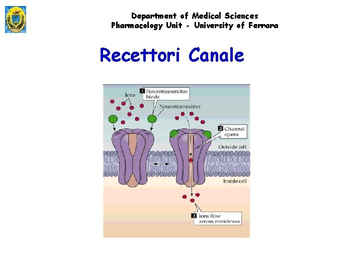
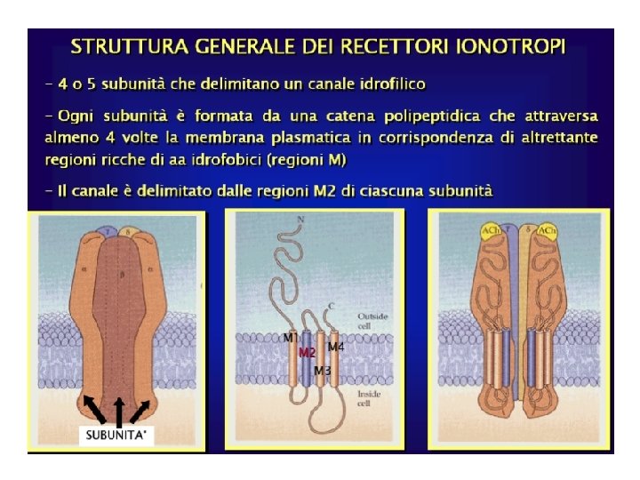
Department of Medical Sciences Pharmacology Unit - University of Ferrara Recettori Canale
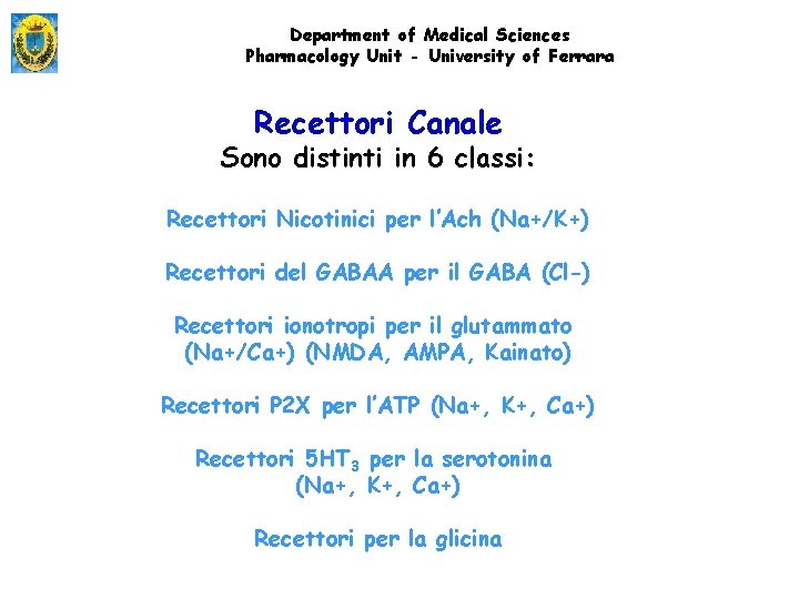
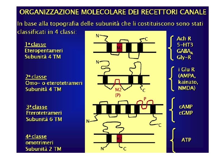
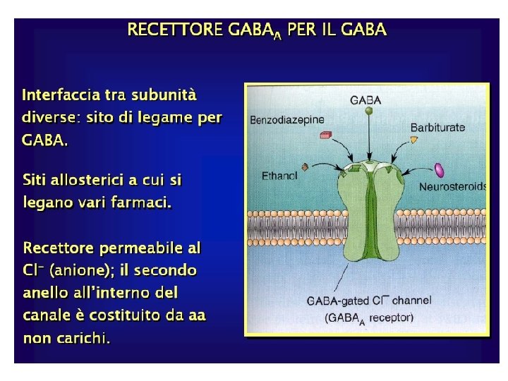
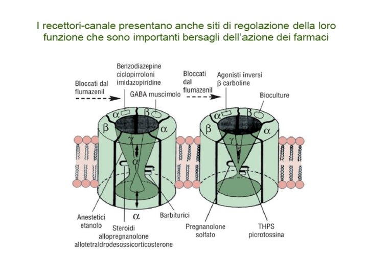
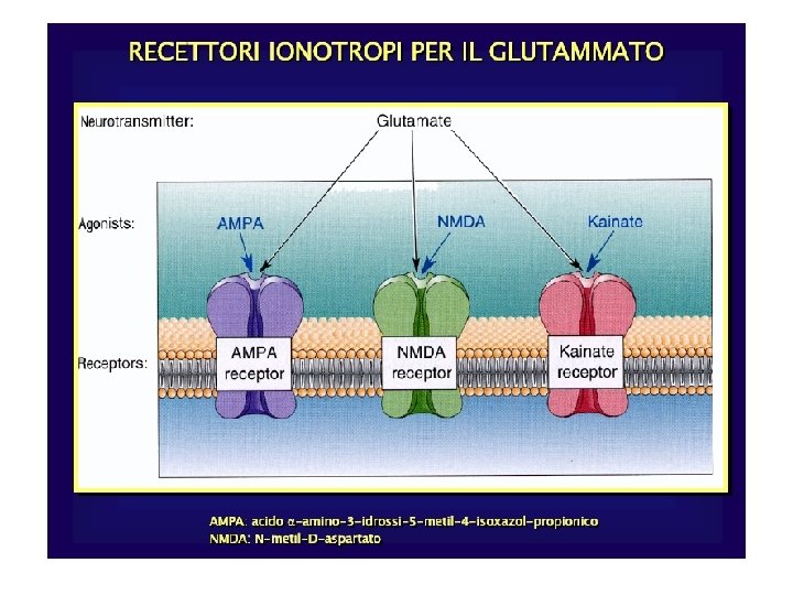
Department of Medical Sciences Pharmacology Unit - University of Ferrara Recettori Canale Sono distinti in 6 classi: Recettori Nicotinici per l’Ach (Na+/K+) Recettori del GABAA per il GABA (Cl-) Recettori ionotropi per il glutammato (Na+/Ca+) (NMDA, AMPA, Kainato) Recettori P 2 X per l’ATP (Na+, K+, Ca+) Recettori 5 HT 3 per la serotonina (Na+, K+, Ca+) Recettori per la glicina
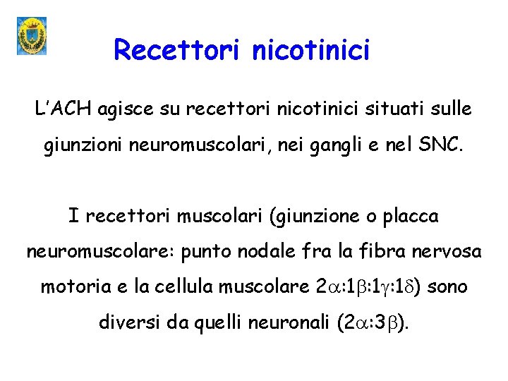
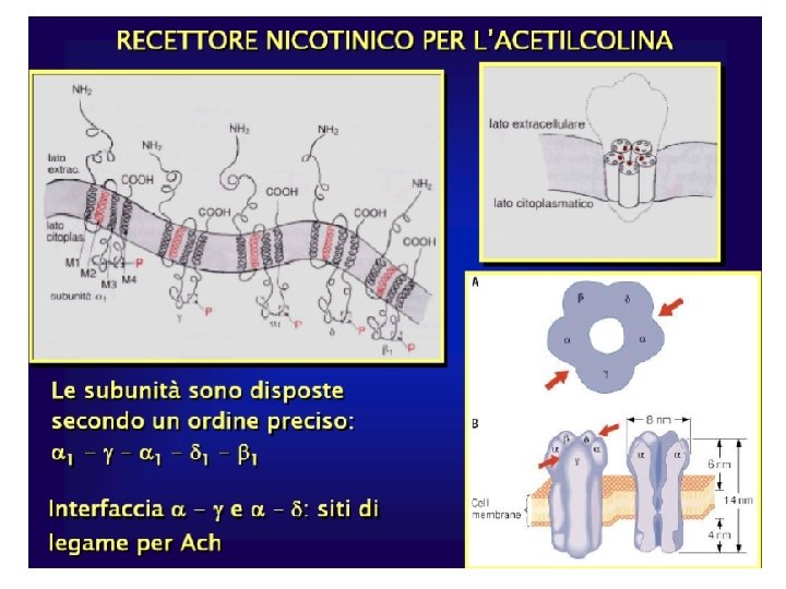
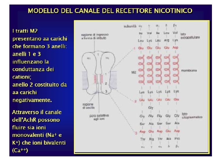
Recettori nicotinici L’ACH agisce su recettori nicotinici situati sulle giunzioni neuromuscolari, nei gangli e nel SNC. I recettori muscolari (giunzione o placca neuromuscolare: punto nodale fra la fibra nervosa motoria e la cellula muscolare 2 : 1 : 1 ) sono diversi da quelli neuronali (2 : 3 ).
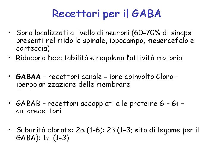
Recettori per il GABA • Sono localizzati a livello di neuroni (60 -70% di sinapsi presenti nel midollo spinale, ippocampo, mesencefalo e corteccia) • Riducono l’eccitabilità e regolano l’attività motoria • GABAA – recettori canale - ione coinvolto Cloro – iperpolarizzazione delle membrane • GABAB – recettori accoppiati alle proteine G – Gi – autorecettori • Subunità clonate: 2 (1 -6): 2 (1 -3; sito di legame per il GABA): 1 (1 -3)
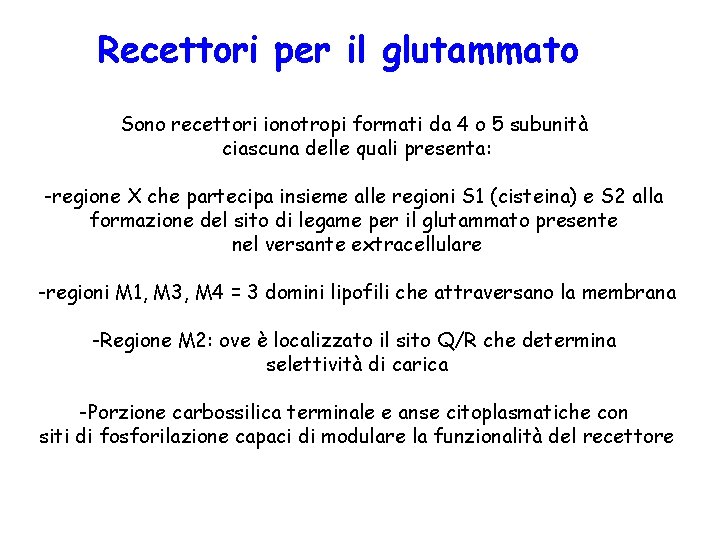
Recettori per il glutammato Sono recettori ionotropi formati da 4 o 5 subunità ciascuna delle quali presenta: -regione X che partecipa insieme alle regioni S 1 (cisteina) e S 2 alla formazione del sito di legame per il glutammato presente nel versante extracellulare -regioni M 1, M 3, M 4 = 3 domini lipofili che attraversano la membrana -Regione M 2: ove è localizzato il sito Q/R che determina selettività di carica -Porzione carbossilica terminale e anse citoplasmatiche con siti di fosforilazione capaci di modulare la funzionalità del recettore
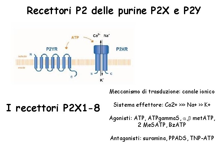
Recettori P 2 delle purine P 2 X e P 2 Y Meccanismo di trasduzione: canale ionico I recettori P 2 X 1 -8 Sistema effettore: Ca 2+ >>> Na+ >> K+ Agonisti: ATP, ATPgamma. S, , met. ATP, 2 Me. SATP, Bz. ATP Antagonisti: suramina, PPADS, TNP-ATP
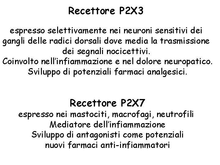
Recettore P 2 X 3 espresso selettivamente nei neuroni sensitivi dei gangli delle radici dorsali dove media la trasmissione dei segnali nocicettivi. Coinvolto nell’infiammazione e nel dolore neuropatico. Sviluppo di potenziali farmaci analgesici. Recettore P 2 X 7 espresso nei mastociti, macrofagi, neutrofili Mediatore dell’infiammazione Sviluppo di antagonisti come potenziali nuovi farmaci anti-infiammatori
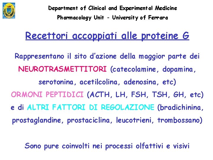
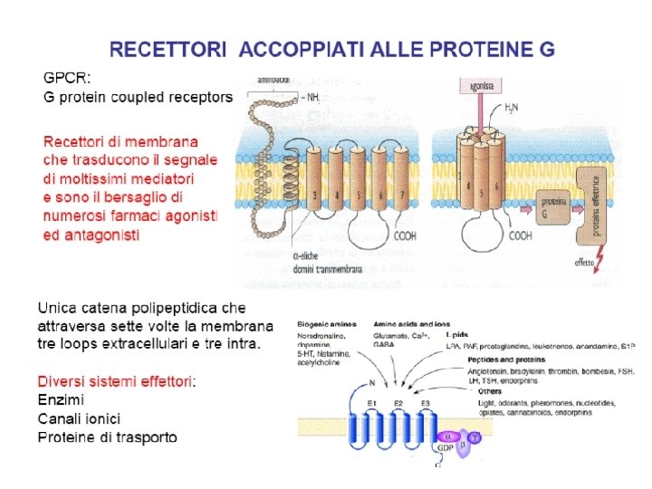
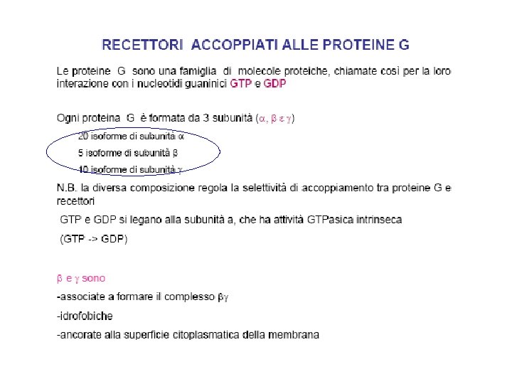
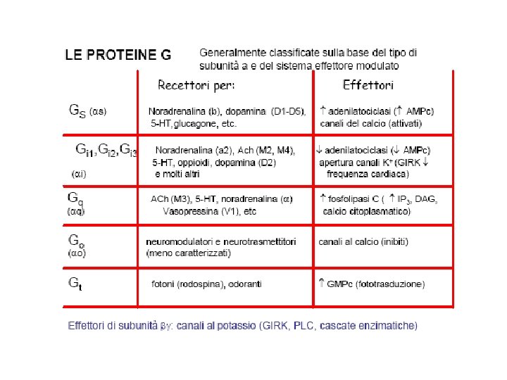
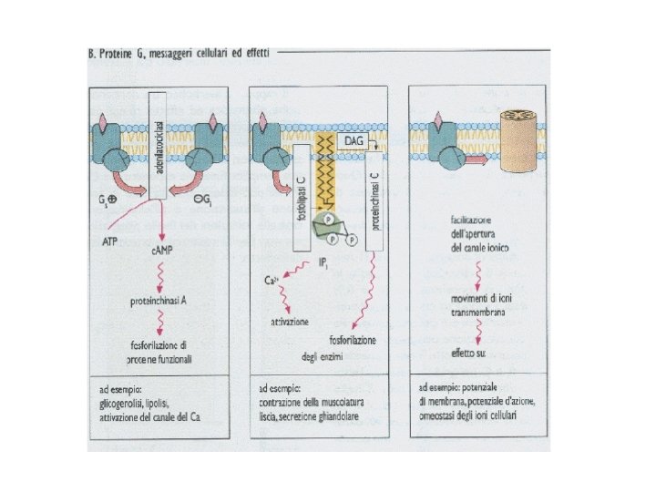
Department of Clinical and Experimental Medicine Pharmacology Unit - University of Ferrara Recettori accoppiati alle proteine G Rappresentano il sito d’azione della maggior parte dei NEUROTRASMETTITORI (catecolamine, dopamina, serotonina, acetilcolina, adenosina, etc) ORMONI PEPTIDICI (ACTH, LH, FSH, TSH, GH, etc) e di ALTRI FATTORI DI REGOLAZIONE (bradichinina, prostaglandine, prostaciclina, leucotrieni, trombossano) Sono pure coinvolti nei processi olfattivi e visivi
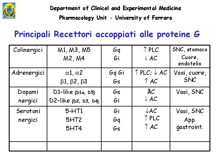
Department of Clinical and Experimental Medicine Pharmacology Unit - University of Ferrara Principali Recettori accoppiati alle proteine G Colinergici M 1, M 3, M 5 M 2, M 4 Gq Gi PLC AC SNC, stomaco Cuore, endotelio Adrenergici 1, 2, 3 Gq Gi Gs PLC; AC Vasi, cuore, SNC Dopami nergici D 1–like (D 1 a, D 5) D 2–like (D 2, D 3, D 4) Gs Gi C A AC Vasi, SNC Serotoni nergici 5 -HT 1 5 HT 2 5 HT 4 Gi Gq Gs AC PLC AC Vasi, SNC App. gastroint.
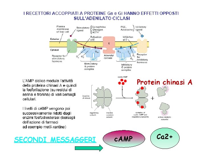
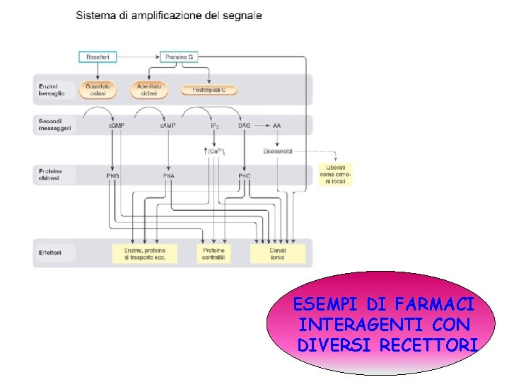
Protein chinasi A SECONDI MESSAGGERI c. AMP
ESEMPI DI FARMACI INTERAGENTI CON DIVERSI RECETTORI
IL CASO DELLA CAFFEINA Caffeine is widely consumed in coffee, tea, soft drinks, chocolate and medicines (200 -300 mg/daily) Caffeine elicits a number of pharmacological effects primarily through antagonism of the effects mediated by ADENOSINE RECEPTOR subtypes Caffeine intake could be associated with a significantly lower risk for Alzheimer’s and Parkinson’s diseases Pentoxifylline suppresses TNF- release in heart failure patients
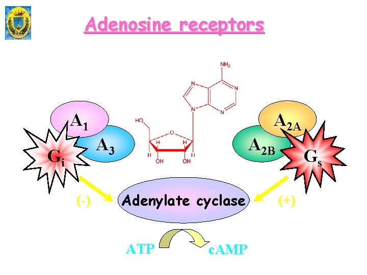
Adenosine receptors A 2 A A 1 A 2 B A 3 Gi (-) Adenylate cyclase ATP c. AMP Gs (+)
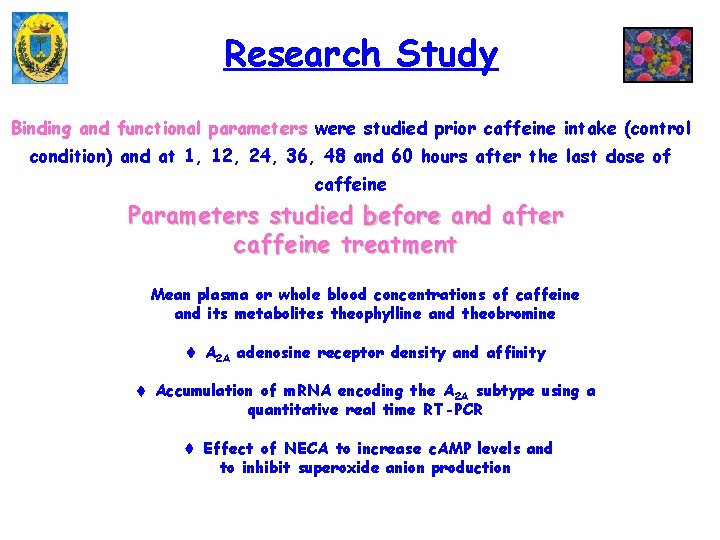
Research Study Binding and functional parameters were studied prior caffeine intake (control condition) and at 1, 12, 24, 36, 48 and 60 hours after the last dose of caffeine Parameters studied before and after caffeine treatment Mean plasma or whole blood concentrations of caffeine and its metabolites theophylline and theobromine A 2 A adenosine receptor density and affinity Accumulation of m. RNA encoding the A 2 A subtype using a quantitative real time RT-PCR Effect of NECA to increase c. AMP levels and to inhibit superoxide anion production
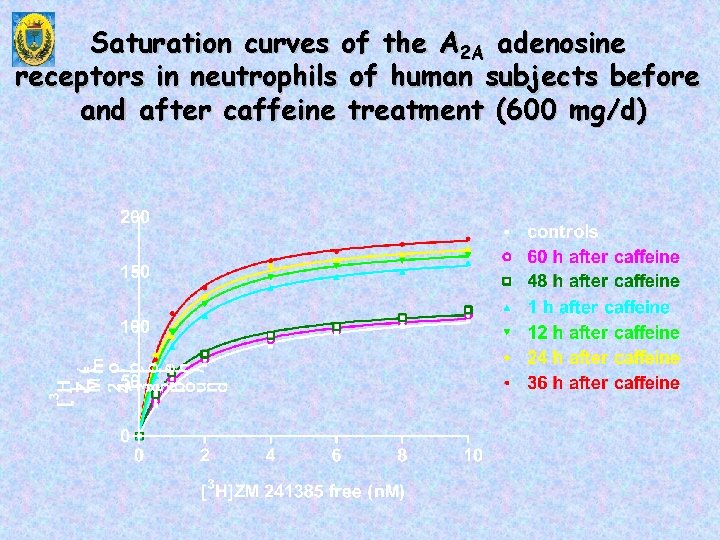
Saturation curves of the A 2 A adenosine receptors in neutrophils of human subjects before and after caffeine treatment (600 mg/d)
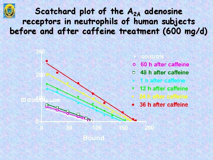
Scatchard plot of the A 2 A adenosine receptors in neutrophils of human subjects before and after caffeine treatment (600 mg/d)
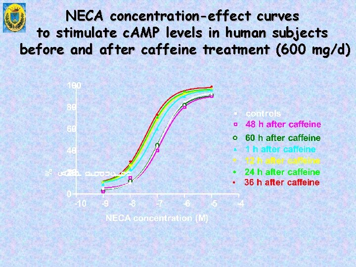
NECA concentration-effect curves to stimulate c. AMP levels in human subjects before and after caffeine treatment (600 mg/d)
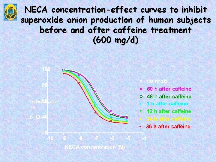
NECA concentration-effect curves to inhibit superoxide anion production of human subjects before and after caffeine treatment (600 mg/d)
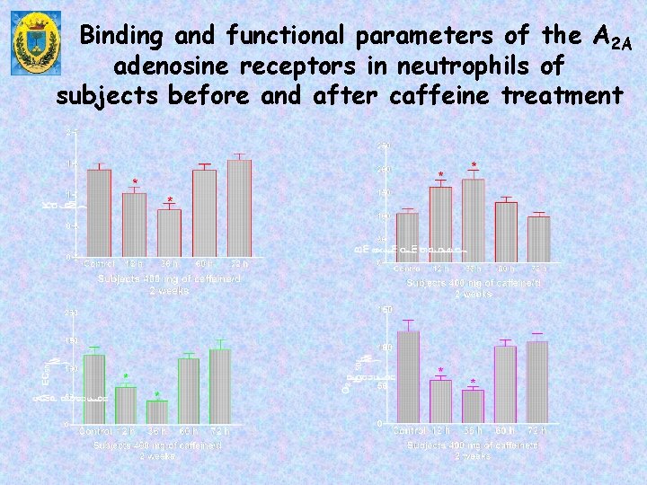
Binding and functional parameters of the A 2 A adenosine receptors in neutrophils of subjects before and after caffeine treatment
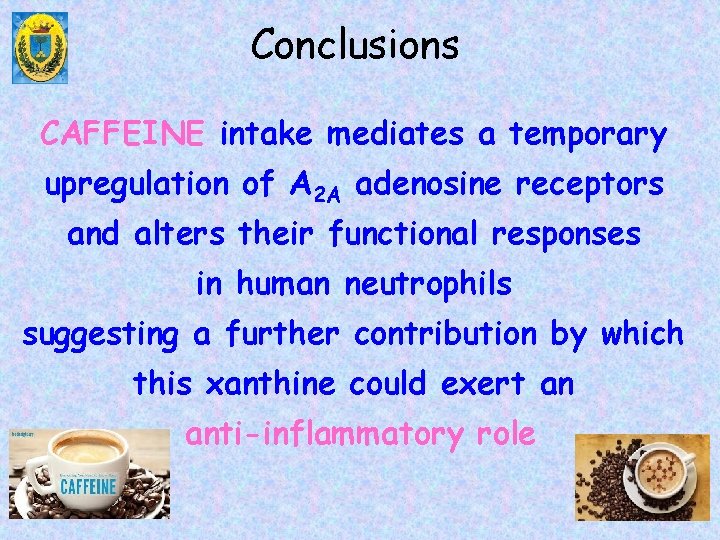
Conclusions CAFFEINE intake mediates a temporary upregulation of A 2 A adenosine receptors and alters their functional responses in human neutrophils suggesting a further contribution by which this xanthine could exert an anti-inflammatory role
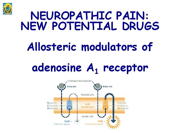
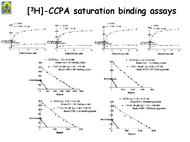
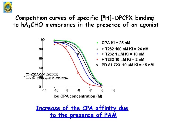
NEUROPATHIC PAIN: NEW POTENTIAL DRUGS Allosteric modulators of adenosine A 1 receptor
[3 H]-CCPA saturation binding assays
Competition curves of specific [3 H]-DPCPX binding to h. A 1 CHO membranes in the presence of an agonist Increase of the CPA affinity due to the presence of PAM
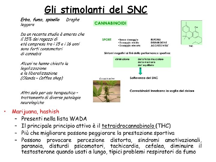
Gli stimolanti del SNC
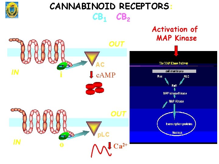
CANNABINOID RECEPTORS: CB 1 CB 2 OUT IN AC G bg i c. AMP OUT IN p. LC G bgo Ca 2+ Activation of MAP Kinase