Dan Cholger Xue Fu and Wei Zheng Expression

- Slides: 1

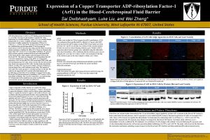
Dan Cholger, Xue Fu, and Wei Zheng* Expression of a Copper Transporter ADP-ribosylation Factor-1 (Arf 1) in the Blood-Cerebrospinal Fluid Barrier Sai Dwibhashyam, Luke Liu, and Wei Zheng* School of Health Sciences, Purdue University, West Lafayette IN 47907, United States Abstract ADP-ribosylation factor 1 (Arf 1) is a GTP binding protein that functions as a secondary messenger in cell signaling to maintain the Golgi architecture for vesicular trafficking. Copper (Cu) is an essential element indispensable for a variety of cellular enzymatic reactions, and its homeostasis is tightly regulated by Cu-transporting ATPases (Cu. ATPases), i. e. , ATP 7 A and ATP 7 B. Evidence from this lab and others has established that elevated intracellular Cu levels prompt the translocation of ATP 7 A from the trans-Golgi network (TGN) towards the plasma membrane to facilitate the efflux of Cu. Moreover, literature data suggest that Arf 1 is involved in Cu intracellular trafficking. The choroid plexus in brain ventricles constitute the blood and cerebrospinal fluid (CSF) barrier (BCB), which regulates material transport between the blood and CSF. This study was designed to test the hypothesis that Arf 1 is present in brain barrier systems. Using q. PCR, we compared the expression of Arf 1 in choroidal Z 310 cells, brain barrier RBE 4 cells, and neuronal dopaminergic N 27 cells. Using a western blot, the Arf 1 proteins were clearly seen expressed in Z 310 cells. Our data clearly showed that m. RNAs encoding Arf 1 were present in all three cell types. Furthermore, confocal images show Arf 1 localization in the Golgi. The function of Arf 1 under toxic metal exposures is currently in progress in this lab. The results by far suggest that Arf 1 is present in the brain barriers and possibly plays an important role in regulating Cu transport between the blood and CSF/brain parenchyma by modulating ATP 7 A and/or ATP 7 B intracellular trafficking. This research is important to our understanding of Cu homeostasis in the central nervous system as affected by environmental exposure and nutritional malfunction. Introduction Copper is important to bodily function. It is required for energy production through its presence in cytochrome c. Copper is also present in superoxide dismutase, which is used for defense against antioxidants. Failure to regulate the amount of copper in cells results in diseases such as Menkes and other psychological disorders such as parkinsonism. This lab has shown that the presence of excess Cu or Mn in the choroidal epithelial cells led to ATP 7 A relocating toward the apical microvilli facing the CSF, but ATP 7 B toward the basolateral membrane facing the blood. However, the existence of Arf 1 in the brain barriers, i. e. , the BCB and blood-brain barrier (BBB), has never been reported; even less is known about the role of Arf 1 in the translocation of Cu-ATPases, albeit the known participation of Arf 1 in subcellular trafficking of Cu. ATPases. The BCB is a unique area for this research for a few reasons. First, The choroid plexus acts as the barrier between the blood and the cerebral spinal fluid (CSF). The choroid plexus secretes about 85 -90% of the cerebral spinal fluid to the brain and regulates trace element’s homeostasis in the brain’s extracellular fluids. Noticeably, copper accumulates extraordinarily high in the sub-brain ventricular zone (SVZ) , which is adjacent to the choroid plexus. The presence of Arf 1 is widely recognized as the key difference maker in copper regulation in cells. Because Arf 1 is extremely important for the individual cell, determining its presence in the choroid plexus would be useful for the scientific community. Contact Information liu 2543@purdue. edu sdwibhas@purdue. edu wzheng@purdue. edu Results Methods q. PCR Lysates were isolated in TRIzol reagent for m. RNA purification. c. DNA was synthesized from purified m. RNA, diluted to 10 ng/µl with DEPC water and used for q. PCR analysis using SYBR Green master mix from Bio-Rad. N 27 and PC 12 cell data was collected with a similar method. B-actin was used as a house-keeping reference gene. Relative m. RNA expression ratios between groups were calculated using the delta-delta cycle time formulation. After confirming that the reference gene was not changed, the cycle time (Ct) values of Arf 1 were normalized with that of the reference gene in the sample to obtain ∆∆Ct values. Figure 2. Co-localization of Arf 1 with Golgi Apparatus in Z 310 Cells and Lead Toxicity Merge Golgi Arf 1 Nucleus Golgi + Arf 1 0 μM Western Blot Proteins were extracted using radioimmunoprecipitation assay buffer, run on a 12% SDS-PAGE gel, and blotted on a polyvinylidene difluoride membrane Confocal Microscopy: Cells were grown on a glass slide using poly-lysine and fixed using 4% paraformaldehyde in PBS p. H 7. 4. They were then treated with antibodies specific for Arf 1. 10 μM Pb Typical confocal microscopic images of Arf 1 with markers of Golgi and nucleus in Z 310 cells. Cells were treated with 10 m. M Pb for 24 hours. Arf 1 signals are overlapped with those of Golgi apparatus. A decreased Arf 1 was evident in Pb-treated cells. Results Figure 3. Expression of Arf 1 in Z 310 Cells by Western Blot and Lead Toxicity Figure 1. Expression of Arf 1 in Z 310, N 27 and PC 12 Cells Control 10 μM 20 μM 50 μM 100 μM Arf 1 α=0. 05 β-Actin Typical Western blot images of Arf 1 and housekeeping protein β-Actin in Z 310 cells. Cells were treated with Pb at various concentrations for 24 hours. Pb exposure inhibited the protein expression of Arft 1 in Z 310 cells. Conclusions and Future Directions Expression of Arft 1 was quantified by q. PCR. Z 310: choroidal epithelial cells. N 27: rat brain-derived dopaminergic neural cells. PC 12: rat adrenal gland phaeochromocytoma-derived nerve cells. The BCB cells express the highest amount of Arf 1 than neuronal cell typies. Data represent mean + SD, n=4. *: p<0. 05. Data from our current studies demonstrate that (1) an intracellular copper regulatory protein Arf 1 is present in the blood-CSF barrier in brain ventricular choroid plexuses, and (2) exposure to toxic metal lead (Pb) causes a decreased expression of Arft 1 in choroidal Z 310 cells. Since Arf 1 is known to regulate the intracellular Cu homeostasis by interacting with Cu-ATPases (i. e. , ATP 7 A and ATP 7 B), altered Arf 1 expression by Pb exposure may disrupt Cu transport by the blood-CSF barrier. Our future study will focus on (1) intracellular Cu levels as affected by Pb treatment; (2) correlation of Arf 1 changes with cellular Cu levels following Pb exposure; and (3) the relationship between the Arf 1 protein and intracellular trafficking of ATP 7 A and ATP 7 B. The study will reveal, for the first time, the molecular mechanism underling copper transport at the blood and CSF interface and help understand Pb-induced neurotoxicity. References: Southon A, Greenough M, Hung YH, Norgate M, Burke R, and Camakaris J (2010). The ADPribosylation factor 1 (Arf 1) is involved in regulating copper uptake. Int J Biochem Cell Biol. 43(1): 146153. Previous research done at Zheng Labs

