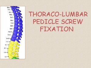
Anterior And Posterior Spinal Arthrodesis with Pedicle Screw


- Slides: 10

Anterior And Posterior Spinal Arthrodesis with Pedicle Screw Instrumentation for Thoracolumbar Kyphosis in Children with Mucopolysaccaridosis Garrido E*, Adams CI * * From the Scottish National Spine Deformity Service Royal Hospital for Sick Children Edinburgh (UK) 5 th International Congress on Early Onset Scoliosis and Growing Spine (ICEOS) Orlando, USA, November 18 -19, 2011

Disclosure • No financial relationships to disclose

Background q Characteristic spinal deformities occur in patients with MPS: Odontoid hypoplasia and/or progressive thoracolumbar kyphosis is frequently present. q Progression of the thoracolumbar kyphosis can lead to spinal cord compression, claudication symptoms and poor trunk balance. q Due to the rarity of Mucopolysaccharidosis relatively little has been published on the treatment of the thoracolumbar gibbus in the last 10 years. q To the current authors knowledge there are no reports of successful instrumentation of the thoracolumbar spine with pedicle screws in children with Mucopolysaccharidosis. Dalvie SS, Noordeen MH, Vellodi A. Anterior instrumented fusion for thoracolumbar kyphosis in mucopolysaccharidosis. Spine 2001 Dec 1; 26(23): E 539 -41. Tandon V, Williamson JB, Cowie RA, Wraith JE. Spinal problems in mucopolysaccaridosis 1 J Bone Joint Surg Br. 1996 Nov; 78(6): 938 -44.

Subjects and Methods q Retrospectively review of three consecutive patients with MPS who underwent single staged anterior and posterior arthrodesis of the thoracolumbar spine with pedicle screw instrumentation q Two had a diagnosis of MPS type I (after Hematopoietic Cell Transplantation) and one MPS type IV (Morquio) q Indications for arthrodesis was progressive thoracolumbar kyphosis > 60º q All patients were ambulant at the time of surgery. q Average at the time of surgery was 37 months (range 30 to 45 months). q Patients were reviewed post-operatively at 3, 6 and 12 months

Radiological Assessment q All patients had standing antero-posterior and lateral radiographs of the whole spine taken at first presentation, pre- and postoperatively and at latest follow-up q The dysplastic vertebrae were located at T 12 and L 1 q Anterior translation of the vertebral body on the dysplastic vertebra in the sagittal plane was expressed as a percentage slip. q The thoracolumbar kyphosis was measured using the cobb method. q Pedicle and spinal canal parameters of the lower thoracic and lumbar vertebrae were measured using CT and MRI Krag MH, Weaver DL, Beynnon BD, Haugh LD. Morphometry of the thoracic and lumbar spine related to transpedicular screw placement for surgical spinal fixation. Spine 1988 Jan; 13(1): 27 -32. Zhang H, Sucato DJ, Nurenberg P, Mc. Clung Morphometric Analysis of Vertebral Growth Using Magnetic Resonance Imaging in the Normal Skeletally Immature Spine (Phila Pa 1976). 2010 Jan 1; 35(1): 76 -82.

Surgical Technique q Intraoperative spinal cord monitoring with multimodal lower SSEP’s was performed q All levels forming part of the thoracolumbar kyphosis on the standing radiograph were included in the fusion. q A thoracolumbar approach was performed through the tenth rib. q A 5 level discectomy. Rib strut grafts are wedged into the disc spaces q All segments of the kyphosis were instrumented posteriorly. q The entry point of the pedicle was marked with needles at the usual landmarks. q Under radiographic control using anteroposterior and lateral projections the optimal orientation of the pedicle screw was selected as to maximise the trajectory of the screw in bone q The pedicle entry point was than opened with a sharp awl and a 2 mm cervicle pedicle finder advanced under radiographic control into the vertebral body. q A spinal jacket was applied for 3 months postoperatively.

Morphometric analysis of thoracolumbar vertebrae Spinal Level Average Pedicle width (mm) Average Pedicle length (mm) Average AP length (mm) Average Interpedicular distance (mm) Average. AP canal diameter (mm) Average Vertebral body width (mm) T 10 4. 375 14. 85 26. 95 13. 55 14 22. 4 T 11 4. 6 13. 975 27. 925 13. 6 15. 15 22. 95 T 12 3. 475 13. 8 26. 3 15. 6 15. 5 24. 35 L 1 4. 125 13. 7 24. 2 16. 9 15. 1 25. 4 L 2 4. 475 15. 55 29. 325 17. 8 14. 25 27 L 3 4. 6 17. 1 26. 95 17. 4 15. 9 27. 1

Results Patient Preoperative Kyphosis (°) Vertebral subluxation preoperatively (%) Postoperative Kyphosis (°) Vertebral subluxation postoperatively (%) 1 64 80 8 20 2 67 60 -10 15 3 70 50 10 15 Average 67 63 3 17

Complications q. No neurological problems occurred. q. One patient required 6 days of Bi. PAP (bilevel positive airway pressure) ventilation due to a left lower lobe collapse. q. No instrumentation failure or junctional problems occured

Conclusion • Single staged anterior release and posterior segmental pedicle screw instrumentation effectively restores spinal alignment in young children with progressive thoracolumbar kyphosis associated with mucopolysaccharidosis.


