General Stool Examination GSE Tishk International University Medical

 specimens (which are more likely to contain")


 → 1. 2376 ♦ Taenia")
 2 or 5 grams of feces. 2.")


- Slides: 10
General Stool Examination GSE Tishk International University Medical Analysis Dep. by: Dr. Hemdad H. Mawlood
Examination of fresh specimens v. Liquid (diarrheic) specimens (which are more likely to contain trophozoites) should be examined within 30 minutes of passage (not within 30 minutes of arrival in the laboratory!) v. Semi liquid or semi solid specimens (which may contain both trophozoites and cysts) should be examined within one hour of passage. v Formed specimens (less likely to contain trophozoites) can be kept for up to one day, with overnight refrigeration if needed, prior to examination.
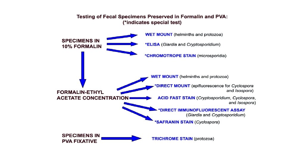
Specimens preserved in PVA are mostly used for keeping cyst , trophozoite and permanent staining with trichrome. Prior to staining, they are processed as follows: 1. Insure that the specimen is well mixed. 2. Prepare a smear using 2 to 3 drops of the specimen then fixed by heating on slide warmer set at 60°C for 5 minutes or air dry completely at room temperature. 3. Keep or Examine it under microscope
Concentration procedure Separate parasites from fecal debris and increase the chances of detecting parasitic organisms when these are in small numbers. They are divided into flotation techniques and sedimentation techniques. Flotation techniques vmost frequently used: (zinc sulfate or sugar solution ) use solutions which have higher specific gravity than the organisms to be floated so that the organisms rise to the top and the debris sinks to the bottom.
v. The disadvantages of most flotation techniques are that the walls of eggs and cysts will often collapse, thus hindering identification. Also, some parasite eggs do not float. ♦ Flotation solution must have a higher specific gravity than parasite egg or oocysts. ♦ Specific gravity of water is 1. 000 and most parasite eggs are 1. 05 1. 24 ♦ Flotation solutions should be > 1. 24 Specific Gravity of Common Parasite
Sp. gr of Parasite Eggs: ♦Physaloptera sp. (stomach worm) → 1. 2376 ♦ Taenia (tapeworm) → 1. 2251 ♦ Trichuris vulpis (whipworm) → 1. 1453 ♦ Toxocara cati (roundworm) → 1. 1005 ♦ Toxocara canis (roundworm) → 1. 0900 ♦ Ancylostoma sp. (hookworm) → 1. 0559
Flotation Technique procedure 1. Weigh out (estimate) 2 or 5 grams of feces. 2. Mix with 10 ml of sugar solution=(sucrose (C 12 H 22 O 11) monosaccharides glucose and fructose)+water. (43 gm sugar +224 ml of distal water =16% of sugar solution 3. Pour through tea strainer into a beaker/fecal cup. 4. Pour solution from beaker/fecal cup into 15 ml centrifuge tube. 5. Fill tube with sugar solution to the top of the tube. DO NOT place a lid on the tube. 7. Centrifuge at 1200 rpm for 5 minutes. Make sure the centrifuge is balanced. 8. Remove the test tube from the centrifuge and fill to the top with sugar solution. 9. Place a lid on the tube and centrifuge again for 10 minutes. 10. Remove lid from tube and place on slide labeled with the name or number. 12. Examine entire coverslip at 10 X. Use 40 X to identify parasites or eggs. 13. Record results.
Sedimentation techniques use solutions of lower specific gravity than the parasitic organisms, thus concentrating the latter in the sediment. Sedimentation techniques are recommended for general diagnostic laboratories because they are easier to perform and less prone to technical errors. The sedimentation technique used at CDC is the formalin-ethyl acetate technique.
Formalin-Ethyl Acetate Sedimentation Concentration 1. Mix the specimen well. 2. Strain 5 ml of the fecal suspension (more or less depending on its consistency) through wetted cheeseclothtype gauze placed over a disposable paper funnel into a 15 ml conical centrifuge tube. (Conical paper cups with the tips cut off are sufficient). 3. Add 0. 85% saline or 10% formalin through the debris on the gauze to bring the volume in the centrifuge tube to 15 ml. Distilled water may be used; however, Blastocystis hominis may be deformed or destroyed. 4. Centrifuge at 500 × g for 10 minutes. 5. Decant supernatant. Add 10 ml of 10% formalin to the sediment and mix thoroughly with wooden applicator sticks. 6. Add 4 ml of ethyl acetate, stopper the tube, and shake vigorously in an inverted position for 30 seconds. Carefully remove the stopper. 7. Centrifuge at 500 × g for 10 minutes. 8. Free the plug of debris from the top of the tube by ringing the sides with an applicator stick. Decant the top layers of supernatant. 9. Use a cotton-tipped applicator to remove debris from sides of the centrifuge tube. 10. Add several drops of 10% formalin to resuspend the concentrated specimen. Proceed with applicable testing.