COMPARISON OF AN INTRAORAL SCANNER AND A EXTRAORAL

- Slides: 1

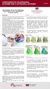
COMPARISON OF AN INTRAORAL SCANNER AND A EXTRAORAL SCANNER ID 23032 Aira Kalniņa, Prof. Una Soboļeva Rīga Stradiņš university, Department of Prosthetic Dentistry Intoduction Results “Impression” is defined as “the reverse, negative copy, or copy of an object's surface; dentist's footprint of teeth and adjacent tissues”. For good fixed dental prosthesis one of most important steps is precise impression. In history gold standard is polyvinyl siloxane, witch is used more then 50 th years. Using the intraoral digital impression, the first prosthesis was made in 1985, and since then there has been tremendous development, especially in recent years. As a digital impression, it is mentioned that there is no need to make a plaster casts, which in turn improves and facilitates the storage of information - there is no need for additional rooms to store plaster models. Digital impression with intraoral scanners are becoming more widespread, existing studies have conflicting results and are often funded by manufacturers. • In the precision analysis, intraoral scans vary from 89. 43 μm (SN 53. 95 μm) from the extraoral scanner. • Significant differences were found in the incisal edge and the prepared margin, but no differences were found between the various comparisons (p = 0. 298). Repeatability showed statistically significant differences: extraoral scans differed by 13. 06 μm (SN=14. 89 μm), intraoral scan - 26. 33 μm (SN=17. 9 μm). There was a statistically significant difference between the subject (p <0. 001). • For all ten digital impressions average time was 130, 5 seconds (min=118; max=152; SN=13, 18). Approximately two minutes. Aim of the project Aim of study was to, in an objective and independent study, determine the accuracy and repeatability of an intraoral scanner, compare it to a an extraoral scanner for obtaining a virtual model which would be used for the manufacturing of fixed dentures. Hypothesis - an intraoral scanner model is as accurate as a model obtained with a conventional polyvinyl siloxane impression and an extraoral scanner. Materials and methods In this study, three teeth dd 13, 12, 21 was prepared according to biomechanical principles. For tooth margins better visibility, in sulcus was placed retraction cord “ 00” Sure Cord Plus (Suredent). Two types of full arch impression were made: o a scan in mouth using an intraoral scanner - 3 Shape Trios, o a polyvinyl siloxane impression (Heraeus Kulzer – Variotime - Easy Putty and Light Flow) and prepared in stone model, scanned with an extraoral scanner (3 Shape 370). Both footprints were saved in the STL file format and the differences were read by the Ortho Analyzer ™ Software program. Data analysis using the ANOVA test was used for the precision analysis. Also working time was checked for each digital impression. Conclusions • The repeatability of both intraoral and extraoral scanners is clinically acceptable, although, the extraoral scanner is slightly more precise. • Another experimental design study should be carried out to determine the accuracy of the various constructions which were made so that it could be decided which of the methods provides a more precise clinical outcome. • With intraoral scanner total working time is shorter then with conventional impression technique. • Clinical recommendation – for both impression techniques prepared tooth should be smooth and without sharp edges, especially should be checked incisal edge and prepared margin.

