ZOONOSES Zoonoses animal diseases which can be transmited

ZOONOSES

Zoonoses • animal diseases, which can be transmited to humans • source of infection is an animal, ethiological agent is bacterium, virus or parasite

Zoonoses Transmission is possible : directly – by biting , with saliva indirectly • ingestion- contamination of hands or food • inhalation of contaminated dust or spray • inoculation by vector ( insect – mosquito, tick, flea) transplacental- the only possible way of transmission from people to people At present we know about 250 zoonoses, 50 of them are common

The most important zoonoses Bacterial ethiology • tularemia • salmonelosis • campylobacteriosis • listeriosis • leptospirosis • bartonelosis (CSD) • borreliosis • brucellosis • anthrax • plague • yersiniosis • ehrlichiosis • Q fever Parasitic ethiology • toxoplasmosis • toxocarosis • leishmaniosis • trichinelosis • echinococcosis Viral ethiology • rabies • tick-born encephalitis Prion ethiology • Creutzfeld- Jakob disease

• • • toxoplasmosis cat scratch disease campylobacteriosis toxocariasis lyssa • • toxocariasis yersiniosis lyssa campylobacteriosis

• leptospirosis • tularemia • lymfocytic choriomeningititis • Hantavirus infection • • ornitosis campylobacteriosis salmonelosis avian flu

TULAREMIA


Tularemia • Etiology agent: Francisella tularensis- small gramnegative coccobacilli, cultivation on common culture medium is difficult • Source of infection: from small rodents to hares and rabits.

Tularemia Epidemiology: widely distributed , esspecially in Northen Hemisphere Transmission is possible via : • cuts on hands during skinning and cutting up the ill animals • aspiration of contaminated dust or aerosol, • ingestion of contaminated fruit or vegetable or undercooked meat • a tick bite IP averages 5 -7 days, ranges from 1 to 21 days. Infectious dose is small, 10 -50 organisms

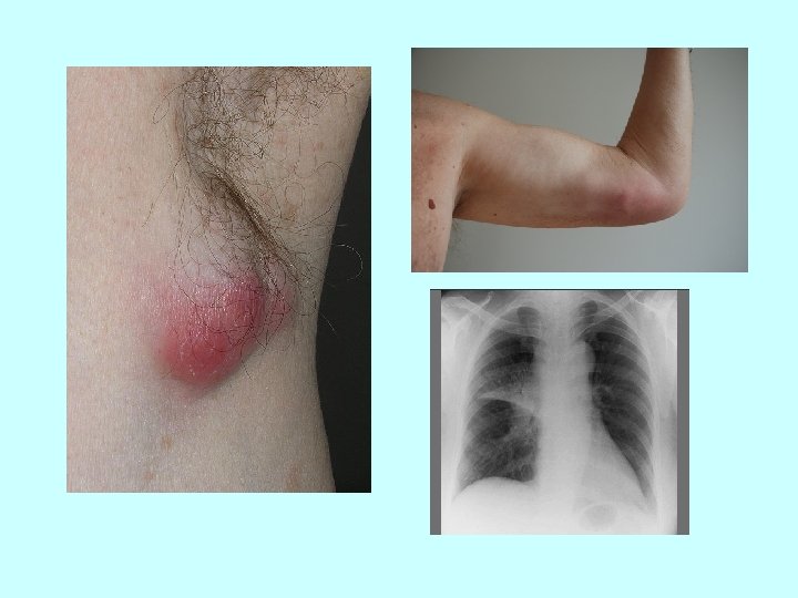
Tularemia Pathogenesis • bacteria spread from the site of entry to the regional lymph nodes • lymph nodes are enlarged and tender, granulomatous inflammation of LN with suppuration, necrosis and colliquation are typical of tularemia
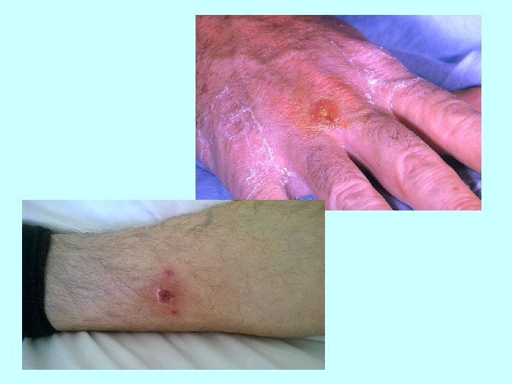

Clinical manifestations: • ulceroglandular tularemia- skin lesion + lymphadenopathy • glandular tularemia – skin lesion (ulcer) is not present • oculoglandular tularemia- the side of entry is conjunctiva, preauricular lymphadenopathy • oralglandular tularemia - exudative unilateral pharyngitis or tonsillitis, cervical lymphadenopathy • typhoidal ( abdominal) tularemia – no prominent lymphadenopathy, nonspecific symptoms - fever with chills, a headache, muscle pain, nausea, vomiting, diarrhea, abdominal pain • pneumonic tularemia – fever, cough with minimal sputum production, pleuritic chest pain Skin rash may be found in up to 35 % of cases.


Tularemia Diagnosis • serologic diagnosis- Ig. M and Ig. G antibodies are positive from the end of the 2 week. • direct cultivation is difficult • ultrasonography of enlarged lymph nodes - typical picture of colliquation Therapy -the most widely used tetracycline + gentamicin, in children under 8 years of age macrolid along with gentamicin, alternative: fluorochinolons -exstirpation of whole suppurative lymph node

TOXOPLASMOSIS

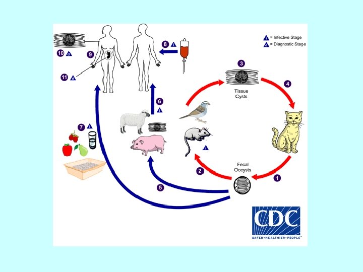

Toxoplasmosis • Etiologic agent: protozoan parasite Toxoplasma gondii – oocyst – bradyzoit – tachyzoit

TOXOPLASMOSIS • Source of infection: members of cat family are the main reservoir • oocyst→ GIT → tachyzoites → blood → neural and muscle tissue → tissue cyst bradyzoites • If a pregnant woman becomes infected , tachyzoites can infect the fetus via the bloodstream

Toxoplasmosis Epidemiology Cats get infected by carnivorism and shed oocyst in their faeces. Human infection may be acquired : • ingestion of undercooked infected meat with bradyzoites • ingestion of the oocyst from fecally contaminated hands or food • transplacental transmission (tachyzoits) • organ transplantation or blood transfusion (tachyzoits, bradyzoits)

TOXOPLASMOSIS Clinical manifestations: 1. Congenital toxoplasmosis 2. Acquired postnatal toxoplasmosis

1. Congenital toxoplasmosis • • • only primary infection in a pregnant woman may result in transplacental transmission most infections in the third trimester only cause seropositivity in the neonate infection in the first trimester may be very consequential The clinical signs of congenital toxoplasmosis : • intrauterine death or abortion • cerebral calcification , hydrocephalus, chorioretinitis ( Sabin trias) • myocarditis, microphtalmus, hepatosplenomegaly, strabism, deafness, blindness • psychomotoric retardation

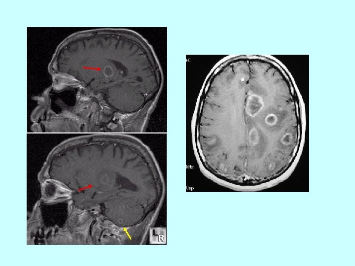
2. Acquired postnatal toxoplasmosis • inapparent or abortive – only nonspecific mild symptoms: fever, tiredness, musclepain • lymphadenopathy • chorioretinitis • brain toxoplasmosis – in immunocompromised hosts , mostly AIDS patients , result from reactivation of latent infection ( bradyzoite cyst). Multiple cerebral lesions on CT

TOXOPLASMOSIS Diagnosis - serologic diagnosis • Ig. G - culminate 6 months after infection, high titers persist months , low titers life- long • Ig. M – disappear within 9 months • Ig. A - dissapear within 6 months - PCR diagnosis is possible

TOXOPLASMOSIS Therapy Toxoplasmosis is a self limiting disease in most cases Treatment is necessary only in : • pregnant women • infected neonates • immunosupressed persons Drugs: sulphonamide + pyrimetamine ( folate inhibitor, its side effect is decreasing of leucocytes in blood- supplementation of folinic acid and monitoring of blood count is necessary) • Spiramycin in pregnancy.

TOXOCARIASIS


TOXOCARIASIS Ethiologic agent : Toxocara canis, Toxocara cati Source : dogs and cats Epidemiology: dogs and cats excrete eggs in their faeces. The larvae invade from small intestine into multiple tissues (the liver, the heart, lungs, the brain, muscles) Human acquire the infection by close contact or by ingestion of contaminated food or soil.

Toxocariasis Clinical features: Many human infections are asymptomatic, with only eosinophilia and positive serology. The two main clinical presentations of toxocariasis are 1) visceral larva migrans (VLM) 2) ocular larva migrans (OLM)
 visceral larva migrans (VLM) which occurs mostly in preschool children,")
TOXOCARIASIS- clinical features a) visceral larva migrans (VLM) which occurs mostly in preschool children, and cause various symptoms including fever, anorexia, weight loss, cough, wheezing, rash, hepatosplenomegaly, and hypereosinophilia b) ocular larva migrans (OLM). The larvae produce various ophthalmologic lesions, intraocular granuloma. In past it could be misdiagnosed as retinoblastoma, resulting in surgical enucleation.
 Treatment: antiparasitic drugs, (mebendazole), usually in combination with antiinflammatory medication")
TOXOCARIASIS Diagnosis: serologic (ELISA) Treatment: antiparasitic drugs, (mebendazole), usually in combination with antiinflammatory medication

LISTERIOSIS


LISTERIOSIS Ethiologic agent: bacterium Listeria monocytogenes Source : widespread in animals, soil, water. Epidemiology: A human may get infected : • ingestion of contaminated food -dairy products (cheeses) or sausages • bathing in contaminated water • contact with ill animal • transplacental transmission

• Intracelular pathogen • GIT→ bacteraemia → CNS, placenta High risk of infection: patients with suppression of cell-mediated immunity

CLINICAL FEATURES OF LISTERIOSIS: 1. Acquired listeriosis : • inapparent or abortive form • submandibular lymphadenopathy • sepsis • purulent meningitis Immunocompromised persons are in a high risk of invasive listeriosis. • newborn may acquire infection intrapartum- severe meningitis or sepsis

Risk factors of listerial infection lymfoma AIDS pregnancy biologic treatment newborns seniors chronic metabolic diseases

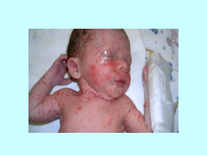
CLINICAL FEATURES OF LISTERIOSIS: 2. Congenital listeriosis : - result of transplacental infection during maternal bacteriaemia, maternal illness is often mild and unrecognized - in early pregnancy it mostly results in fetal death - affected infants may be born prematurely, with sepsis or severe disease granulomatosis infantiseptica granulomatous infiltration of parenchymal organs, papular rash

LISTERIOSIS Diagnosis • culture of blood, CSF, sputum, amniotic fluid • serology Therapy: ampicilin, in serious cases in combination with gentamicin Cotrimoxazol Vancomycin, meropenem Resistance to cephalosporines!!

LEPTOSPIROSIS
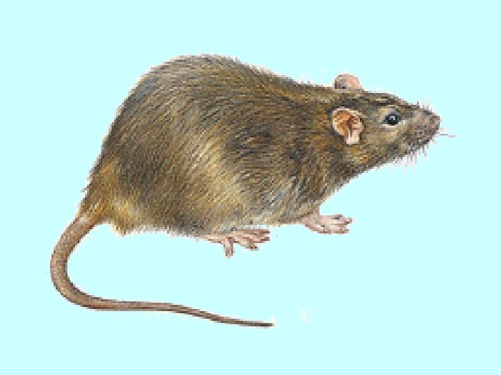

LEPTOSPIROSIS Ethiologic agent: Leptospira grippotyphosa, Leptospira icterohaemorrhagiae, Leptospira sejroe Source: rats and cattle.

LEPTOSPIROSIS Epidemiology: • trasmission via animal urine or contaminated water and food • the site of entrance is damaged skin, conjunctiva or mucous • risk- group persons are farmers, vets, sewer workers, or people bathing in contaminated water or fishing • incidence is increasing after floods. IP 1 -3 weeks

Clinical manifestations of leptospirosis : • Infected people may be asymptomatic or experience of mild course • disease has mostly 2 periods: 1. leptospiremia - flu like syndrome ( fever, musclepain, a headache)

LEPTOSPIROSIS 2. damage to the organs – the liver, kidneys, the heart, an immunopathologic reaction • • icteric form – the most serious, Weil´s disease. Often complicated by renal failure, hepatitis, aseptic meningitis, DIC, severe trombocytopenia , SPHS (Severe Pulmonary Haemorrhagic Syndrom) anicteric form – aseptic meningitis, damage to the liver and kidneys is mild Complications: myocarditis, iridocyclitis

LEPTOSPIROSIS Diagnosis: serology, specific antibodies appear in the second week of disease PCR Therapy: antibiotics ( PNC, AMPI )

Zoonoses – dif. dg Fever + lymphadenopathy regional - tularemia - bartonellosis - yersiniosis generalized - toxoplasmosis - listeriosis - brucelosis - leishmaniosis - Lyme disease (1. stage) Fever + meningeal symptoms listeriosis TBE Lyme disease (2. stage) leptospirosis ehrlichiosis lymfocytic choriomeningitis

Zoonoses – dif. dg Fever + gastrointestinal symptoms • • • salmonellosis campylobacteriosis tularemia (abdominal form) yersiniosis bird flu Fever + respiratory symptoms • • • tularemia (pulmonary form) legionellosis ornitosis Q - fever bird flu

• Thank you for your attention.
- Slides: 54