ZOO 405 Week 4 ZOO 405 by Rania

ZOO 405, Week 4 ZOO 405 by Rania Baleela is licensed under a Creative Commons Attribution-Non. Commercial-Share. Alike 3. 0 Unported License
 • Genetic diseases • Diagnosis")
This week • Viruses (cont. ) • Genetic diseases • Diagnosis

• > 30 cellular oncogenes were discovered: prefixed with a ‘c’ (e. g. c-myc, c-erb, c-fos) • oncogenes carried by viruses are prefixed with a ‘v’ (e. g. v-myc and v-ras)
 1964")
virus Year method of discovery virus classification malignancy other disease Epstein–Barr virus (EBV) 1964 cell culture and electronmicroscopy Herpesviridae ds. DNA 7 types 3 hepatitis B virus (HBV) 1965 serologic screening Hepadnaviridae ds. DNA-RT hepatocellular hepatitis cirrhosis human Tlymphotropic virus 1 (HTLV-1) 1980 cell culture, RT assay and electromicroscopy Retroviridae ss. RNA-RT, positive strand adult T-cell leukaemia (ATL) 2 human genital papillomavirus (HPV) 1983 DNA hybridization Papillomaviridae ds. DNA cervical carcinoma squamous cell head and neck ca. squamous cell anal cancer vulvar cancer hepatitis C virus (HCV) 1989 c. DNA library screening Flaviviridae ss. RNA-RT, positive strand hepatocellular carcinoma rare lymphomas? Kaposi sarcoma herpesvirus (KSHV/HHV 8) 1994 representational difference analysis (RDA) Herpesviridae ds. DNA 3 Merkel cell polyomavirus (MCV) 2008 digital transcriptome subtraction (DTS) Polyomaviridae ds. DNA Merkel cell carcinoma hepatitis cirrhosis

2. Polyoma Viruses ds. DNA, ~5 kb, circular genome, 40 -50 nm in diameter, naked icosahedral, normally cause lytic infection BUT transform when they are incomplete • Simian virus 40 (SV 40) - juvenile hamster sarcomas, transformation • Polyoma - mouse leukemia, in vitro transformation • Human polyomas - infects the respiratory system, kidneys, or brain- basically not cancerous • Merkel cell polyomavirus (MCV)=> Merkel cell carcinoma (rare skin cancer) SV 40 has been found in bone and a rare lung cancer patients

Malignant transformation occurs only if SV 40 becomes integrated into the host genome. This rare event assures that the SV 40 genome is replicated during cell division, and the progeny cells maintain the malignant phenotype (top panel of accompanying figure). Gazdar et al. , 2002, SV 40 and human tumours: myth, association or causality? Nature Reviews Cancer 2, 957 -964

3. Adenoviruses (ds. DNA, linear genome, 36 to 38 kb, 90– 100 nm in size, naked icosahedral) Adenovirus can be involved in RETINOBLASTOMA and may be in some other rare cancers

• Highly oncogenic in animals • 47 distinct serotypes and 93 varieties • In human they infect the ocular, respiratory or GI epithelium • Adenoviral infection is highly complex.
) induce tumours with high")
Oncogenesis • Subgroup A (e. g. adenovirus 12 (Ad 12)) induce tumours with high frequency and short latency, • Subgroup B (such as Ad 3 and Ad 7) are weakly oncogenic. • Subgroup C (e. g. Ad 2 & Ad 5), D, E and F are nononcogenic. • All human adenoviruses studied can transform rodent cells in culture, however, only cells transformed by viruses of subgroup A and B are oncogenic in newborn rodents.
.")
Oncogenes Schematic diagram of the linear adenovirus genome (E=Early genes, L= Late genes).
 Linear ds. DNA, encode 100 -200 genes, icosahedral, enveloped")
4. Herpes viruses (Epstein-Barr Virus) Linear ds. DNA, encode 100 -200 genes, icosahedral, enveloped • Considerable evidence for role in human cancer. • Some very tumorigenic in animals • Viral DNA found in small proportion of tumor cells: “hit and run” • Most people have antibodies against EBV

Herpes Viruses • Epstein-Barr Virus 1. Burkitt’s Lymphoma 2. Nasopharyngeal cancer 3. Infectious mononucleosis 4. Transforms human B-lymphocytes in vitro Herpes papilloma

EBV • Is associated with multiple types of human cancers • EBV infection is associated with undifferentiated nasopharyngeal carcinoma (NPC) • NPC is endemic in the southern Chinese population. • A strong association between NPC risk and the HLA locus at chromosome 6 p has been identified • EBV infection in NPC is clonal in origin, strongly suggesting that NPC develops from the expansion of a single EBV-infected cell. • Genomic study of NPC has identified multiple somatic mutations in the upstream negative regulators of NF-κB signalling. Philos Trans R Soc Lond B Biol Sci. 2017 Oct 19; 372(1732). pii: 20160270. doi: 10. 1098/rstb. 2016. 0270. Epstein-Barr virus infection and nasopharyngeal carcinoma.
 Causes Hepatitis B,")
5. Hepatitis B Virus • • • 8 known genotypes (A-H) Causes Hepatitis B, Can lead to liver cirrhosis Can lead to hepatocellular carcinoma Suspected to increase the risk of pancreatic cancer. • Vast public health problem • In Sudan: exposure to the virus from 47%– 78%, • Prevalence: in central Sudan= 6. 8%, in southern Sudan= 26% (Mudawi et al. , 2008) and in Nyala= 6. 25% (Abou et al. , 2009).
 Viral cancers are biological accidents • (b)")
Common features of oncogenic viruses • (a) Viral cancers are biological accidents • (b) Absence of virion production from tumour cells • retained in a near silent state (latency) (either producing viral oncoproteins or initiating insertional mutagenesis that drive tumour cell proliferation) • (c) Viral cancers occur as chronic infections • human cancer viruses are capable of prolonged, persistent infections • (d) Target cell specificity of oncogenic viruses • reflect viral cell tropism • (e) Immune control of viral tumours • increased incidence among immunosuppressed persons • (f) All viral cancers have non-infectious cofactors • Age, genetics, environmental factors

Anti-oncogenes

Oncogenes Mutations in a proto-oncogene are dominant “gain of function” mutations However other oncogenic genes show recessive mutations Anti-Oncogenes • Loss of function mutations • Retinoblastoma • p 53

Proto-oncogenes Dominant mutations Heterozygote Homozygote Allele 1 Allele 2 Normal Mutant Binds under special circumstances Mutant always binds Always binds Function gained

Anti-Oncogenes Recessive mutations Mutation growth Rb Gene Mutant Rb Rb Rb protein Heterozygote Rb Binds and controls cell cycle Homozygote Function lost No binding - Growth continues
 has normal regulatory function in many cells Involved in Retinoblastoma")
Retinoblastoma gene (p. Rb) has normal regulatory function in many cells Involved in Retinoblastoma (cancer of the retina) Lung carcinomas Breast carcinomas

Tumor suppressor Rb • Rb binds to transcription factor E 2 F and prevents gene expression of proteins needed to go to S phase

Retinoblastoma Adenovirus E 1 A p. Rb Gene p. Rb protein Rb 105 k. D Rb Rb Stops replication Cell cycle continues

Tumor suppressor p 53 • P 53 halts progression when DNA damaged – to give cell time to repair or – triggers apoptosis of damaged cell by activating Bcl-2 causing mitochondria to release cytochrome C and activate caspase system • If damaged (mutated) cell moves to S phase then it may replicate

p 53 • Inactivated by 1. deletion 2. point mutation • In a series of colorectal cancers all showed: 1. Allele 1: partial or complete deletion 2. Allele 2: Point mutation

p 53 P 53 gene Hepatitis C P 53 DNA Stops replication P 53 gene Papilloma P 53 Papilloma proteolysis replication

How should these proteins be similar?
 translocation Burkitt's lymphoma myc 8 to 14 Acute myeloblastic")
HOWEVER Disease cellular protooncogenes (C-onc) translocation Burkitt's lymphoma myc 8 to 14 Acute myeloblastic leukemia mos 8 to 21 Chronic myelogenous leukemia abl 9 to 22 Acute promyelocytic leukemia fes 15 to 17 Acute lymphocytic leukemia myb 6 deletion Ovarian cancer myb 6 to 14 Viruses are not always the culprit!

e. g. Antimitotic Agents

Mitosis Prophase Metaphase Anaphase Telophase Antimitotic agents bind to microtubuli Supression of microtubuli dynamics Metaphase arrest
 from Vinca rosea (Catharantus roseus) Madagaskar Perivinkle Binds to microtubuli-")
1. Vinca alkaloids (Indols) from Vinca rosea (Catharantus roseus) Madagaskar Perivinkle Binds to microtubuli- Supression of microtubuli dynamics. Metaphase arrest Depolymerization of microtubuli high conc.
 NIH")
2. Taxanes First isolated from bark of Western / Pacific yew (Taxus brevifolia) NIH screening of plant extracts 1960 s Mecanism ≈ Vinca alkaloids, different binding sites
 seeds Binds to microtubuli - metaphase arrest,")
3. Colchicine From Meadow-saffron, Colchicum autumnale (Tidløs) seeds Binds to microtubuli - metaphase arrest, too toxic to be used in cancer treatment. Used to treat gout (podagra)

e. g. Photodynamic therapy Metylaminolevulinat Metvix® Photo. Cure

GENETIC DISORDERS

1. Sickle Cell Anemia People suffering from SCD have different form of Hb. • • Hemoglobin (Hb) is the O 2 carrying protein = 146 AA. An inherited, chronic disease (monogenic). Cells function abnormally and cause small blood clots.

Sickle cell anemia Glutamic to valine Normal to SCD Normal allele: T @ locus 2 in codon 6 Mutant allele: A @ locus 2 in codon 6 POINT MUTATION
 • A mutation in the cystic fibrosis transmembrane conductance regulator")
2. Cystic Fibrosis (CF) • A mutation in the cystic fibrosis transmembrane conductance regulator (CFTR) gene (at 7 q 31. 2). • CFTR gene is required to regulate components of sweat, digestive juices and mucus. • The most common mutation, ΔF 508, is a deletion of 3 bp= loss of AA phenylalanine (F) @ position 508 on the protein= Affects respiration. • Common among Caucasians (1 in 20 are carriers).

Cystic fibrosis has an autosomal recessive pattern of inheritance
.")
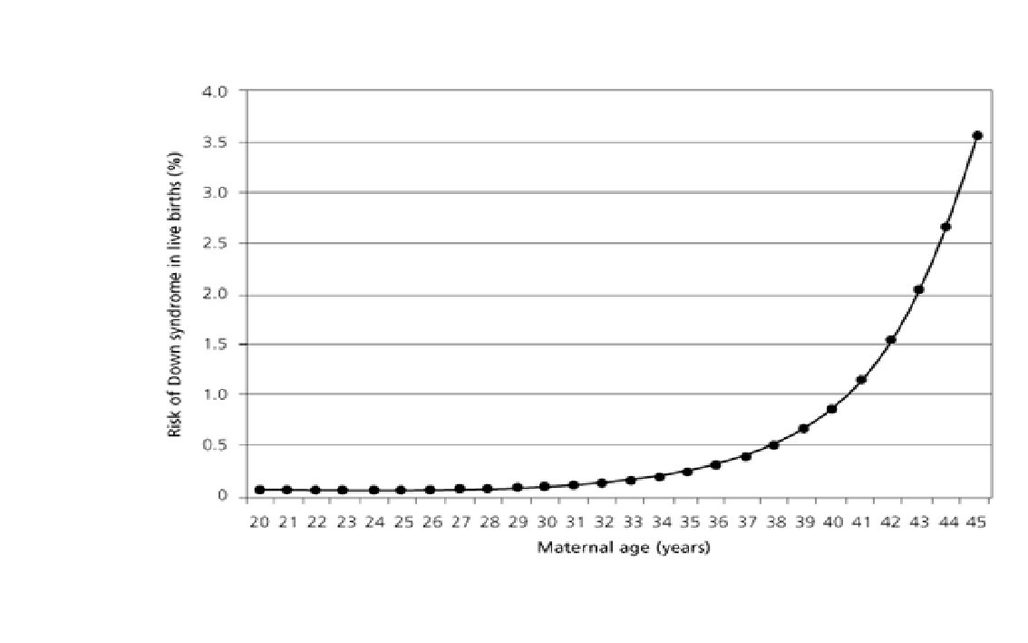
3. Down’s Syndrome • Individual has a trisomy (3 x 21 st chromosomes).

Genetics • Trisomy 21, in ~ 94 %, The frequency of trisomy increases with increasing maternal age. • Robertsonian translocation involving chromosome 21 in approx. 3 -4 %, not related to maternal age. • Trisomy 21 mosaicism in 2 to 3 % cases
 • Occurs due to non-disjunction of the X chromosome.")
4. Kleinfelter’s syndrome (or Klinefleter’s) • Occurs due to non-disjunction of the X chromosome. • The sperm containing both X & Y combines with an egg containing the X, results in a male child (XXY). • The egg may contribute the extra X chromosome.

5. Turner’s • Associated with underdeveloped ovaries, short stature, and only affects women= one copy of x • Individuals are sterile

7. Hemophilia X-linked recessive • The oldest known hereditary bleeding disorder. • Affects males much more frequently (1 in 10, 000) than females (1 in 100, 000) • Patient can bleed to death with small cuts. • About 70% of hemophilia patients have <1% of the normal amount and, thus, have severe hemophilia.

A royal disease pedigree chart

8. Huntington’s Disease/chorea autosomal dominant • A brain disorder (results in loss of mental and physical control). • Caused by the length of a repeated section of a gene exceeding a normal range • Chorea means "dance-like movements" and refers to the uncontrolled motions often associated with the disease.
 autosomal-recessive hereditary disease • Mutations in structure of the phenylalanine hydroxylase")
9. Phenylketonuria (PKU) autosomal-recessive hereditary disease • Mutations in structure of the phenylalanine hydroxylase gene => deficiency in the hepatic enzyme phenylalanine hydroxylase (PAH)=> Patients cannot consume aspartame.
 monogenic mutation • A defect on chromosome 21 in")
10. Amyotrophic Lateral Sclerosis (ALS) monogenic mutation • A defect on chromosome 21 in the gene encoding superoxide dismutase => protein that is toxic to motor nerve cells is associated with approximately 20% of familial cases of ALS. • No known cause in ~95% of cases. • Might be caused by environmental pollutants. • A common first symptom is a painless weakness in a hand, foot, arm or leg, other early symptoms include speech swallowing or walking difficulty.

11. Albinism X-linked, recessive • Patients are unable to produce skin or eye pigments and thus are light-sensitive
 Monogenic, autosomal dominant • Caused by the presence")
12. Achondroplasia (a. k. a. dwarfism) Monogenic, autosomal dominant • Caused by the presence of a faulty allele in a person's genome. • Achondroplasia is caused by a mutation in the fibroblast growth factor receptor gene 3 (fgfr 3). • Fgfr 3 is an inhibitor that regulates bone growth. • In cases of achondroplasia, the fgfr 3 gene is too aggressive, negatively impacting bone growth. • If 2 alleles are present, the result is fatal.

Prenatal diagnosis of genetic diseases Developed in 1966 Provide informed choices to couples at risk of having a child with abnormality

Early Prenatal Screening What are we screening for? – Most associate prenatal screening with aneuploidy, commonly Trisomy 21, 18, 13, monosomy X But there is a lot more than aneuploidy: – Congenital defects – Post dates screening – Complex congenital cardiac defects

Methods of prenatal diagnosis 1. Invasive 2. (i. e. with risk to the foetus or mother): • Amniocentesis • Chorionic villus sampling 2. Non-invasive (i. e. with no risk to the foetus or mother): • Maternal serum/blood screen • Ultrasonography • Isolation of foetal cells from maternal circulation

1. Invasive methods of prenatal diagnosis a. Amniocentesis • Aspiration of 10 -20 ml of amniotic fluid through the abdominal wall under ultrasound guidance around the 16 weeks of gestation. • Result in ~ 14 days to more • Risk of abortion 0. 5 -1%

b. Chorionic villus sampling • Enables diagnosis in first trimester (10 -11 week of gest. ) under ultrasound guidance by transcervical or transabdominal aspiration of chorionic villi (foetal cells). • Results can be obtained in 1 -3 days. • Higher risk of abortion (2 -3%) and limb abnormalities if carried before the 9 weeks of gestation.

2. Non-invasive methods of prenatal diagnosis Maternal serum AFP • Mostly done around the 15 -20 weeks of gestation. • A standard practice to offer screening for Down‘s and Edward syndromes using a blood sample obtained from the mother. • It can diagnose between 60 -70% of Down‘s sy.

Ultrasound to detect Dawn syndrome

Preimplantation genetic diagnosis

Prenatal treatment • In most situations the diagnosis of prenatal abnormalities has a subsequent option of termination of the pregnancy. • With the advent of gene therapy prenatal diagnosis will, in time, lead to effective treatment in utero.
- Slides: 59