Yersinia Pestis Yersinia Y pestis Y pseudoctuberculosis Y

Yersinia Pestis

Yersinia Y. pestis ¡ Y. pseudoctuberculosis ¡ Y. entercolitica ¡

Yersinia pestis ¡ ¡ Discovered by yersin and Kitasato Short plump ovoid gram negative bacillus. In Giemsa stained smears it shows bipolar staining (safety pin appearance). It is capsulated, nonmotile.

Cultural characters ¡ ¡ Aerobic and facultative anaerobic Optimum temperature for growth is 27°C Nutrient agar: transparent colonies Blood agar with sodium azide: dark brown colonies due to absorption of hemin pigments

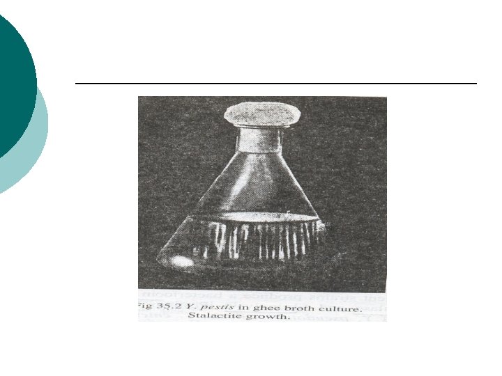
Ghee broth ¡ Y. pestis when grown in a flask with oil or ghee floated on top, a characteristic growth occurs which hangs down from under surface of ghee, resembling stalactites (stalactite growth)

Virulence factors Fraction-I or F-I: Envelope protein which inhibts phagocytosis. ¡ V and W proteins also inhibit phagocytosis. ¡ Plague toxin: This is an endotoxin ¡

Historical Impacts ¡ ¡ ¡ The first plague was in 542 B. C. and lasted almost 60 years. The second and most severe pandemic was in the 14 th century, also known as the Black Death. The final and most recent pandemic occurred in 1894.

Transmission of the Plague is a zoonotic disease ¡ Fleas found on rodents( xenopsiella cheopis) carry disease ¡ Man to man transmission is through the droplets from coughing ¡

Diseases caused by Yersinia Pestis ¡ ¡ ¡ Bubonic plague: based on the lymphatic system. Septicemic plague: Centers in the bloodstream. Pneumonic Plague: Centers in the lungs.

Clinical feautres ¡ ¡ Bubonic Plague l Constitutional symptoms l Lymphadenopathy ¡ “bubo”: inguinal lymphnodes enlarged ¡ May suppurate and drain Septicemic Plague l Same terminal event of bubonic plague l Massive involvement of blood vessels leads to hemorrhages in skin and mucosa: BLACK DEATH

Primary pneumonic plague Organisms inhaled Lobular or lobar pneumonia Pulmonary necrosis Bacteremia Multiorgan seeding, failure Sepsis ¡ Highly infectious form of plague

Laboratory diagnosis Samples - blood, sputum, aspirated bubo fluid, splenic tissue on postmortem ¡ Microscopy– Gram stain and Methylene blue stain: bipolar staining bacilli ¡

Laboratory diagnosis o Culture – Blood agar, Ghee broth o Animal inoculation: Guinea pigs and rats injected subcutaneously with bubo fluid. Animals die within 2 -5 days. o Antigen detection- F 1 glycoprotein in bubo fluid and sputum o Serology –Antibodies to F 1 glycoprotein appear at the end of 1 st week of illness, and remain positive for several years o Rapid tests (PCR, DFA, etc) at reference labs

Treatment and Prevention Tetracycline is drug of choice. ¡ Vaccines: Two types 1. Killed vaccine: Haffkine’s vaccine. Virulent strain, Whole bacterial cell vaccine, 2000 million organisms/ml ¡ v 2. Dosage: . 05 ml S/C followed by 1 ml after 7 -14 days Live vaccine: Avirulent strain of Y. pestis: Otten’s tjiwidej strain and Girard's 76 strain

Yersinia pseudotuberculosis Motile at 22 o. C. ¡ Tuberculosis like lesions ¡

Yersinia enterocolitica Causes 3 types of diseases: ¡ Self limiting gastroenteritis ¡ Mesentric adenitis ¡ Systemic disease c/by bacteraemia, meningitis, arthalgia ¡

Tularemia ¡ ¡ Francisella tularensis -Intracellular gram neg. coccobacillus. It is also known as rabbit fever due to the fact that it may be transmitted to hunters and others who may have exposure to infected rabbits

Tularemia Human infection may occur by handling or eating infected meat or drinking contaminated water Several forms: ¡ Ulcers ¡ Nodes ¡ Pneumonic ¡ usually ulceroglandular disease ¡

Pasteurella multocida ¡ ¡ ¡ Non motile, gram negative rod resembling yersinia. Normal inhabitant of respiratory tract in animals like dogs cattle etc. Human infections rare, but may cause wound sepsis following animal bites
- Slides: 20