XRays in Medicine Discovery of XRays Wilhelm Conrad


 1")


- Slides: 23
X-Rays in Medicine
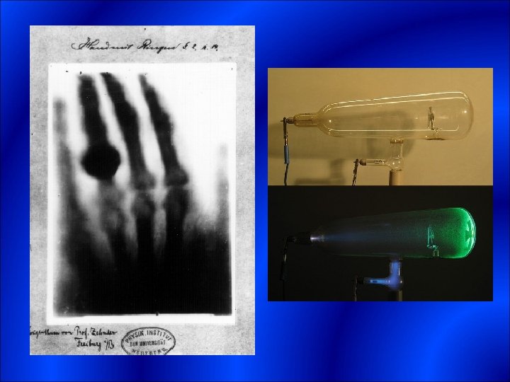
Discovery of X-Rays -Wilhelm Conrad Roentgen-1895 -Electrical discharges in a vacuum tube -Caused a phosphoric screen to fluoresce -Formed an image of bone -First x-ray photograph of wife’s hand/ring
X-rays in Medicine One year later British doctors were already using x- ray imaging. Prolonged exposure to x-rays caused tissue burns. Wounds were abnormal- took time before appearing.
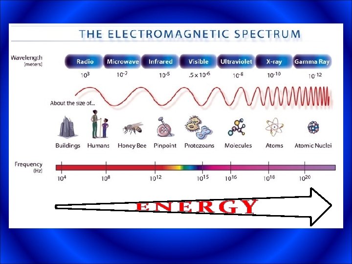
PHOTONS -The energy of a light-wave is carried by packets called photons -The energy of a photon depends on the light’s wavelength -Smaller wavelengths have larger energies
ENERGY Photon energies are usually given in units of electron volts (e. V) 1 e. V = 1. 602 x 10 -19 Joules Need 56, 000, 000, 000 e. V to cook an egg.
X-Ray Tube
X-Ray Tube
X-Ray Tube
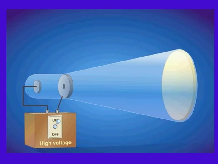
X-Ray Tube X-ray tube consists of the following major parts 1 - Filament: made from tungsten wire which is heated electrically at a potential difference of 8 to 12 V to a high temperature so that electrons ‘boiled off’ from its surface. These electrons form the tube beam. The filament is located within a negatively charged nickel cathode which is shaped so that a precise beam geometry is obtained.
X-Ray Tube 2 - Cathode: this houses the filament and is manufactured from nickel. The filament is located within a cup having sharp contoured edges which electrostatically focus the electron beam. The cathode cup is independently supplied with a high negative voltage 3 - Anode: is manufacture from tungsten (high melting metal). Dental units and small mobile X-ray units use stationary or fixed anode design. The most efficient design however uses a rotating-disk anode which enables higher X-ray output owing to more effective cooling.
Rotary X-Ray Tube
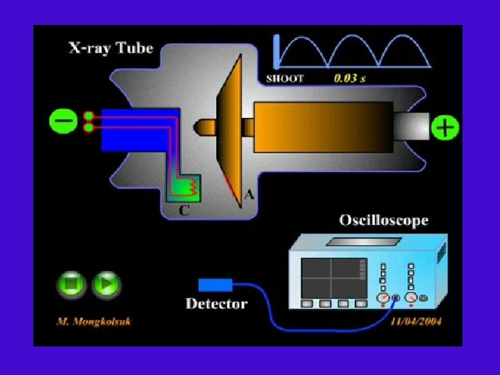
How X-Ray Tube Works
Diagnostic X-Ray Spectra
Therapeutic X-Ray Spectra
X-ray Image Two Forks, Ball pin, and teeth brush in the intestine
X-Ray image of a Gun
Production of Diagnostic X-Rays �In general, the X-ray is produced through bombarding a heavy metal target (high Z-metal) by high speed electrons (20 Ke. V– 150 Ke. V) 1 - Characteristic X-rays. When an accelerated electron collides with a K-shell or Lshell electrons (K and L shells are very close to the nuclide) So the K or L shell electrons will leave their orbits to new orbits, when the electron comes back to its original orbit it will loose its energy in the form of electromagnetic waves (X-rays) with discrete spectrum.
Production of Diagnostic X-Rays 2 - Bremsstrahlung X-Ray. The other way of producing X-ray, when the electron pass close to the nucleus they attracted by the positive charge of the nucleus (electric force). The high speed electron start to accelerate changing their velocity (magnitude or direction or both). When they so they start emitting electro magnetic waves (X-rays) with continuous spectrum called Bremsstrahlung.
Production of Diagnostic X-Rays � 99% of electron energy is transformed into heat and light when electrons collides with the target. � 1% of electron energy is transformed into X-Rays. � 0. 5% remaining of electron energy after inherent filtration. � 0. 1% remaining of electron energy after added filtration (available X-rays for Imaging).