Xray spectroscopy Repetition MENA 3100 OBK 03 05

X-ray spectroscopy Repetition MENA 3100, OBK, 03. 05. 17

XRF is not in the curriculum, but the text is written so that it is difficult to define sections that can be excluded. Paragraphs that only treat XRF can be skipped.

X-ray spectroscopy, n. The art of using the spectroscope; that branch of science which involves the use of the spectroscope. In mod. use, the investigation of spectra by any of various instruments. spectrum, n. 3 a. The coloured band into which a beam of light is decomposed by means of a prism or diffraction grating. Also, a dark band containing bright lines produced similarly; such a (coloured or dark) band, or the pattern of lines in it, as characteristic of the light source; hence, the pattern of absorption or emission of light or other electromagnetic radiation over any range of wavelengths exhibited by a body or substance. 3 b. The entire range of wavelengths (or frequencies) of electromagnetic radiation, from the longest radio waves to the shortest gamma rays of which the range of visible light is only a small part; any one part of this larger range. 3 c. An actual or notional arrangement of the component parts of any phenomenon according to frequency, energy, mass, or the like. Cf. mass spectrum n. , power n. 1

X-ray spectroscopy Have to detect the X-rays: Energy-dispersive spectroscopy EDS Wavelength-dispersive spectroscopy Shame! EELS detect the energy of electrons! WDS Sorry for this, Ole B Electron energy loss spectroscopy EELS (X-ray fluorescence spectroscopy) XRF
 Johan Taftø: Spectroscopy with incident x-rays and")
Curriculum Chapter 6 (− XRF were possible) Johan Taftø: Spectroscopy with incident x-rays and with incident electrons On the course web page Øystein Prytz’ lecture notes Posted as Week 06: Spectroscopy on our web page

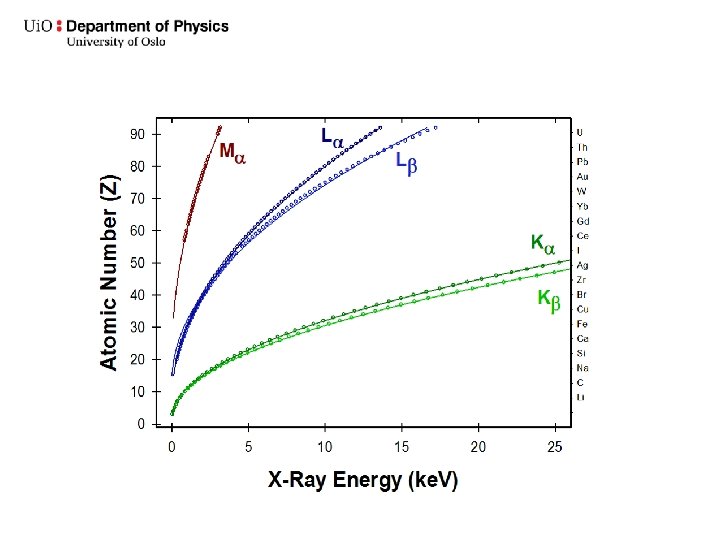
Copper K lines

Siegbahn notation

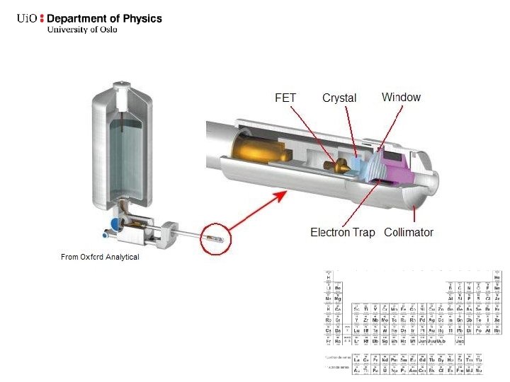
The fluorescence yield The probability for generating a characteristic X-ray is given by the fluorescence yield w The probability of generating an Auger electron is the 1 - w.

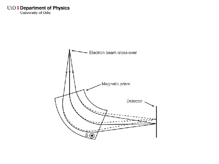
Wavelength-dispersive spectroscopy WDS Sequential
 On a TEM")
Microprobe (Mikrosonde) On a TEM
 Energy dispersive X-ray analysis (EDXA)")
Energy-dispersive spectroscopy Energy-dispersive X-ray spectroscopy (EDS, EDX, or XEDS) Energy dispersive X-ray analysis (EDXA) Energy dispersive X-ray microanalysis (EDXMA) Energy dispersive analysis of X-rays (EDAX)
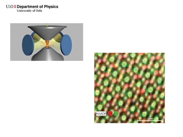
 detectors Silicon drift detectors SDD Simultaneous")
Energy-dispersive spectroscopy EDS Si(Li) detectors Silicon drift detectors SDD Simultaneous
 detectors An electron-hole pair is created for every 3. 76 e. V of")
Si(Li) detectors An electron-hole pair is created for every 3. 76 e. V of incoming X radiation The energy of a Ni Kα X-ray photon is 7, 471 ke. V Will produce a current of 1 966 electrons

Signal vs. noise Dead time

Sum peak Escape peak

Accelerating voltage: Exceed the critical ionization energy of the element by a factor of 1. 5 to 3 to efficiently excite the X-ray line Interaction volume
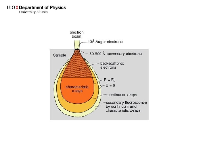

Dictates the working distance

Energy dispersive spectroscopy EDS Characteristic X-rays Qualitative analysis

Ce Fe Sr
 La. Nb. O 4 100 La. Nb. O 4 5 m")
Line scan a) La. Nb. O 4 100 La. Nb. O 4 5 m Ni. O 100 m Atomic % 80 60 La. L 40 Ni. O 20 0 0 10 20 distance (µm) 30

Spot analysis

Quantitative analysis

The Cliff-Lorimer equations Binary system TEM

The ratios are just the starting point ZAF

Flat samples Not too small!
 D. B. Williams and C.")
The post column Electron Energy Loss spectrometer/energy filter (EELS) D. B. Williams and C. B. Carter (2009)
")
Sketch of an EELS spectrum • Zero energy loss peak (ZLP, 0 e. V) • Low loss region. Valence band excitations, dominated by plasmons (collective excitations), dielectric properties ( 0 -50 e. V) • Core loss region, single electron transitions, bonding and density of R. Brydson (2001) states, progressively weaker signals ( 50 - 2000 e. V)
 • Electronic structure")
Uses of the core-loss features • Composition analysis and mapping (EFTEM) • Electronic structure and bonds (ELNES), comparison with DOS • Structure and atomic coordination (EXELFS)

The low loss region • The low loss region contains both collective excitations and single electron events • Plasmons • Valence band/interband excitations (VEELS) • Also sometimes «core» loss events 34

Summary • From Øystein The main energy transfer processes are: – Single electron excitations – Bremsstrahlung – Collective excitations (plasmons) • • Differentiate between primary and secondary processes Nomenclature • • What determines the characteristic X-ray energies How can X-ray energies be measured? – EDS vs WDS • The thin film approximation in TEM – What approximationis made? – What are the limits to this approximation and what happens when it breaks down? – Difference between the Cliff-Lorimer method and the ZAF method • Electron Energy Loss Spectroscopy (EELS) – The EELS spectrometer – Typical features in the EELS spectrum and their uses – Monochromation
- Slides: 35