
Xray Phase Contrast Imaging Vincent Revol Zrich 04
- Slides: 29
X-ray Phase Contrast Imaging Vincent Revol Zürich, 04. 06. 2009 Copyright 2009 CSEM | Ph. D Seminar, 4 th June 2009| V. Revol | Page 0
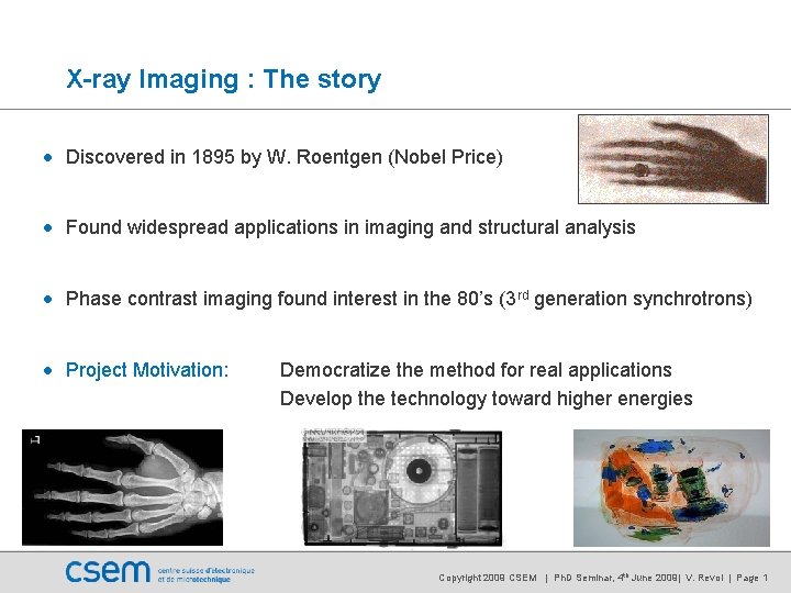
X-ray Imaging : The story • Discovered in 1895 by W. Roentgen (Nobel Price) • Found widespread applications in imaging and structural analysis • Phase contrast imaging found interest in the 80’s (3 rd generation synchrotrons) • Project Motivation: Democratize the method for real applications Develop the technology toward higher energies Copyright 2009 CSEM | Ph. D Seminar, 4 th June 2009| V. Revol | Page 1
Outline of the talk • Physical Principle • What is phase contrast imaging? • Operating Principle • How can we measure the phase with X-rays? • Set Up Presentation • How is it implemented? • Results • How does it looks like? • Outline • What are the challenges? Copyright 2009 CSEM | Ph. D Seminar, 4 th June 2009| V. Revol | Page 2
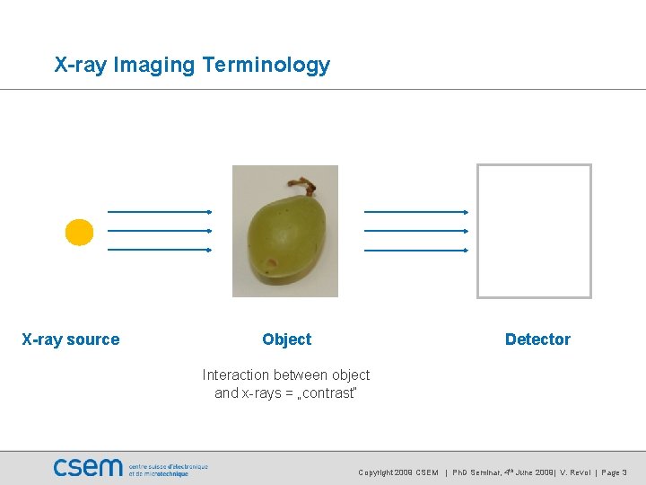
X-ray Imaging Terminology X-ray source Object Detector Interaction between object and x-rays = „contrast“ Copyright 2009 CSEM | Ph. D Seminar, 4 th June 2009| V. Revol | Page 3
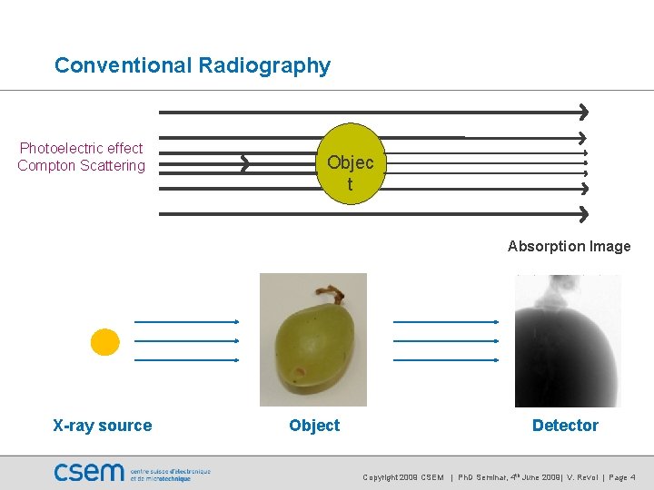
Conventional Radiography Photoelectric effect Compton Scattering Objec t Absorption Image X-ray source Object Detector Copyright 2009 CSEM | Ph. D Seminar, 4 th June 2009| V. Revol | Page 4
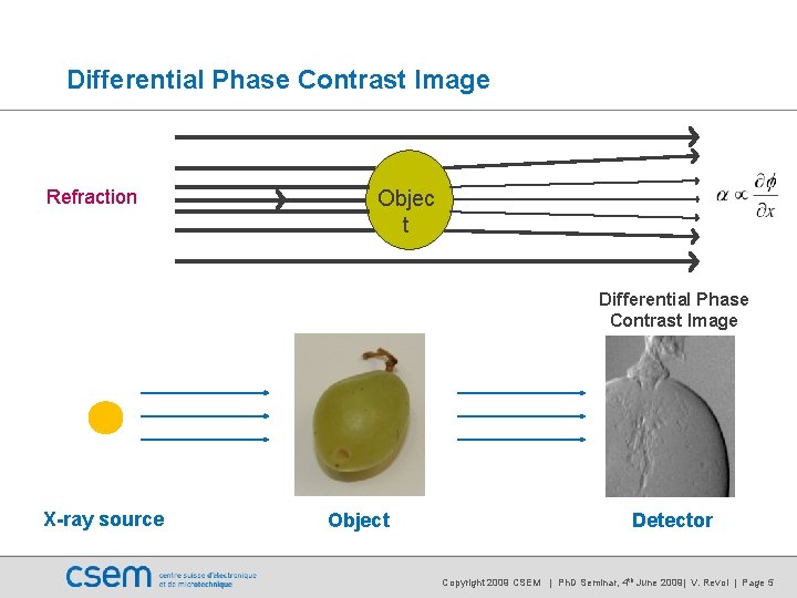
Differential Phase Contrast Image Refraction Objec t Differential Phase Contrast Image X-ray source Object Detector Copyright 2009 CSEM | Ph. D Seminar, 4 th June 2009| V. Revol | Page 5
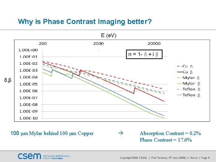
Why is Phase Contrast Imaging better? E (e. V) n = 1 - d + i b δ β δ β δ, β 100 μm Mylar behind 100 μm Copper Absorption Contrast = 0. 2% Phase Contrast = 17. 6% Copyright 2009 CSEM | Ph. D Seminar, 4 th June 2009| V. Revol | Page 6
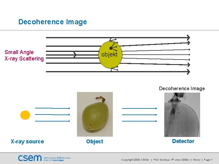
Decoherence Image Small Angle X-ray Scattering objekt Decoherence Image X-ray source Object Detector Copyright 2009 CSEM | Ph. D Seminar, 4 th June 2009| V. Revol | Page 7
Outline of the talk • Physical Principle • What is phase contrast imaging? • Operating Principle • How can we measure the phase with X-rays? • Set Up Presentation • How is it implemented? • Results • How does it looks like? • Outline • What are the challenges? Copyright 2009 CSEM | Ph. D Seminar, 4 th June 2009| V. Revol | Page 8
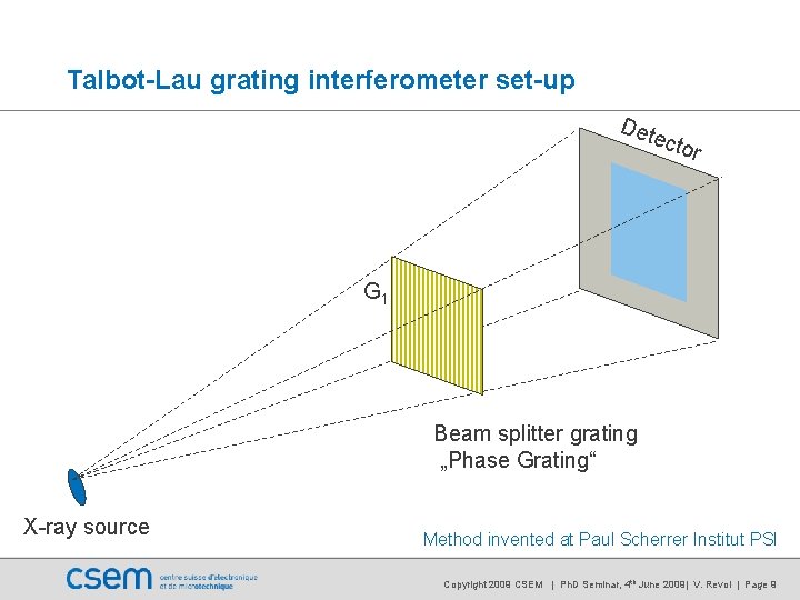
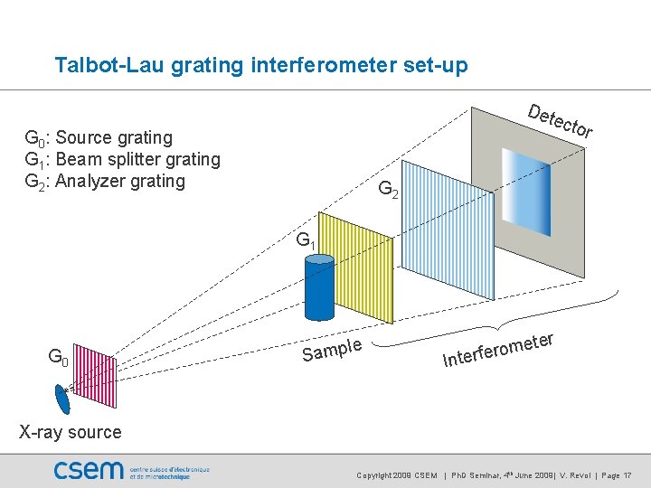
Talbot-Lau grating interferometer set-up Det ecto r G 1 Beam splitter grating „Phase Grating“ X-ray source Method invented at Paul Scherrer Institut PSI Copyright 2009 CSEM | Ph. D Seminar, 4 th June 2009| V. Revol | Page 9
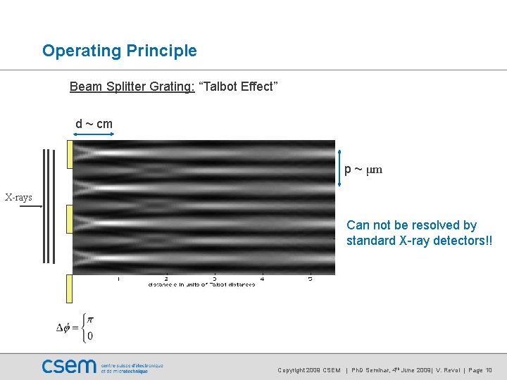
Operating Principle Beam Splitter Grating: “Talbot Effect” d ~ cm p ~ μm X-rays Can not be resolved by standard X-ray detectors!! Copyright 2009 CSEM | Ph. D Seminar, 4 th June 2009| V. Revol | Page 10
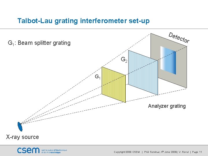
Talbot-Lau grating interferometer set-up Det ecto G 1: Beam splitter grating r G 2 G 1 Analyzer grating X-ray source Copyright 2009 CSEM | Ph. D Seminar, 4 th June 2009| V. Revol | Page 11
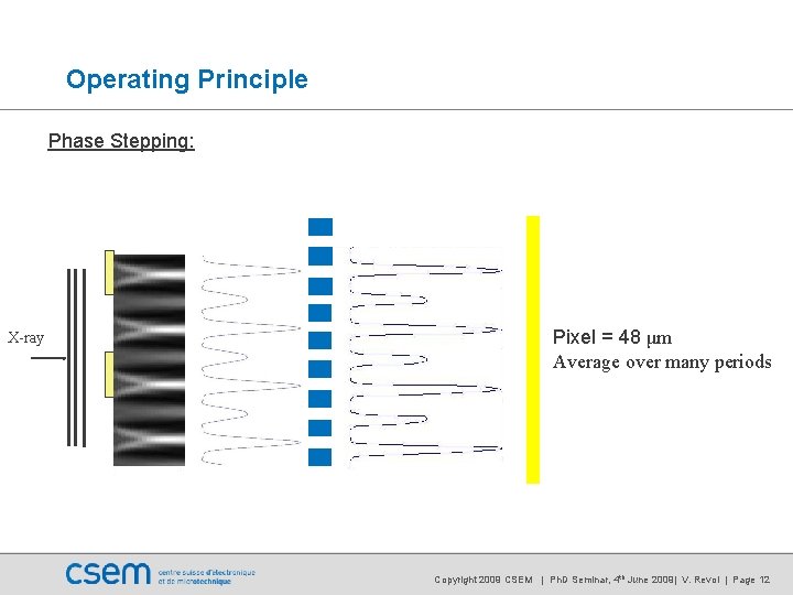
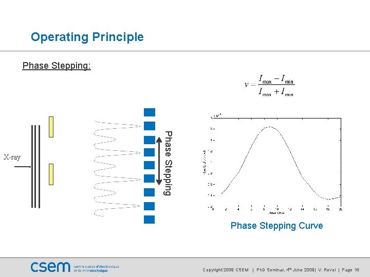
Operating Principle Phase Stepping: X-ray Pixel = 48 μm Average over many periods Copyright 2009 CSEM | Ph. D Seminar, 4 th June 2009| V. Revol | Page 12
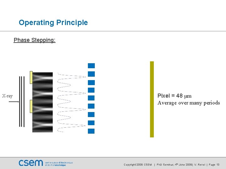
Operating Principle Phase Stepping: X-ray Pixel = 48 μm Average over many periods Copyright 2009 CSEM | Ph. D Seminar, 4 th June 2009| V. Revol | Page 13
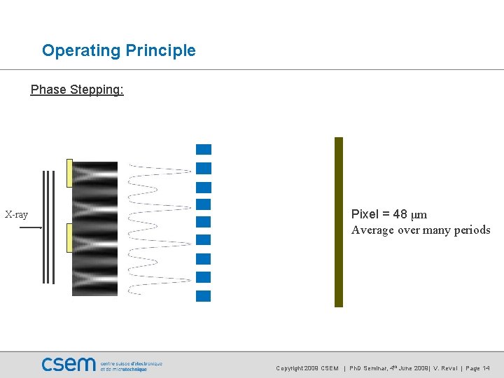
Operating Principle Phase Stepping: X-ray Pixel = 48 μm Average over many periods Copyright 2009 CSEM | Ph. D Seminar, 4 th June 2009| V. Revol | Page 14
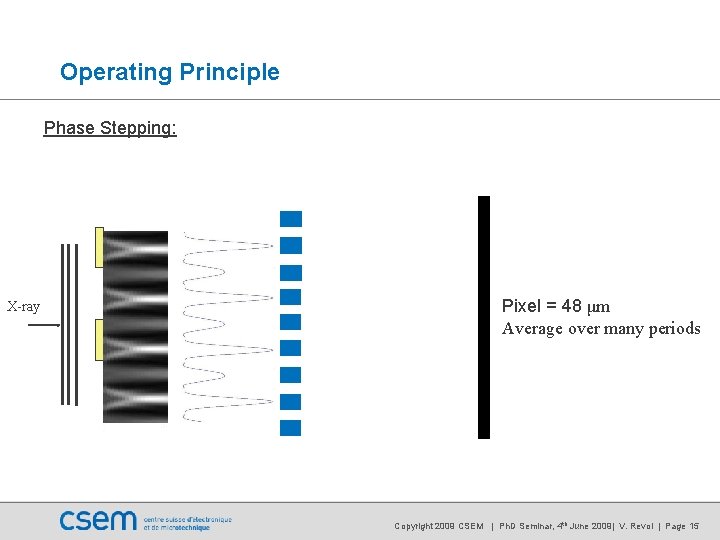
Operating Principle Phase Stepping: X-ray Pixel = 48 μm Average over many periods Copyright 2009 CSEM | Ph. D Seminar, 4 th June 2009| V. Revol | Page 15
Operating Principle Phase Stepping: Phase Stepping X-ray Phase Stepping Curve Copyright 2009 CSEM | Ph. D Seminar, 4 th June 2009| V. Revol | Page 16
Talbot-Lau grating interferometer set-up Det ecto G 0: Source grating G 1: Beam splitter grating G 2: Analyzer grating r G 2 G 1 G 0 le Samp er In met o r e f r te X-ray source Copyright 2009 CSEM | Ph. D Seminar, 4 th June 2009| V. Revol | Page 17
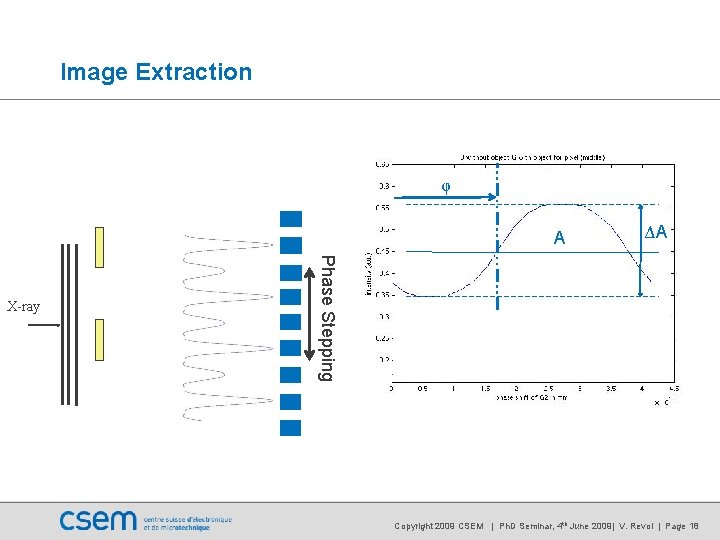
Image Extraction φ A Phase Stepping X-ray ΔA Copyright 2009 CSEM | Ph. D Seminar, 4 th June 2009| V. Revol | Page 18
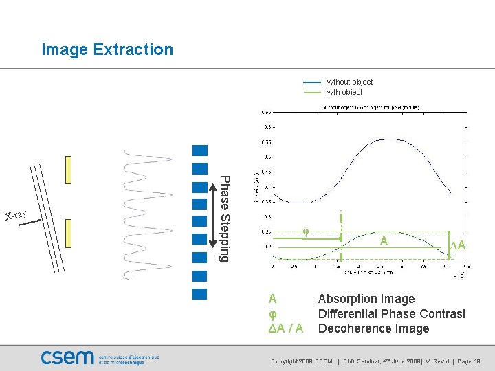
Image Extraction without object with object Phase Stepping X-ray φ A φ ΔA / A A ΔA Absorption Image Differential Phase Contrast Decoherence Image Copyright 2009 CSEM | Ph. D Seminar, 4 th June 2009| V. Revol | Page 19
Outline of the talk • Physical Principle • What is phase contrast imaging? • Operating Principle • How can we measure the phase with X-rays? • Set Up Presentation • How is it implemented? • Results • How does it looks like? • Outline • What are the challenges? Copyright 2009 CSEM | Ph. D Seminar, 4 th June 2009| V. Revol | Page 20
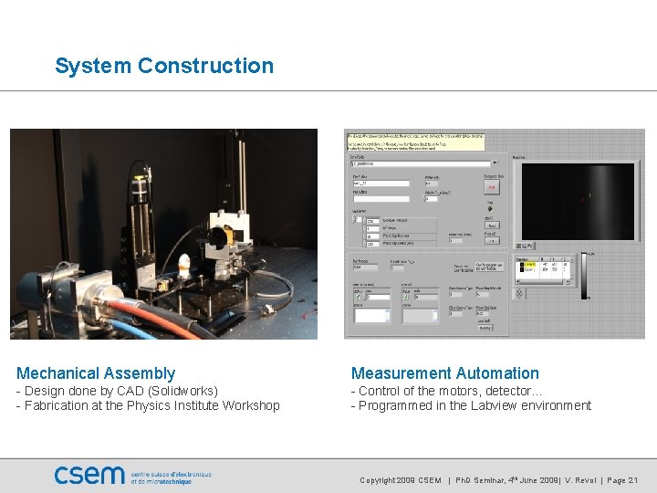
System Construction Mechanical Assembly Measurement Automation - Design done by CAD (Solidworks) - Fabrication at the Physics Institute Workshop - Control of the motors, detector… - Programmed in the Labview environment Copyright 2009 CSEM | Ph. D Seminar, 4 th June 2009| V. Revol | Page 21
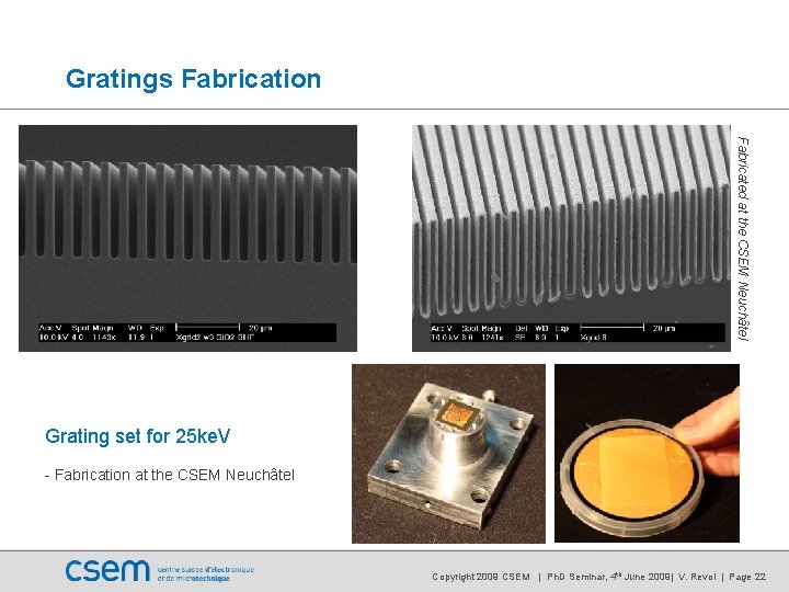
Gratings Fabrication Fabricated at the CSEM Neuchâtel Grating set for 25 ke. V - Fabrication at the CSEM Neuchâtel Copyright 2009 CSEM | Ph. D Seminar, 4 th June 2009| V. Revol | Page 22
Outline of the talk • Physical Principle • What is phase contrast imaging? • Operating Principle • How can we measure the phase with X-rays? • Set Up Presentation • How is it implemented? • Results • How does it looks like? • Outline • What are the challenges? Copyright 2009 CSEM | Ph. D Seminar, 4 th June 2009| V. Revol | Page 23
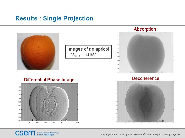
Results : Single Projection Absorption Images of an apricot Vtube = 40 k. V Differential Phase Image Decoherence Copyright 2009 CSEM | Ph. D Seminar, 4 th June 2009| V. Revol | Page 24
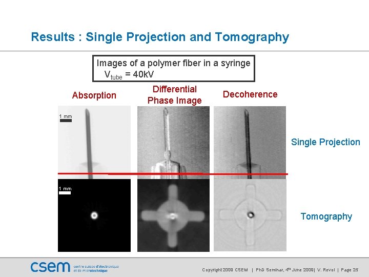
Results : Single Projection and Tomography Images of a polymer fiber in a syringe Vtube = 40 k. V Absorption Differential Phase Image Decoherence 1 mm Single Projection 1 mm Tomography Copyright 2009 CSEM | Ph. D Seminar, 4 th June 2009| V. Revol | Page 25
Outline of the talk • Physical Principle • What is phase contrast imaging? • Operating Principle • How can we measure the phase with X-rays? • Set Up Presentation • How is it implemented? • Results • How does it looks like? • Outline • What are the challenges? Copyright 2009 CSEM | Ph. D Seminar, 4 th June 2009| V. Revol | Page 26
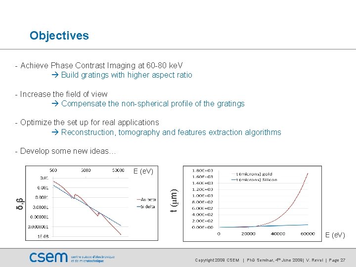
Objectives - Achieve Phase Contrast Imaging at 60 -80 ke. V Build gratings with higher aspect ratio - Increase the field of view Compensate the non-spherical profile of the gratings - Optimize the set up for real applications Reconstruction, tomography and features extraction algorithms - Develop some new ideas… t (μm) δ, β E (e. V) Copyright 2009 CSEM | Ph. D Seminar, 4 th June 2009| V. Revol | Page 27
Thank you for your attention! Copyright 2009 CSEM | Ph. D Seminar, 4 th June 2009| V. Revol | Page 28