Xray Interpretation Skills Dr Hisham Alsanawi Orthopaedic Consultant


- Slides: 68

X-ray Interpretation Skills Dr. Hisham Alsanawi Orthopaedic Consultant and Assistant Professor

Medical Decision Making is a Triad • History – from patients/records • Physical Examination • Confirming Studies – Imaging, Labs, etc.

Imaging • • • X-ray Ultrasound CT Scan MRI Nuclear Medicine

X-RAY • • Radiation Source Patient Exposed Capture Image Interpret Image

X-RAY • Ionizing Radiation • Radiation damages cells

X-RAY • Patient Blocks Transmission of Radiation – Soft tissues Less – Bones More

X-RAY • Capture Image – Films – Digital

X-RAY • Interpret Image – Radiologist – Orthopaedist

X-RAY • Best for: – Hard tissue – Bones – Often combined with other imaging

OBJECTIVES • Review a systematic approach to interpreting orthopedic x-rays • Review the language of fracture description

ABCs APPROACH �Pre ABC: identify pt, read provided info �A ◦ Adequacy ◦ Alignment �B ◦ Bones �C ◦ Cartilage �S ◦ Soft Tissues �Apply ABCs approach to every orthopedic film you evaluate

ADEQUACY • All x-rays should have an adequate number of views. – Minimum of 2 views—AP and lateral – 3 views preferred – Joint above and joint below • All x-rays should have adequate penetration


ALIGNMENT • Alignment: Anatomic relationship between bones on x-ray – Bone alignment vs other side – Bone alignment relative to proximal and distal bones • Normal x-rays should have normal alignment • Fractures and dislocations may affect the alignment on the x-ray

BONES 1. Identify bone 2. Examine the whole bone for 1. Discontinuity fractures 2. Change in bone shadow consistency change in density 3. Describe bone abnormality 1. Location 2. Shape


CARTILAGE • Cartilage – joint spaces on x-rays – you cannot actually see cartilage on x-rays • Widening of joint spaces signifies ligamentous injury and/or fractures • Narrowing of joint spaces arthritis


SOFT TISSUES • Soft tissues implies to look for soft tissue swelling and joint effusions • These can be signs of – Trauma – occult fractures – Infection – Tumors

REVIEW: ABCs �A ◦ Assess adequacy of x-ray which includes proper number of views and penetration ◦ Assess alignment of x-rays �B ◦ Examine bones throughout their entire length for fracture lines and/or distortions �C ◦ Examine cartilages (joint spaces) for widening �S ◦ Assess soft tissues for swelling/effusions

EXAMPLE # 1

EXAMPLE # 1… �This x-ray demonstrates a lateral elbow x-ray. �There is swelling anteriorly which is displaced known as a pathologic anterior fat pad sign �There is swelling posteriorly known as a posterior fat pad sign �Both of these are signs of an occult fracture although none are visualized on this x-ray �Remember, soft tissue swelling can be a sign of occult fracture!

EXAMPLE # 2…WHERE ARE THE FRACTURES?

EXAMPLE # 2… • If you follow ABCs, you will notice there is are problems with alignment on this x-ray (A) • (B)…You will notice there are fracture lines through the 2 nd, 3 rd, and 4 th metacarpals • These are 2 nd, 3 rd, and 4 th, midshaft metacarpal fractures. • A teaching point: Notice the ring on this film. Always remove rings of patients with fractured extremities because swelling may preclude removal later.

LANGUAGE OF FRACTURES • Important for use to describe x-rays in medical terminology. • Improves communication with orthopedic consultants

LANGUAGE OF FRACTURES • Things you must describe (clinical and x-ray): – Open vs Closed fracture – Anatomic location of fracture – Fracture line – Relationship of fracture fragments – Neurovascular status

OPEN VS CLOSED �Must describe to a consultant if fracture is open or closed �Closed fracture ◦ Simple fracture ◦ No open wounds of skin near fracture �Open fracture ◦ Compound fracture ◦ Cutaneous (open wounds) of skin near fracture site. Bone may protrude from skin ◦ Open fractures are open complete displaced and/or comminuted

OPEN FRACTURES • • Orthopedic emergency Requires emergency orthopedic consultation Bleeding must be controlled Management – IV antibiotics – Tetanus prophylaxis – Pain control – Surgery for washout and reduction

ANATOMIC LOCATION • Describe the precise anatomic location of the fracture • Include if it is left or right sided bone • Include name of bone • Include location: – Proximal…Mid…Distal – To aid in this, divide bone into 1/3 rds

FOR EXAMPLE. . WHERE IS THIS LOCATED?

EXAMPLE… • This is a closed L distal femur fracture. • The main thing I want you to take from this example is the description of location

ANATOMIC LOCATION • Besides location, it is helpful to describe if the location of the fracture involves the joint space —intra-articular

INTRA-ARTICULAR FRACTURE OF BASE 1 ST METACARPAL

FRACTURE LINES • Next, it is imperative to describe the type of fracture line • There are several types of fracture lines

FRACTURE LINES

FRACTURE LINES • A is a transverse fracture • B is an oblique fracture • C is a spiral fracture • D is a comminuted fracture • There is also an impacted fracture where fracture ends are compressed together

WHAT TYPE OF FRACTURE LINE IS THIS? ? ?

ANS: TRANSVERSE FRACTURE • Transverse fractures occur perpendicular to the long axis of the bone. • To fully describe the fracture, this is a closed midshaft transverse humerus fracture.

ANOTHER EXAMPLE OF FRACTURE LINE…

ANS: SPIRAL FRACTURE • Spiral fractures occur in a spiral fashion along the long axis of the bone • They are usually caused by a rotational force • To fully describe the fracture, this is a closed distal spiral fracture of the fibula

ONE MORE EXAMPLE…

ANS: COMMINUTED FRACTURE • Comminuted fractures are those with 2 or more bone fragments are present • Sometimes difficult to appreciate on x-ray but will clearly show on CT scan • To fully describe the fracture, this is a closed R comminuted intertrochanteric fracture

FRACTURE FRAGMENTS • Terms to be familiar with when describing the relationship of fracture fragments – Alignment – Angulation – Apposition – Displacement – Bayonette apposition – Distraction – Dislocation

ALIGNMENT/ANGULATION • Alignment is the relationship in the longitudinal axis of one bone to another • Angulation is any deviation from normal alignment • Angulation is described in degrees of angulation of the distal fragment in relation to the proximal fragment—to measure angle draw lines through normal axis of bone and fracture fragment

20 DEGREES OF ANGULATION

OTHER TERMS • Apposition: amount of end to end contact of the fracture fragments • Displacement: use interchangeably with apposition • Bayonette apposition: overlap of fracture fragments • Distraction: displacement in the longitudinal axis of the bones • Dislocation: disruption of normal relationship of articular surfaces

DESCRIBE FRACTURE FRAGMENTS

ANSWER • This is a closed midshaft tibial fracture…. But how do we describe the fragments? • This is an example of partial apposition; note part of the fracture fragments are touching each other • Alternatively you can describe this as displaced 1/3 the thickness of the bone • Remember aposition and displacement are interchangeable—we tend to describe displacement • Final answer: Closed midshaft tibial fracture with moderate (33%) displacement

ANOTHER ONE…

ANSWER • There are 2 fractures on this film • Closed distal radius fracture with complete displacement. Also there is an ulnar styloid fracture which is also displaced • The displacement is especially prominent on the lateral view highlighting the importance of multiple views. • There may be intra-articular involvement as joint space is close by • Remember, remove all jewelry from extremity fractures

BAYONETTE APPOSITION

DISLOCATION

DISLOCATION • Note the dislocation on the previous slide; the articular surfaces of the knee no longer maintain their normal relationship • Dislocations are named by the positioin of the distal segemnt • This is an Anterior knee dislocation

NEUROVASCULAR STATUS • Finally when communicating a fracture, you will want to describe if the patient has any neurovascular deficits • This is determined clinically

LANGUAUGE OF FRACTURES • To review, when seeing a patient with a fracture and the x-ray, describe the following: – Open vs closed fracture – Anatomic location of fracture (distal, mid, proximal) and if fracture is intra-articular – Fracture line (transverse, oblique, spiral, comminuted) – Relationship of fracture fragments (angulation, displacement, dislocation, etc) – Neurovascular status

DESCRIBE THIS R MIDDLE PHALANX FRACTURE

ANSWER • Oblique fracture of midshaft of R 4 th middle phalanx with minimal displacement and no angulation • Remember to comment if open vs closed & neurovascular status

DESCRIBE TO ORTHO ATTENDING…

ANSWER • This one is a bit more challenging! • R midshaft tibia fracture displaced ½ the thickness of the bone without angulation; also there is bayonette appositioning of the fracture fragments • R midshaft fibular fracture with complete displacement and • Also comment if the fracture is open vs closed & neurovascular status
 Dr hisham khalil
 Vbpm orthopaedic
 Rch clavicle fracture
 Charcot's joint
 Orthopaedic surgery south east london
 Cappagh hospital hip replacement
 Professor abudu royal orthopaedic hospital
 Miss sally tennant
 Stephen brennan bon secours
 Phong tran surgeon
 Peter worlock
 How does interpretation b differ from interpretation a
 Xray training
 Plain film kub
 V/q scan pulmonary embolism
 Homework
 Xray lara
 Mdcis
Mdcis Silicon valley xray
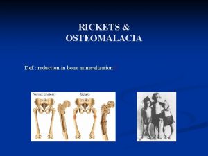
 Rickets def
 Picker xray
 Bga xray
 Xray laser
 Xray lithography
 Xray file cabinet
 Properties of xray
 Pnuemothorax xray
 Xray technique chart
 Thumbprinting on xray
 Sza xray
 Xray laser
 Xray laser
 Xray waves examples
 Prehistoric era
 Mmc xray
 Foreshortening and elongation
 Signo rigler
 Xray photoelectron spectroscopy
 Cvs x ray
 Ellis s curve pleural effusion
 Acanthioparietal
 Inertial confinement fusion lasers
 Triboluminescence x ray
 Yxlon xray
 Xray mastoid townes view
 Gimp xray
 Cavernous sinus thrombosis
 Emulsion peel x ray
 جريد
جريد Jfk xray
 Vena cava syndrome
 Xray laser
 Xray telescope
 Xray neck lateral view
 Epiglottitis
 Spectrum xray
 Hemothorax xray
 Who discovered xray
 Modified waters
 Atom xray
 Omegascans
 Zenker's diverticulum xray
 Xray laser
 Xray laser
 Xray searches
 The purpose of a lead foil sheet in the film packet is
 Darkroom tiles
 Talipes equinovarus xray
 Grid controlled x ray tube


