
XRAY CRITERIA 1 Lesion location 2 Lesion size




- Slides: 18
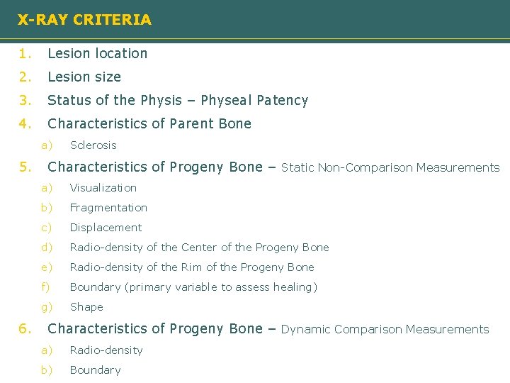
X-RAY CRITERIA 1. Lesion location 2. Lesion size 3. Status of the Physis – Physeal Patency 4. Characteristics of Parent Bone a) 5. 6. Sclerosis Characteristics of Progeny Bone – Static Non-Comparison Measurements a) Visualization b) Fragmentation c) Displacement d) Radio-density of the Center of the Progeny Bone e) Radio-density of the Rim of the Progeny Bone f) Boundary (primary variable to assess healing) g) Shape Characteristics of Progeny Bone – Dynamic Comparison Measurements a) Radio-density b) Boundary
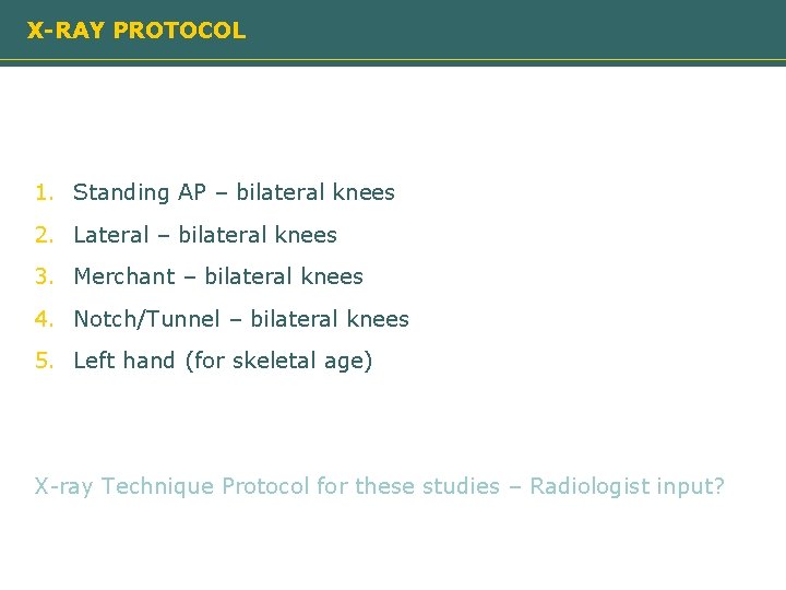
X-RAY PROTOCOL 1. Standing AP – bilateral knees 2. Lateral – bilateral knees 3. Merchant – bilateral knees 4. Notch/Tunnel – bilateral knees 5. Left hand (for skeletal age) X-ray Technique Protocol for these studies – Radiologist input?
CRITERION 3 – STATUS OF THE PHYSIS / PHYSEAL PATENCY Open Closing 1 – Open 2 – Closing 3 – Closed 1 – Open 2 – Closing 3 – Closed Assessment should consider only the femoral physis. Closing’ Definition: - Must be able to see both some areas of the physis that appear to be ‘closed’ and some areas that are clearly still ‘open’.
CRITERION 1 – LOCATION OF THE LESION AP Lateral Pat 1 2 3 4 – – Medial femoral condyle Lateral femoral condyle Patella Trochlea 1 2 3 4 T – – A Post Anterior Condyle Posterior Condyle Patella Trochlea LATERAL VIEW: Project line off posterior femoral shaft. If posterior to line = Posterior. If between posterior femoral line and Blumenstadts line then =Anterior. If anterior to Blumenstadts then=Trochlear. Pick center of lesion surface predominant compartment
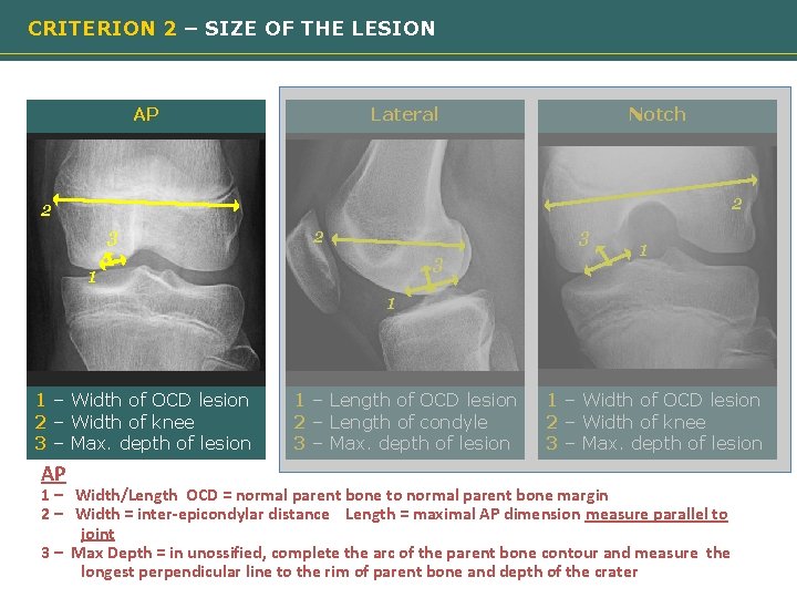
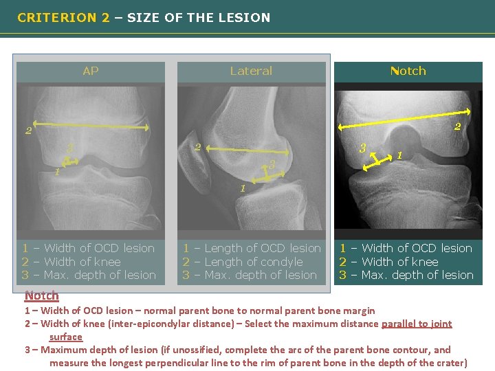
CRITERION 2 – SIZE OF THE LESION AP Lateral Notch 2 2 3 3 1 Need image 1 1 – Width of OCD lesion 2 – Width of knee 3 – Max. depth of lesion AP 1 – Length of OCD lesion 2 – Length of condyle 3 – Max. depth of lesion 1 – Width of OCD lesion 2 – Width of knee 3 – Max. depth of lesion 1 – Width/Length OCD = normal parent bone to normal parent bone margin 2 – Width = inter-epicondylar distance Length = maximal AP dimension measure parallel to joint 3 – Max Depth = in unossified, complete the arc of the parent bone contour and measure the longest perpendicular line to the rim of parent bone and depth of the crater
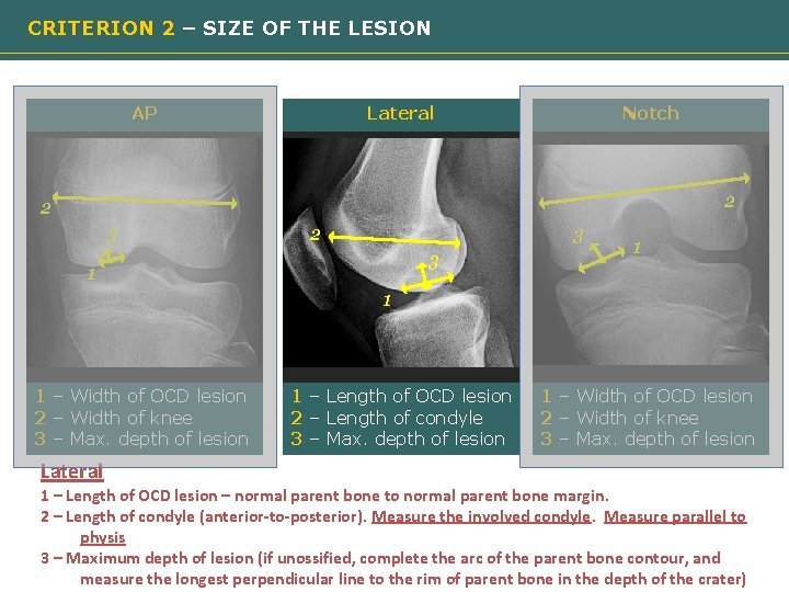
CRITERION 2 – SIZE OF THE LESION AP Lateral Notch 2 2 3 3 1 Need image 1 1 – Width of OCD lesion 2 – Width of knee 3 – Max. depth of lesion Lateral 1 – Length of OCD lesion 2 – Length of condyle 3 – Max. depth of lesion 1 – Width of OCD lesion 2 – Width of knee 3 – Max. depth of lesion 1 – Length of OCD lesion – normal parent bone to normal parent bone margin. 2 – Length of condyle (anterior-to-posterior). Measure the involved condyle. Measure parallel to physis 3 – Maximum depth of lesion (if unossified, complete the arc of the parent bone contour, and measure the longest perpendicular line to the rim of parent bone in the depth of the crater)
CRITERION 2 – SIZE OF THE LESION AP Lateral Notch 2 2 3 3 1 Need image 1 1 – Width of OCD lesion 2 – Width of knee 3 – Max. depth of lesion Notch 1 – Length of OCD lesion 2 – Length of condyle 3 – Max. depth of lesion 1 – Width of OCD lesion 2 – Width of knee 3 – Max. depth of lesion 1 – Width of OCD lesion – normal parent bone to normal parent bone margin 2 – Width of knee (inter-epicondylar distance) – Select the maximum distance parallel to joint surface 3 – Maximum depth of lesion (if unossified, complete the arc of the parent bone contour, and measure the longest perpendicular line to the rim of parent bone in the depth of the crater)
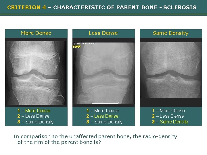
CRITERION 4 – CHARACTERISTIC OF PARENT BONE - SCLEROSIS More Dense Less Dense Need image 1 – More Dense 2 – Less Dense 3 – Same Density Need image 1 – More Dense 2 – Less Dense 3 – Same Density In comparison to the unaffected parent bone, the radio-density of the rim of the parent bone is?
CRITERION 5 a – CHARACTERISTIC OF PROGENY BONE - VISUALIZATION Visualized Not-visualized Need image 1 – No 2 – Yes Is the progeny bone visualized ON ALL VIEWS? Evaluate all views to determine presence of ossification. If ossification not visualized on any view and appears as a “black hole”, check NO (Not-visualized)
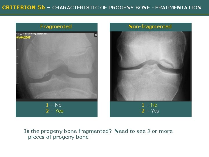
CRITERION 5 b – CHARACTERISTIC OF PROGENY BONE - FRAGMENTATION Fragmented Non-fragmented Need image 1 – No 2 – Yes Is the progeny bone fragmented? Need to see 2 or more pieces of progeny bone
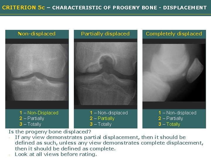
CRITERION 5 c – CHARACTERISTIC OF PROGENY BONE - DISPLACEMENT Non-displaced Partially displaced Need image 1 – Non-Displaced 2 – Partially 3 – Totally 1 – Non-displaced 2 – Partially 3 – Totally Completely displaced Need image 1 – Non-displaced 2 – Partially 3 – Totally Is the progeny bone displaced? - If any view demonstrates partial displacement, then it should be defined as such, unless any view demonstrates complete displacement, then it should be defined as complete. - Look at all views before rating.
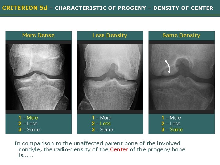
CRITERION 5 d – CHARACTERISTIC OF PROGENY – DENSITY OF CENTER More Dense Less Density Need image 1 – More 2 – Less 3 – Same Density Need image 1 – More 2 – Less 3 – Same In comparison to the unaffected parent bone of the involved condyle, the radio-density of the Center of the progeny bone is……
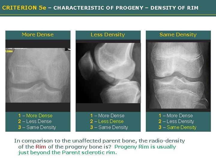
CRITERION 5 e – CHARACTERISTIC OF PROGENY – DENSITY OF RIM More Dense Less Density Need image 1 – More Dense 2 – Less Dense 3 – Same Density Need image 1 – More Dense 2 – Less Density 3 – Same Density In comparison to the unaffected parent bone, the radio-density of the Rim of the progeny bone is? Progeny Rim is usually just beyond the Parent sclerotic rim.
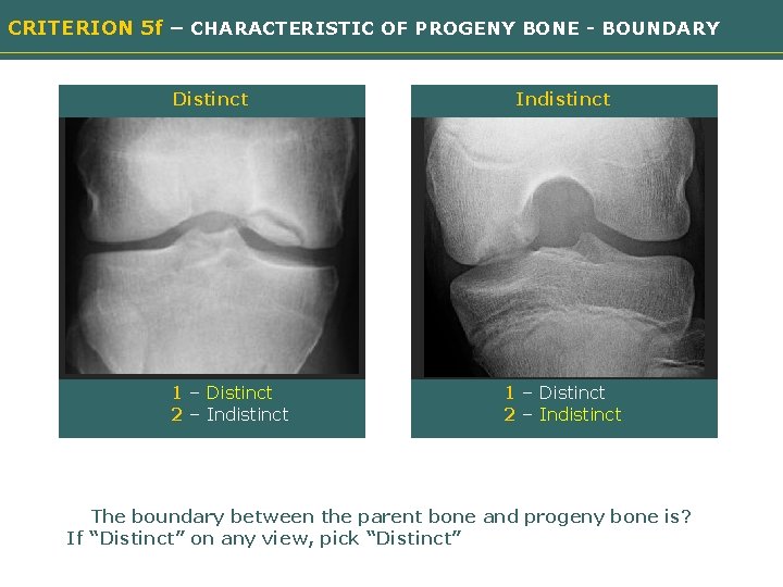
CRITERION 5 f – CHARACTERISTIC OF PROGENY BONE - BOUNDARY Distinct Indistinct Need image 1 – Distinct 2 – Indistinct The boundary between the parent bone and progeny bone is? If “Distinct” on any view, pick “Distinct”
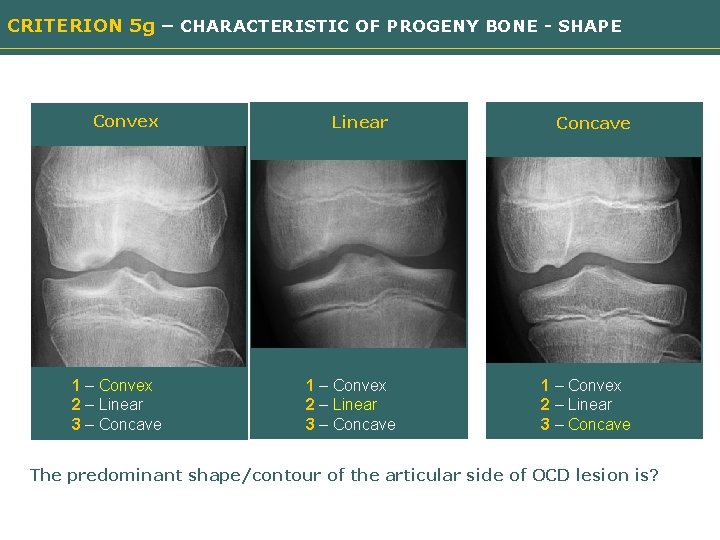
CRITERION 5 g – CHARACTERISTIC OF PROGENY BONE - SHAPE Convex Linear Need image 1 – Convex 2 – Linear 3 – Concave Need image 1 – Convex 2 – Linear 3 – Concave The predominant shape/contour of the articular side of OCD lesion is?
END
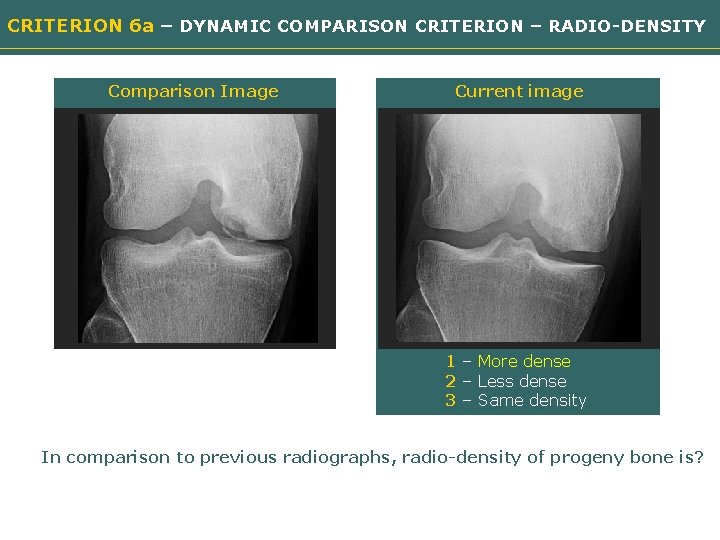
CRITERION 6 a – DYNAMIC COMPARISON CRITERION – RADIO-DENSITY Comparison Image Current image Need image 1 – More dense 2 – Less dense 3 – Same density In comparison to previous radiographs, radio-density of progeny bone is?
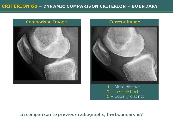
CRITERION 6 b – DYNAMIC COMPARISON CRITERION – BOUNDARY Comparison Image Current image Need image 1 – More distinct 2 – Less distinct 3 – Equally distinct In comparison to previous radiographs, the boundary is?