X ray is an electromagnetic radiation X ray


 for a mobile C-arm fluoroscopy system")


 for")


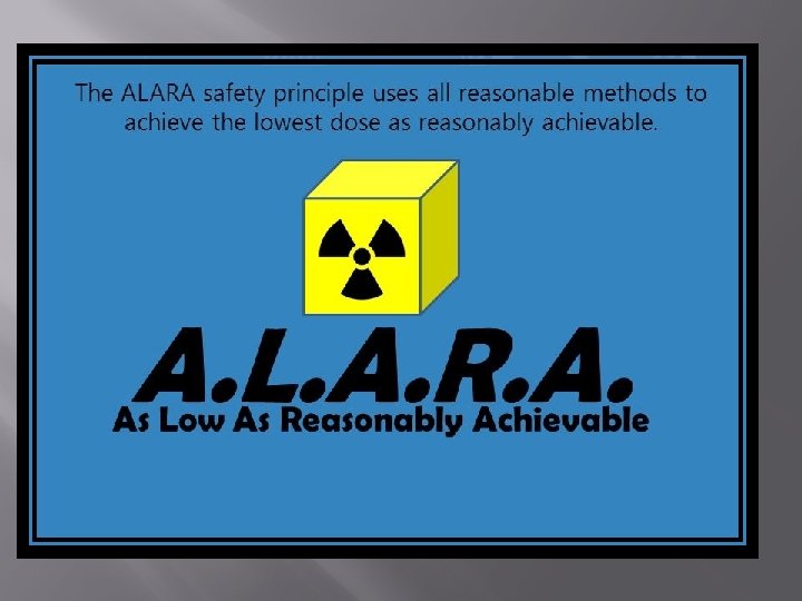
 principle Minimize time Use shielding Maximize")


")


- Slides: 50
X ray is an electromagnetic radiation
X ray production A m & k concepts Vp are key
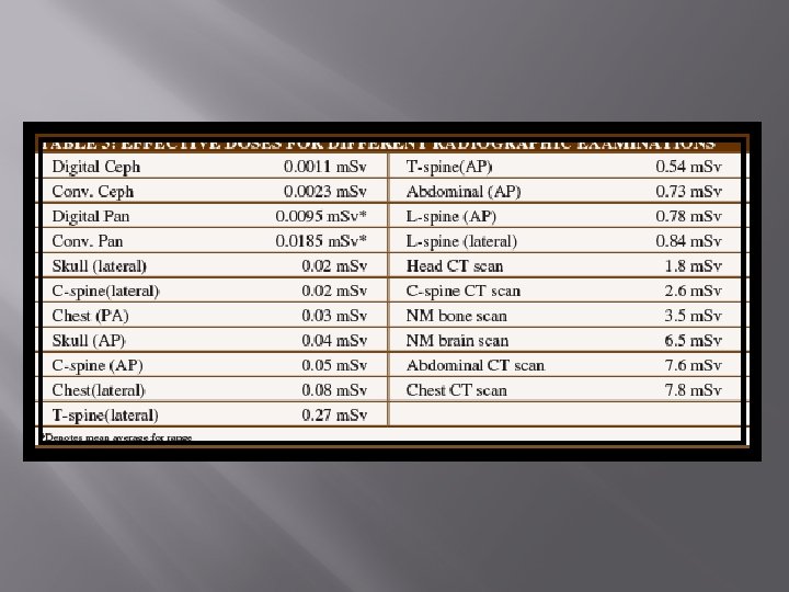
dose k. Vp m. As image
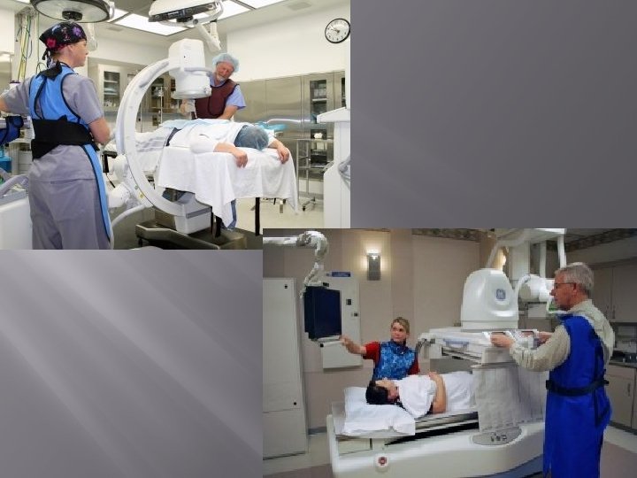
Flouroscopy system
Thoracic fluoroscopy using handheld fluorescent screen, 1909. No radiation protection is used, as the dangers of x-rays were not yet recognised
Surgical operation during World War I using a fluoroscope to find embedded bullets
Pedscope!!!
Biological effects
Biological effects deterministic Early Nausea diarrhea skin lesions hair loss bleeding cytopenia infertility ………. late cataract stochastic late Somatic cancer Hereditary Genetic teratogenic
Biological effects, quantification REM , Sivert 1 Si = 100 Rem Rontgen RAD , Gray 1 Gy = 100 RAD
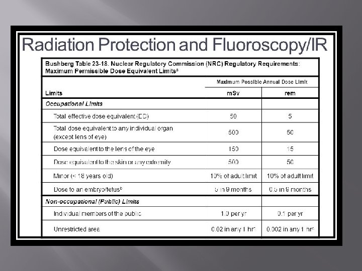
Radiation protection TDS
Typical occupational skin absorbed dose rates near fixed fluoroscopic equipment in the absence of protective aprons and drapes: (a) over-couch and (b) under-couch X-ray tube.
Typical isodose curves (in μGy/min) for a mobile C-arm fluoroscopy system
Collimation effect
Back scattered radiation
standing in relation to the X ray tube during a fluoroscopic procedure
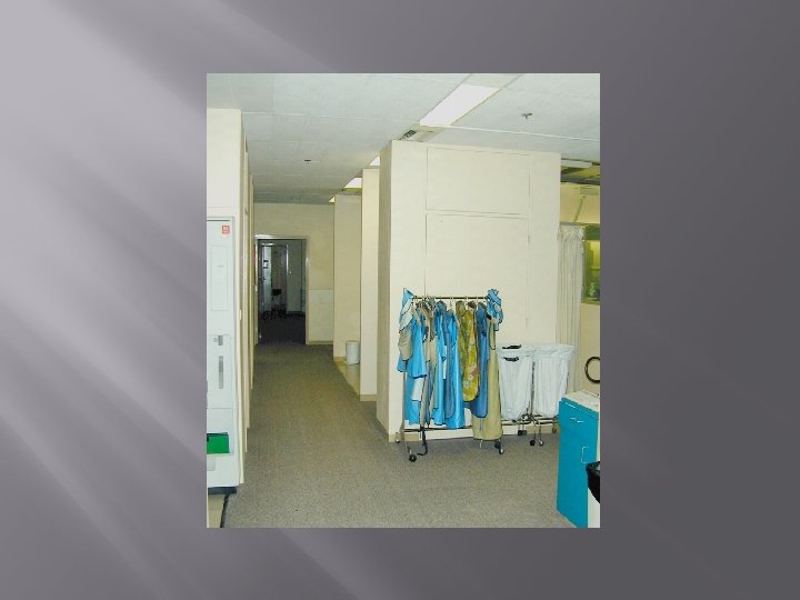
shielding
10 PEARLS: RADIATION PROTECTION OF PATIENTS IN FLUOROSCOPY
1. Maximize distance between the X ray tube and the patient to the extent possible
2. Minimize distance between the patient and the image receptor
3. Minimize fluoroscopy time Keep records of fluoroscopy time and DAP/KAP (if available) for every patient
4. Use pulsed fluoroscopy with the lowest frame rate possible to obtain images of acceptable quality
5. Avoid exposing the same area of the skin in different projections Vary the beam entrance port by rotating the tube around the patient
6. Larger patients or thicker body parts trigger an increase in entrance surface dose (ESD)
7. Oblique projections also increase ESD Be aware that increased ESD increases the probability of skin injury
8. Avoid the use of magnification Decreasing the field of view by a factor of two increases dose rate by a factor of four
9. Minimize number of frames and cine runs to clinically acceptable level Documentation should be performed with last image hold whenever possible and not with cine images
10. Use collimation Collimate the X ray beam to the area of interest
10 PEARLS: RADIATION PROTECTION OF STAFF IN FLUOROSCOPY Reducing patient dose always results in staff dose reduction
1. Use protective devices! q. Advisable skirt type lead apron to distribute weight q 0. 25 mm lead equivalence but with overlap on front to make it 0. 5 mm on the front and 0. 25 mm on the back Lead glass eyewear with side protection q (Provides >90% protection) Thyroid protection
2. Make good use of time-distance -shielding (TDS) principle Minimize time Use shielding Maximize distance as much as clinically possible
3. Use ceiling suspended screens, lateral shields and table curtains � � They provide more than 90% protection from scattered radiation in fluoroscopy Mobile floor shielding is advisable when using cine acquisition
4. Keep hands outside the primary beam unless totally unavoidable Hands inside the central area of the primary beam will increase exposure factors (k. V, m. A) and doses to patient and staff
5. Only 1 -5% of radiation falling on the patient’s body exits the other side Stand on the side of the transmitted beam (i. e. by the detector), which contains only 1 -5% of the incident radiation and its respective scatter
6. Keep X ray tube under the patient table and not over it Undercouch systems provide better protection from scattered dose
7. Use personal dosimetry- Use at least two dosimeters � � � One inside the apron at chest level One outside the apron at neck or eye level Additional finger ring dosimeter for procedures requiring hands close to primary beam
8. Update your knowledge about radiation protection
9. Address your concerns about radiation protection to radiation protection specialists (medical physicists)
10. REMEMBER �Quality control testing of fluoroscopy equipment enables safe and stable performance �Know your equipment! Using the equipment’s features appropriately will help reduce doses to patients and staff �Use injector devices