Write Unit 07 Skeletal System Write Latin Root


 • • •")


 • Label the bones in your packet")


 anteriorly • Retraction: Movement of the")


 Epiphyseal plate (hyaline cartilage) Joint between first rib and")

 Articular")


- Slides: 62
Write Unit 07 Skeletal System
Write. Latin Root Word Definition Peri- Around Osteo Bone Physis Growth Dia- Through Medulla Pith or marrow -blast Build -clast destroy Articular Small connecting part Condyle Rounded process Crest Ridge-like projection Foramen Opening in a bone (hole) Fossa Passageway Process Prominent projection Trochanter Relatively large process Tubercle Small, knoblike process
Write 7. 1 Introduction Structure and Function of the skeletal system
Listen
Write Skeletal System Organs Functions • Bones • Joints • Provide points of attachment for muscles • Protect and support • House blood producing cells • Store inorganic salts
Write 7. 2. 1 Bone Structure Axial and Appendicular
Listen
Listen Axial Skeleton Appendicular Skeleton
Listen
Color on your handout Axial Skeleton Appendicular Skeleton
Write 7. 2. 2 Bone Structure Bone Overview
Write 7. 2. 3 Bone Structure 4 Types of Bone
Listen
Listen
Listen
Listen
Listen
Write Types of Bones • Long Bones • Longer than they are wide • Ex. Femur, Humerus • Short Bones • About as long as they are wide • Carpals and tarsals • Irregular Bones • Oddly shaped; usually have foramen(holes) for nerves • Vertebrae, sacrum • Flat Bones • Flat and used mostly for protection • Skull, sternum
Listen Long Short Flat Irregular Humerus Carpals Cranium Vertebrae Radius Tarsals Sternum Sacrum Ulna Patella Ribs Metacarpals Scapula Femur Ilium Tibia Ischium Fibula Metatarsals Phalanges
Directions • Get a text book • Go to page 128 and read 7. 2 Bone structure • Use your text book to fill out the Long Bone Structure handout • On the back of the skeleton I gave you
Cranium Mandible Cervical Vertebrae Thoracic Vertebrae Lumbar Vertebrae Ilium Sacrum Ischium Clavicle Scapula Sternum Ribs Humerus Radius Ulna Carpals Metacarpals Phalanges Femur Patella Tibia Fibula Calcaneus Tarsals Metatarsals Phalanges
Write Bone Tissue • Matrix • Collagen Fibers • Hydroxyapatite (Mineral) • • • Cells calcium phosphate calcium carbonate calcium fluoride calcium hydroxide and citrate. • Osteocytes • Osteoblasts • Osteoclasts “Super. That’s how milk makes you feel. The calcium helps your bones grow strong, so even if you’re not from Krypton™ you can have bones of steel”
Listen
Write Two Types of Bone Compact Spongy • Weight Bearing • Support • Made up of Osteons • Allows for infiltration of blood vessels and nerves • Red Marrow Creation
Write Central Canal Lamellae Osteocytes Canaliculi *little canals Lacunae Osteon
y g n o Sp one B ct a p m Co one B Osteon Central Canal Periosteum Nerve Central Canal Blood Vessel Perforating Canaliculi Osteocyte Lacunae
Bone Growth Write Epiphyseal Plate Secondary Ossification Center Primary Ossification Center Cartilage 1. Periosteum begins to form 2. Cartilage breaks down and spongy bone is built 3. Bone is hooked up to blood supply 4. Primary and secondary ossification centers form spongy and compact bone 5. Primary and secondary ossification centers continue to grow until they’ve turned the epiphyseal plate into the epiphyseal line
Listen
Write Bone Cell Types • Osteoblasts • Lay down bone matrix • Osteoclasts • Destroy bone matrix • Osteocytes • Manage osteoblasts and osteoclasts
Write Bone Remodeling • Resorption • Osteoclasts dissolve bones • Ossification • Osteoblasts build new bone
Assignment Osteoporosis Article • http: //anatomyandphysiologyi. com/osteoporosis/ • http: //www. niams. nih. gov/health_info/bone/Bone _Health/Exercise/default. asp • Assignment: • In your own words, describe what happens to people who have osteoporosis (make sure to use the previously covered terminology). You should also cover who is at risk, symptoms and ways you can treat/prevent it.
Watch Can we Grow Bones? • http: //ed. ted. com/lessons/how-to-grow-a-bonenina-tandon
Daily Warm Up 1. What is the periosteum? 2. Which part of the long bone is made up of compact bone? 3. Your bones start out as ______ 4. What is the epiphyseal line?
Daily Warm Up 1. What is the periosteum? Tissue that surrounds the bone 2. Which part of the long bone is made up of compact bone? Diaphysis Cartilage 3. Your bones start out as ______ 4. What is the epiphyseal line? Remnant of the epiphyseal plate, a slice of cartilage between the primary and secondary ossification centers that was converted into bone.
Bone Packet Reading • 7. 7 (142) • Label the bones in your packet • Vertebral Column • 7. 8 (147) • Study the bones in your packet • Ribs • 7. 10 (148) • • Humerus Radius Ulna Carpals • 7. 11 (151) • Pelvis • 7. 12 (154) • Tibia • Fibula • Tarsals • Be prepared for a quiz by the end of class
Straight Line To Pinky 1. 2. 1. 2. 3. 4. 5. 6. 7. 8. Scaphoid Lunate Triquetrium 3. Pisiform Hamate Capitate Trapezoid Trapezium (by the thumb) L S T P H C TT Here Comes The Thumb
Daily Warm Up 1. What are three cells found in bone tissue and what do they do? 2. What is an osteon? 3. The tissue that connects bone to bone is called? 4. The tissue that connects muscle to bone is called? 5. Describe how the ulna attaches onto the Humerus.
Daily Warm Up 1. What are three cells found in bone tissue and what do they do? Osteocyte: Manager Osteoblast: Builder Osteoclasts: Destroyer 2. What is an osteon? Unit that builds up compact bone 3. The tissue that connects bone to bone is called? Ligament 4. The tissue that connects muscle to bone is called? Tendon 5. Describe how the ulna attaches onto the Humerus. The olecranon and coronoid processes of the ulna hook into the olecranon and coronoid fossa of the humerus
Daily Warm Up Tarsals Word Bank Triquetrium Lunate Pisiform Scaphoid Calcaneus Talus 1. 2. 3. Cuboid 4. Lateral 5. Cuneiform Intermediate 6. Cuneiform 7. Medial Navicular Cuneiform Calcaneus Cuboid Intermediate Cuneiform Lateral Cuneiform Medical Cuneiform Navicular Talus Carpals Word Bank Capitate Hamate Lunate Pisiform Scaphoid Trapezium Trapezoid Triquetrium Hamate Capitate 10 9. 8. 11. 12. 13. 14. 15. Trapezium Trapezoid
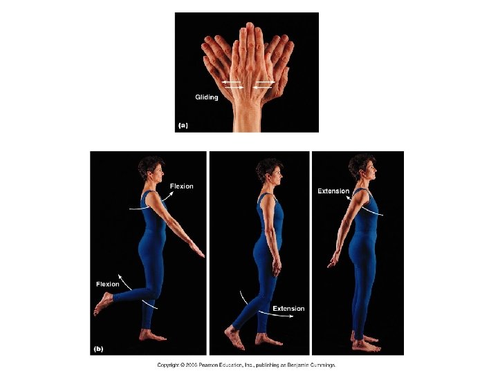
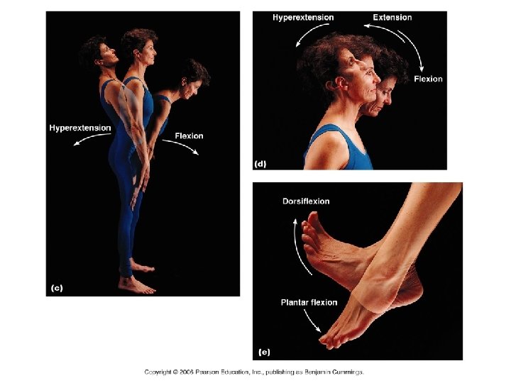
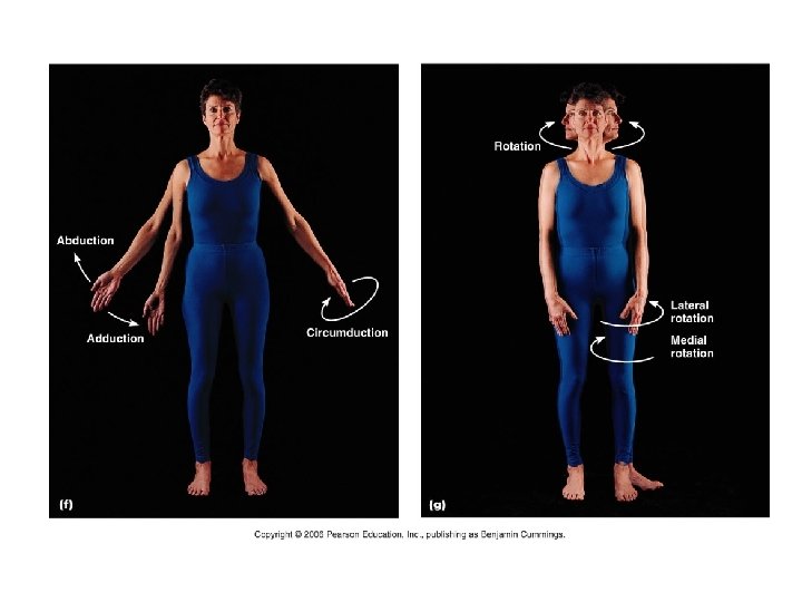
Body Movements • Adduction: Movement toward the midline • Abduction: Movement away from the midline • Flexion: Decreases the angle between two body parts • Extension: Increases the angle between two body parts – usually to 180 degrees • Hyper Extension: Increases the angle between two body parts – usually past 180 degrees • Dorsiflexion • Bends foot towards the ankle • Plantar flexion • Bends foot towards the ground
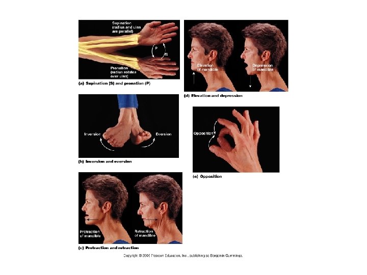
Body Movements • Rotation • Medial Rotation: rotation towards the midline • Lateral rotation: rotation away from the midline • Supination: palm up • Pronation: Palm down
Body Movements • Protraction: Movement of body (jaw) anteriorly • Retraction: Movement of the body (jaw) posteriorly • Eversion: turning sole outward; weight on inner edge of the foot • Inversion: turning the sole inward Elevation: movement upward, as in shrugging the shoulders Depression: movement downward
Write Articulations/Joints • Articulation: Joint
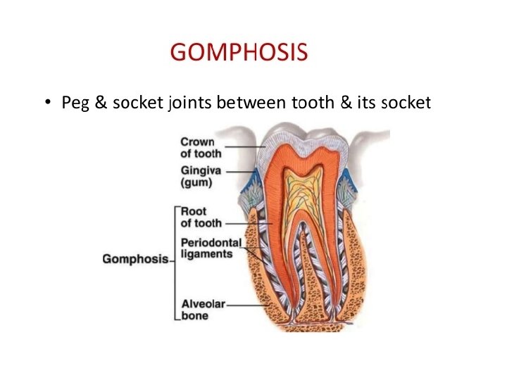
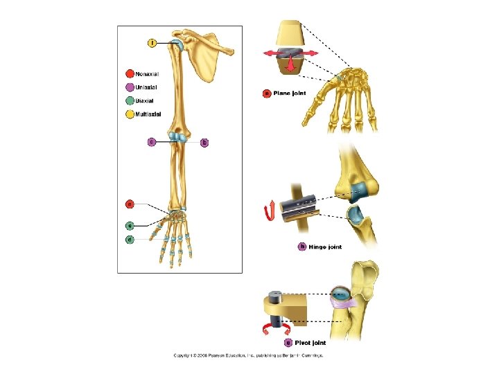
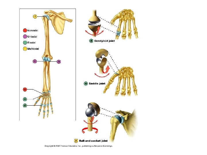
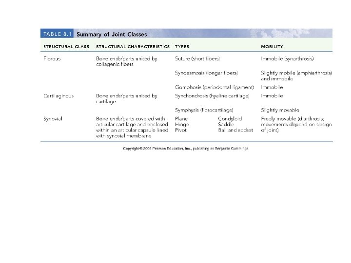
Write Types of joints 1. Fibrous joints 1. 2. 3. Sutures Syndesmoses Gomphosis 2. Cartilaginous joints 3. Synovial joints 1. 2. 3. 4. 5. 6. Plane (gliding) Hinge Pivot Condyloid Saddle Ball and socket
Listen Human Anatomy and Physiology, 7 e by Elaine Marieb & Katja Hoehn Sutures Copyright © 2007 Pearson Education, Inc. , publishing as Benjamin Cummings.
Listen Syndesmosis
Write 1. Fibrous joints • Characteristics • Joined by fibrous tissue. • No joint cavity. • Seldom movement. • Types • Sutures • Interlocked by connective tissues and irregular edges between bones (skull). • Syndesmoses • Articulated bones connected by ligaments and bones that do not interlock. (Fibula and Tibia) • Gomphosis: tooth secured by periodontal ligament.
Cartilaginous joints Synchondroses Sternum (manubrium) Epiphyseal plate (hyaline cartilage) Joint between first rib and sternum (immovable) (a) (b) Fibrocartilaginous intervertebral disc Body of vertebra (c) Symphyses Human Anatomy and Physiology, 7 e by Elaine Marieb & Katja Hoehn Copyright © 2007 Pearson Education, Inc. , publishing as Benjamin Cummings.
2. Cartilaginous joints • Characteristic • Articulating bone ends are connected by a plate of cartilage.
General structure of a synovial joint Periosteum Ligament Joint cavity (contains synovial fluid) Articular (hyaline) cartilage Fibrous capsule Synovial membrane Articular capsule (b) (a) Human Anatomy and Physiology, 7 e by Elaine Marieb & Katja Hoehn Copyright © 2007 Pearson Education, Inc. , publishing as Benjamin Cummings.
3. Synovial joints • Characteristics • Synovial Fluid
Friction-reducing structures: Bursae and tendon sheaths Coracoacromial ligament Subacromial bursa Acromion of scapula Coracoacromial ligament Subacromial bursa Fibrous articular capsule Cavity in bursa containing Glenoid cavity synovial fluid containing synovial fluid Hyaline cartilage Tendon sheath (b) Synovial membrane Fibrous capsule Tendon of long head of biceps brachii muscle Humerus (a) Human Anatomy and Physiology, 7 e by Elaine Marieb & Katja Hoehn Copyright © 2007 Pearson Education, Inc. , publishing as Benjamin Cummings.
Types of Synovial Joints • Types of synovial joints • Hinge • Elbow • Pivot • forearm • Ball and socket • Shoulder and hip • Ellipsoid • wrist • Saddle • Thumb • Plane • Carpals and Tarsals https: //www. youtube. com/watch? v=0 c. Yal_hitz 4
Unit 07 Review • Axial and Appendicular Skeleton • Bone Types • Long, Short Flat, and Irregular • Parts of a Long Bone • Osteon Anatomy and Function • Bone Growth • Osteocytes, Osteoblasts, Osteoclasts, Resorption and Ossification • Three Types of Joints • Know movement and where to find them • Difference between tendons and ligaments • Gross Anatomy of the Skeleton • Body Movements