Write down 3 neurones and describe their structure

§ Write down 3 neurones and describe their structure and function. (you could do this as a table – or when you revisit your notes, you could re-write it as a table!) § Sensory neurones have long axons and transmit nerve impulses from receptors to an intermediate or motor neurone. The dendron carries impulse to the cell body and the axon that carries it away from the cell body. § Motor neurones have long axons and many short dendrites. They transmit nerve impulses to effectors (muscles and glands) all over the body. § Intermediate neurones (also called connector neurones or relay neurones) are usually much smaller cells. Transmit impulses between 2 neurones.
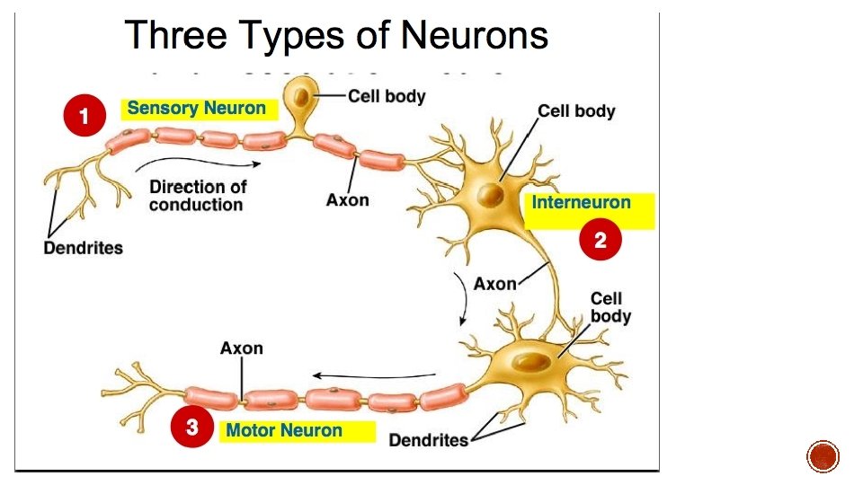

A dendron carries impulses towards a cell body Axons carry nerve impulses away from a cell body § Which of these are the correct structural and functional features of each of the neurones? Sensory neurone Motor neurone Relay neurone Short dendrites Long dendron No dendron Short axon Long axon Impulse carried from CNS receptor to CNS to effector Short dendrites Long dendron Short axon Long axon Impulse carried from receptor to CNS Impulse carried within CNS between neurones Impulse carried from CNS to effector Impulse carried within CNS between neurones

The autonomic nervous system is the part of the nervous system responsible for control of the bodily functions not consciously directed, e. g. breathing, the heartbeat, and digestive processes. The somatic nervous system is the part of the peripheral nervous system associated with skeletal muscle voluntary control of body movements. It consists of afferent nerves or sensory nerves, and efferent nerves or motor nerves.

Location of the reaction The light-dependent reaction takes place in the thylakoid membranes of the chloroplasts. Situated in these membranes are light-harvesting systems called photosystems. There are two types, photosystem I (PSI) and photosystem II (PSII). Both have chlorophyll at their centres. photosystem light chlorophyll 7 of 36 © Boardworks Ltd 2009

Light-dependent reaction 8 of 36 © Boardworks Ltd 2009

Light-dependent reaction: summary Cyclic photophosphorylation Non-cyclic photophosphorylation photolysis 9 of 36 © Boardworks Ltd 2009

Products of the light-dependent reaction that pass into the light-independent reaction: l reduced NADP l ATP Products of the light-dependent reaction that leave the plant: l oxygen Products of the light-dependent reaction that are re-used in another part of the light-dependent reaction: 10 of 36 l H+ ions l electrons © Boardworks Ltd 2009

Gross structure of the kidney Cortex Renal Vein Medulla Renal Artery Ureter 11 of 36 Medullary Pyramid Renal Pelvis © Boardworks Ltd 2009

Ultrastructure of the Kidney 12 of 36 © Boardworks Ltd 2009

Ultrastructure of the Kidney 13 of 36 © Boardworks Ltd 2009

Answers: Ultrastructure of the Kidney PCT Bowman’s Capsule Cortex Medullary pyramid Capillaries DCT Renal Artery Renal Vein Renal Pelvis 14 of 36 Ureter Venule Arteriole Loop of Henle Collecting Duct © Boardworks Ltd 2009

Answers Bowman’s Capsule Glomerulus Distal Convoluted Tubule Proximal Convoluted Tubule Loop of Henle Collecting Duct 15 of 36 Urine collects in the pelvis © Boardworks Ltd 2009

The PCT �Proximal Convoluted Tubule – the first section of kidney tubule after the Bowman’s Capsule!
 �Cells specialised")
Selective Reabsorption �Most reabsorption occurs in the proximal convoluted tubule (~85% filtrate) �Cells specialised in PCT for reabsorption: � Microvilli for increased surface area � Co-transporter proteins to transport glucose or amino acids (by facilitated diffusion) � Sodium-potassium pumps � Cytoplasm containing lots of mitochondria See Figure 2 on page 55 of textbook

Structure of the PCT • Cells of the PCT have microvilli on the inside which increase surface area and improve efficiency of selective reabsorption… 18 of 36 © Boardworks Ltd 2009

Selective Reabsorption 2 1. Sodium-potassium pumps remove sodium ions from cells lining PCT and into blood (to lower conc. of Na in the cells) 2. Sodium ions + glucose or amino acids transported into cells lining PCT from PCT lumen by facilitated diffusion 3. As concentrations of glucose or amino acids rise in tissues, they move by diffusion into blood 4. Reabsorption of glucose and amino acids reduces water potential in cell so water enters cells and then reabsorbed into blood by osmosis 5. Larger molecules reabsorbed by endocytosis 19 of 36 © Boardworks Ltd 2009

OVERVIEW

Collecting Duct • As the fluid passes into the collecting duct it has high water potential • It then passes through the salty medulla and water is removed from the filtrate until the urine/medulla are isotonic. • This is then affected by A. D. H

The effect of ADH on the collecting duct wall Osmoreceptors – loss of water causes them to shrink. This stimulates neurosecretory cells which causes passage of an action potential Leads to secretion of ADH

• • Day 1: Lining is shed to start new cycle Day 1: FSH is released – egg development Follicle cells release oestrogen This stimulates LH release • Day 14: high LH level causes egg to be released and travels down oviduct • Cells in ovary release progesterone • - lining thickens more • - LH and FSH release reduced • Uterus now ready TO RECEIVE FERTILISED EGG • PROGESTERONE MAINTAINS WALL AND PREVENTS MORE EGG DEVELOPMENT • If egg not fertilised, • Progesterone and oestrogen levels fall AND MENSTRUATION OCCURS • LOW PROGESTERONE AND OESTROGEN CAUSES FSH RELEASE FROM PITUITARY AND NEXT EGG DEVELOPS oestrogen
- Slides: 30