
WRIST HAND PTA 106 Unit 3 presentation by
WRIST & HAND PTA 106 Unit 3 presentation by: Lisa, Bobbi, Deanna, Chris

Surface Anatomy
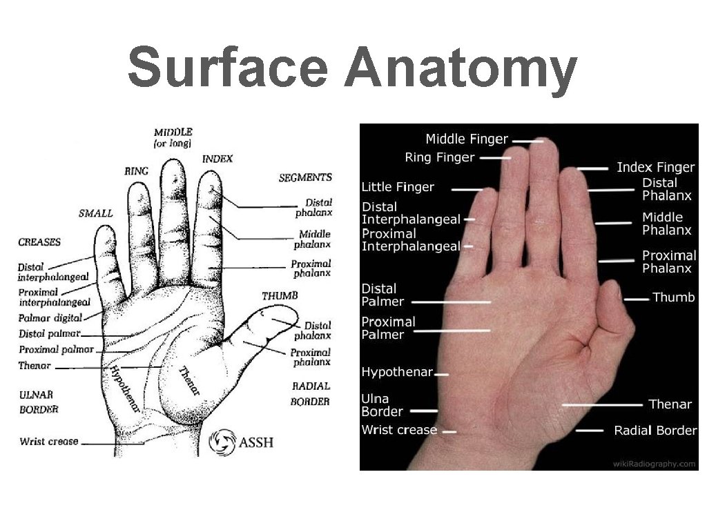
Surface Anatomy
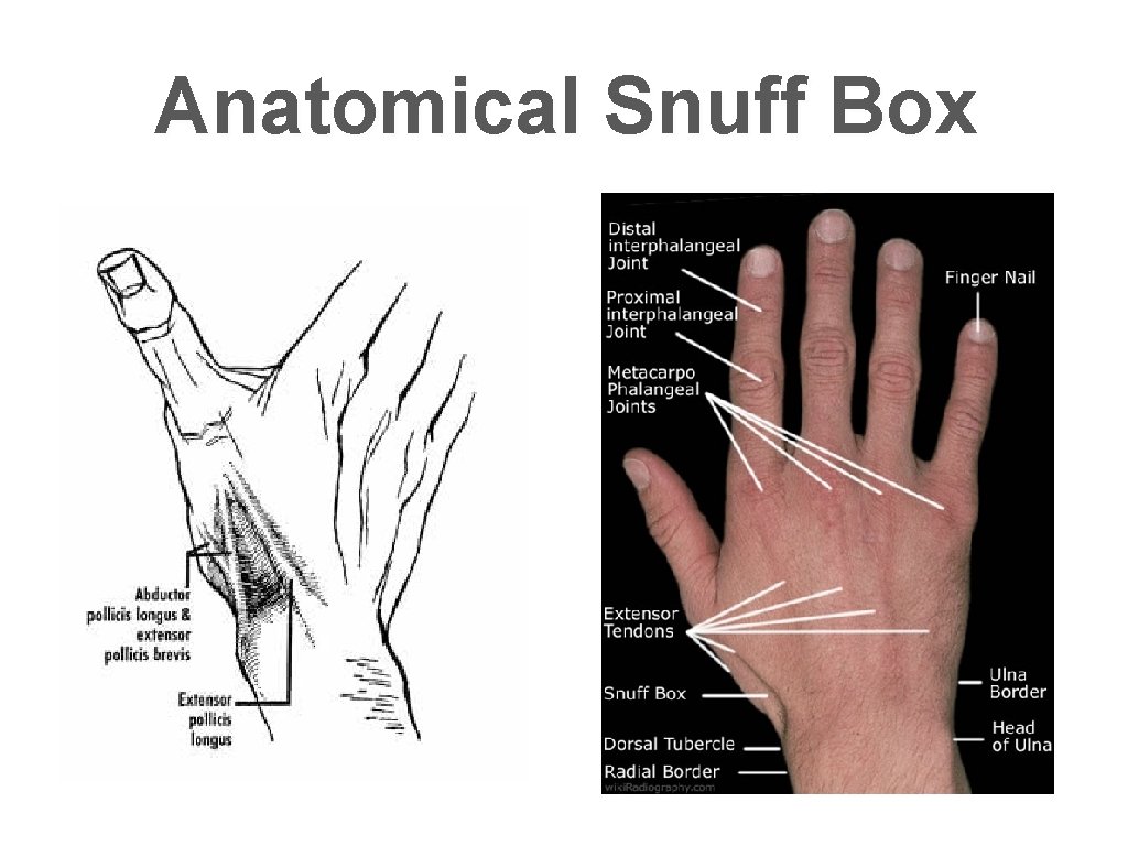
Anatomical Snuff Box
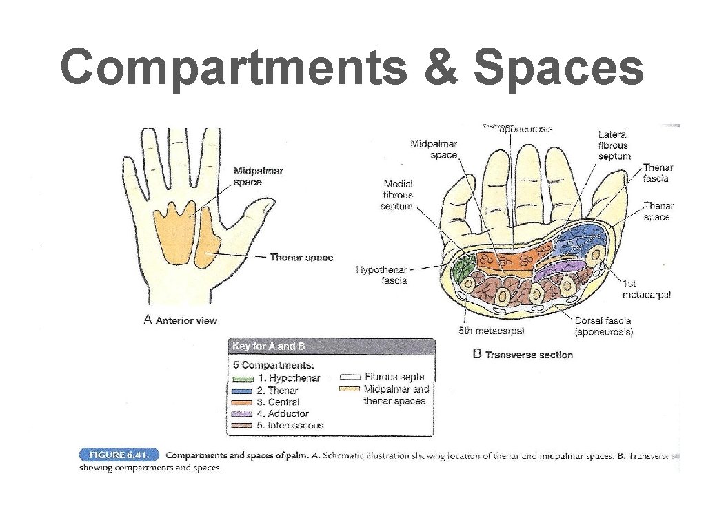
Compartments & Spaces
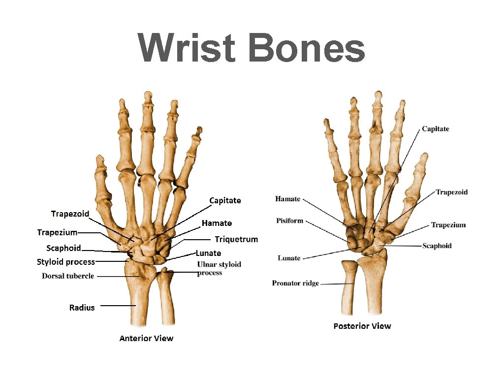
Wrist Bones
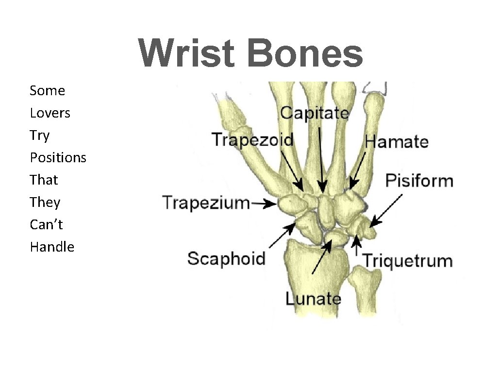
Wrist Bones Some Lovers Try Positions That They Can’t Handle
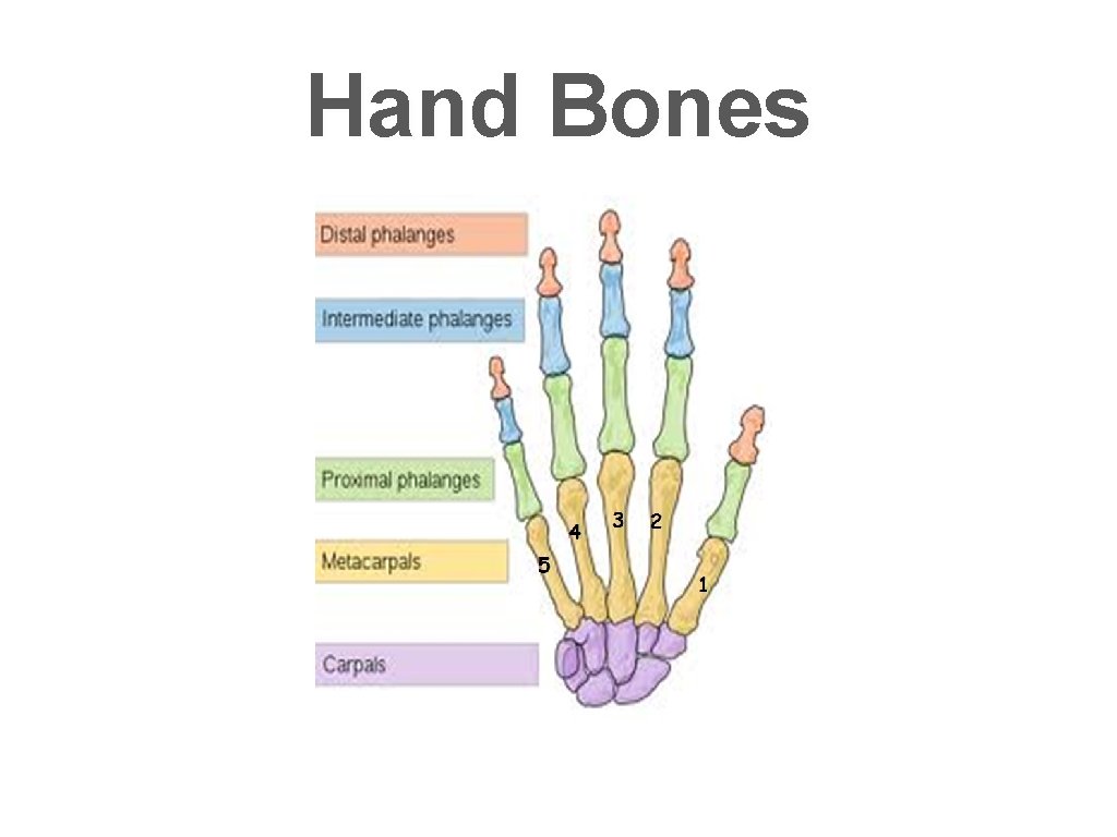
Hand Bones

Cartilage Articular Cartilage Is smooth shiny material that covers the bones of the hand wrist. It absorbs forces of impact and provides for low friction motion.
Articular Capsule Synovial Membrane Fibrous Layer • Is the inner membrane of a joint made up of loose connective tissue. • Secretes synovial fluid which services to lubricate the joint and reduce the friction between bones in joints. • Supplies nutrients to the joint • Formed by dense irregular connective tissue attached to the periosteum surface of joints. • Allows for movement. • Provides stability
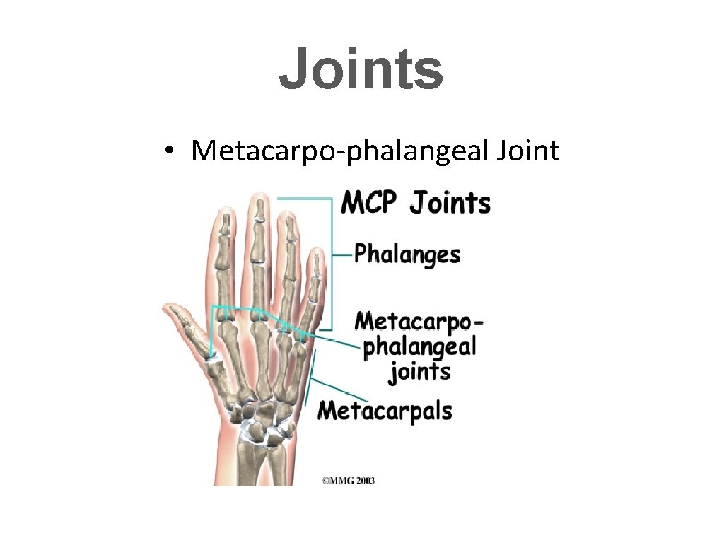
Joints • Metacarpo-phalangeal Joint
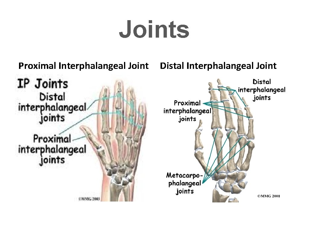
Joints Proximal Interphalangeal Joint Distal Interphalangeal Joint
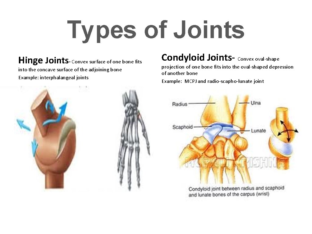
Types of Joints Hinge Joints- Convex surface of one bone fits into the concave surface of the adjoining bone Example: interphalangeal joints Condyloid Joints- Convex oval-shape projection of one bone fits into the oval-shaped depression of another bone Example: MCPJ and radio-scapho-lunate joint
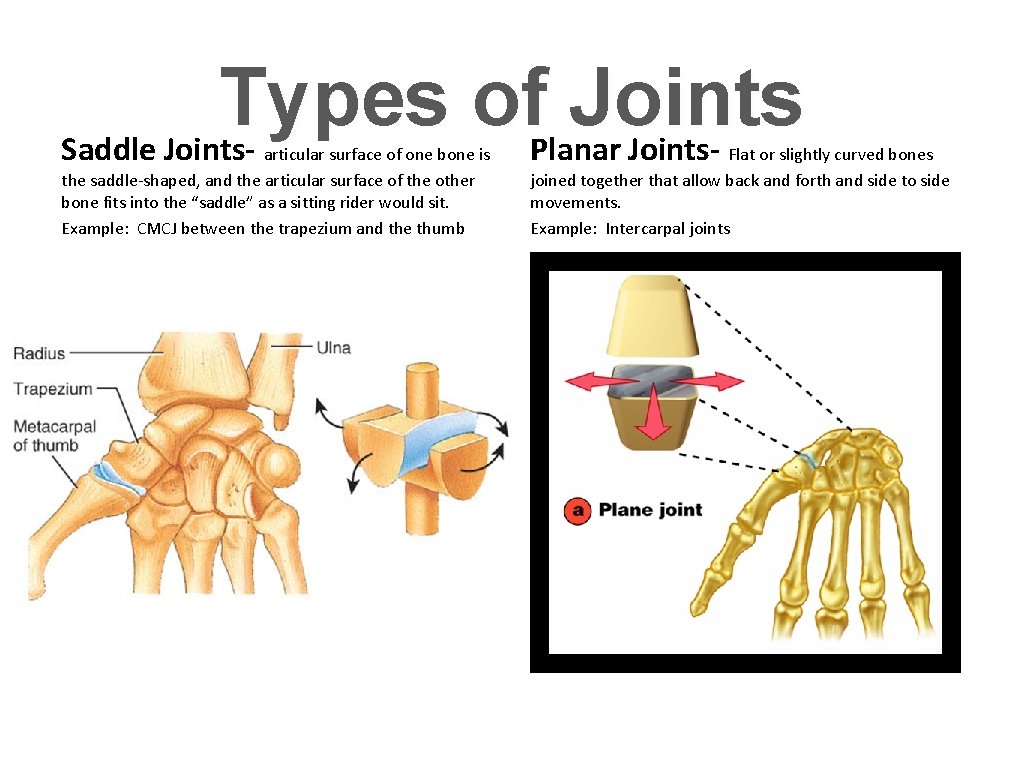
Types of Joints Saddle Joints. Planar Jointsarticular surface of one bone is the saddle-shaped, and the articular surface of the other bone fits into the “saddle” as a sitting rider would sit. Example: CMCJ between the trapezium and the thumb Flat or slightly curved bones joined together that allow back and forth and side to side movements. Example: Intercarpal joints
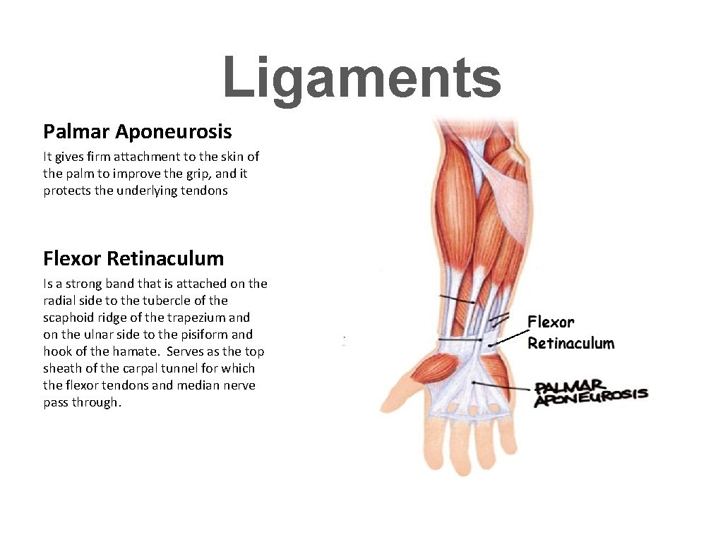
Ligaments Palmar Aponeurosis It gives firm attachment to the skin of the palm to improve the grip, and it protects the underlying tendons Flexor Retinaculum Is a strong band that is attached on the radial side to the tubercle of the scaphoid ridge of the trapezium and on the ulnar side to the pisiform and hook of the hamate. Serves as the top sheath of the carpal tunnel for which the flexor tendons and median nerve pass through.
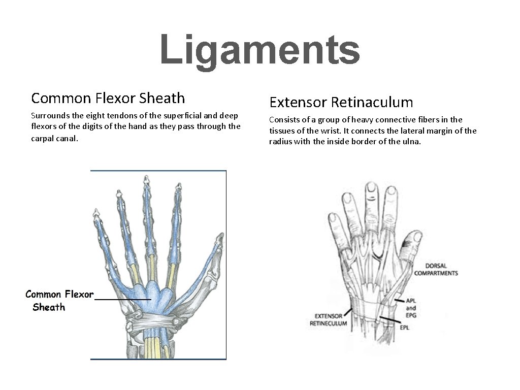
Ligaments Common Flexor Sheath Surrounds the eight tendons of the superficial and deep flexors of the digits of the hand as they pass through the carpal canal. Extensor Retinaculum Consists of a group of heavy connective fibers in the tissues of the wrist. It connects the lateral margin of the radius with the inside border of the ulna.
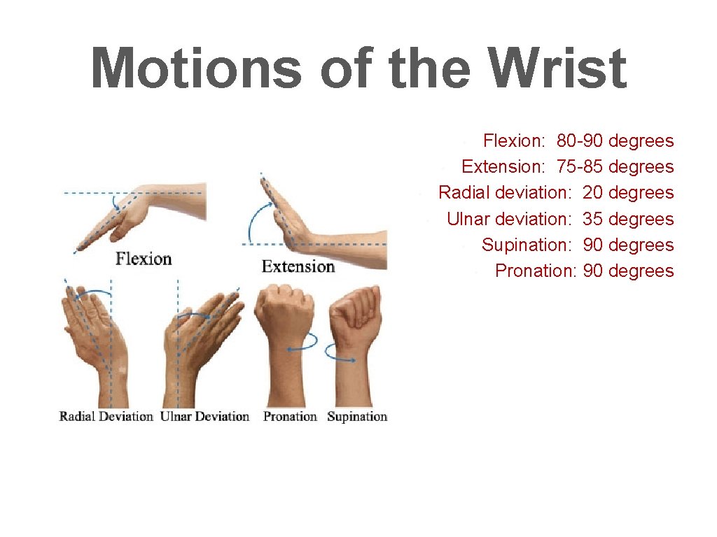
Motions of the Wrist Flexion: 80 -90 degrees • Extension: 75 -85 degrees Radial deviation: 20 degrees Ulnar deviation: 35 degrees • Supination: 90 degrees • Pronation: 90 degrees • • •
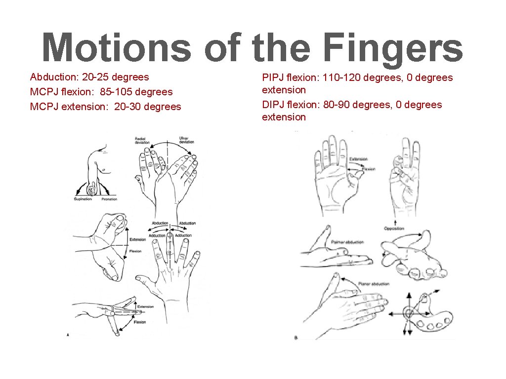
Motions of the Fingers Abduction: 20 -25 degrees MCPJ flexion: 85 -105 degrees MCPJ extension: 20 -30 degrees PIPJ flexion: 110 -120 degrees, 0 degrees extension DIPJ flexion: 80 -90 degrees, 0 degrees extension
Range of Motion of the Thumb • Thumb: CMCJ flexion: 60 -70 degrees Flexion at MCPJ: 85 -105 degrees Abduction: 70 -80 degrees Opposition- combined motion of abduction, flexion, and rotation of the thumb.
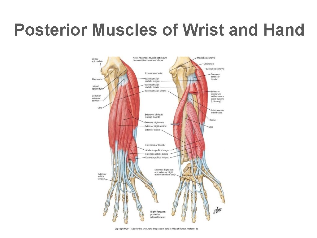
Posterior Muscles of Wrist and Hand
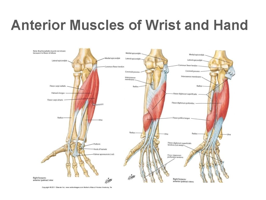
Anterior Muscles of Wrist and Hand
Nerve, Artery/ Veins of the Hand Wrist Debrief:
The Sinistra Nervous Family
Central Nervous System Aka “The Boss”
The “Under Boss”
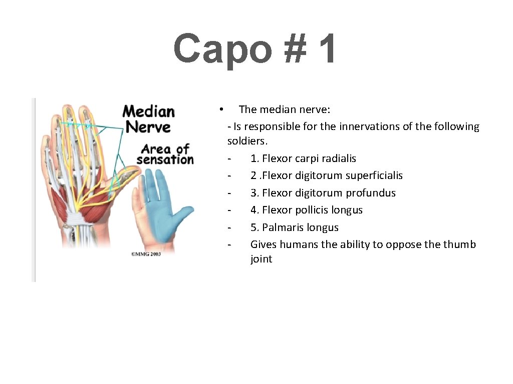
Capo # 1 • The median nerve: - Is responsible for the innervations of the following soldiers. 1. Flexor carpi radialis 2. Flexor digitorum superficialis 3. Flexor digitorum profundus 4. Flexor pollicis longus 5. Palmaris longus Gives humans the ability to oppose thumb joint
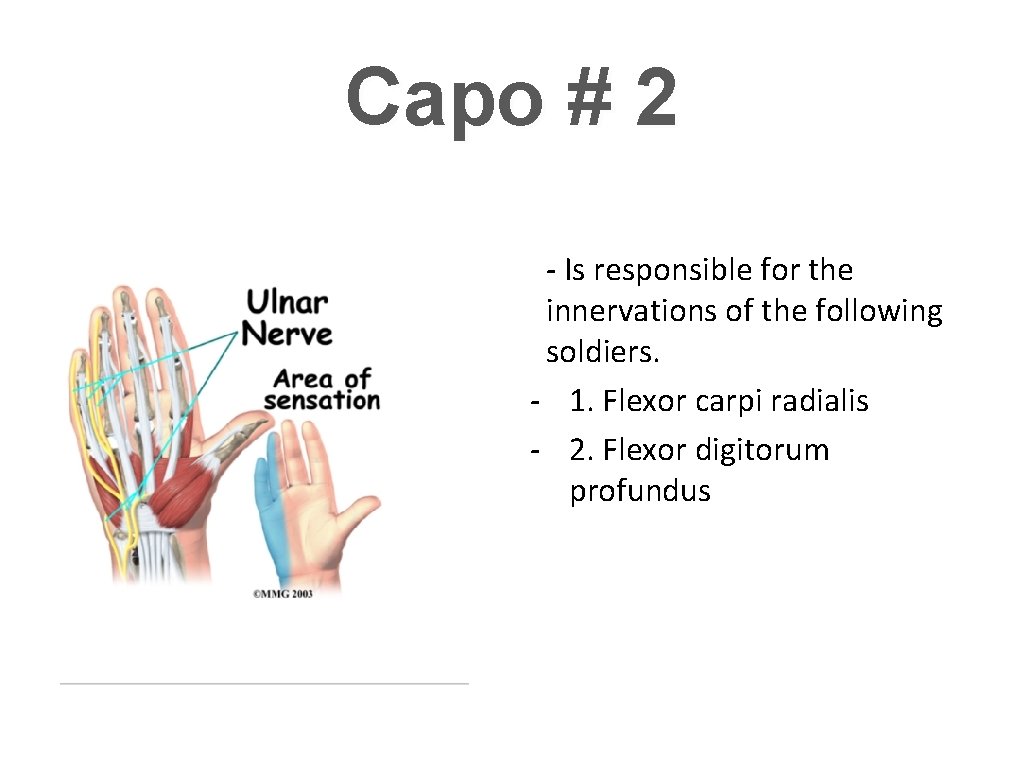
Capo # 2 - Is responsible for the innervations of the following soldiers. - 1. Flexor carpi radialis - 2. Flexor digitorum profundus
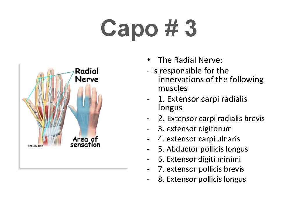
Capo # 3 • The Radial Nerve: - Is responsible for the innervations of the following muscles - 1. Extensor carpi radialis longus - 2. Extensor carpi radialis brevis - 3. extensor digitorum 4. extensor carpi ulnaris 5. Abductor pollicis longus 6. Extensor digiti minimi 7. extensor pollicis brevis 8. Extensor pollicis longus
Other known associates Dorsal cutaneous branch Vaga and Palmer cutaneous branch Winfield
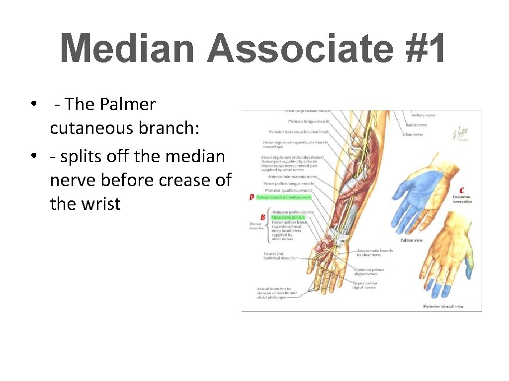
Median Associate #1 • - The Palmer cutaneous branch: • - splits off the median nerve before crease of the wrist
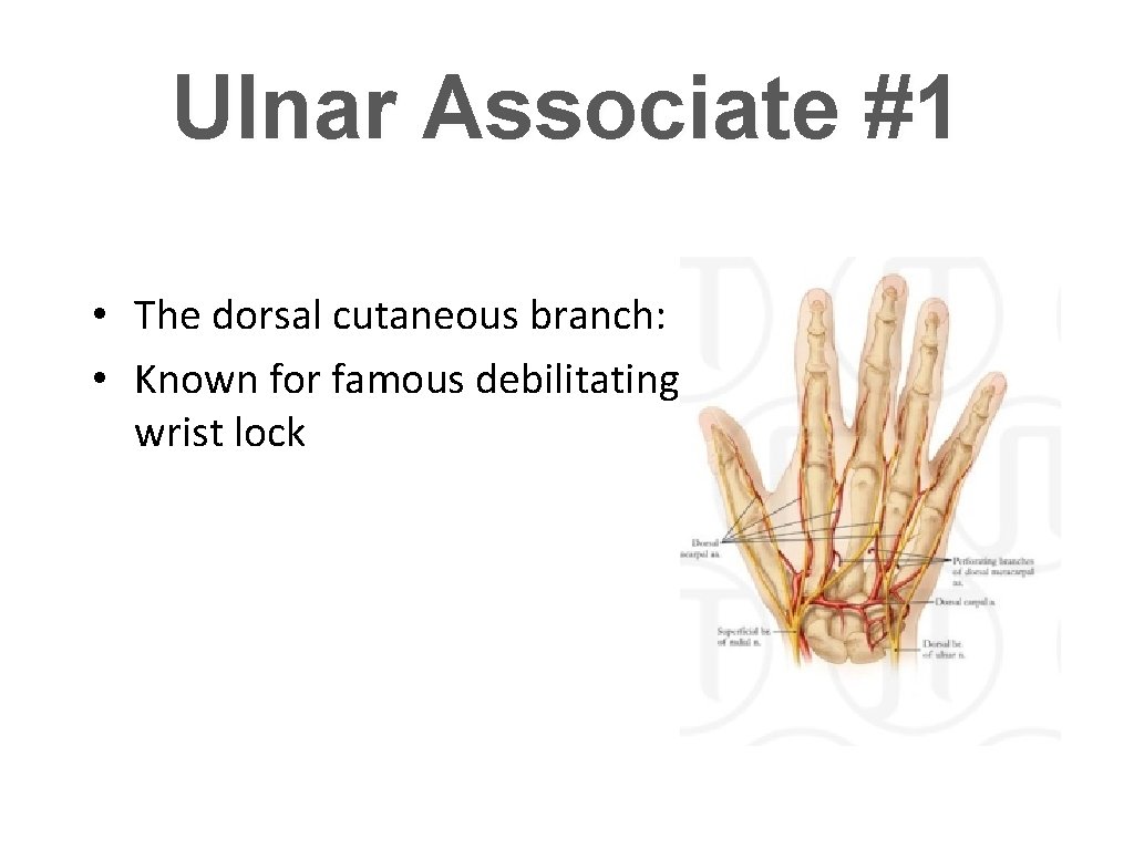
Ulnar Associate #1 • The dorsal cutaneous branch: • Known for famous debilitating wrist lock
There Will Be Blood
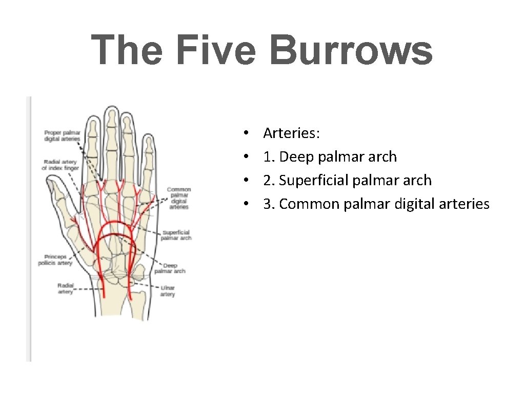
The Five Burrows • • Arteries: 1. Deep palmar arch 2. Superficial palmar arch 3. Common palmar digital arteries
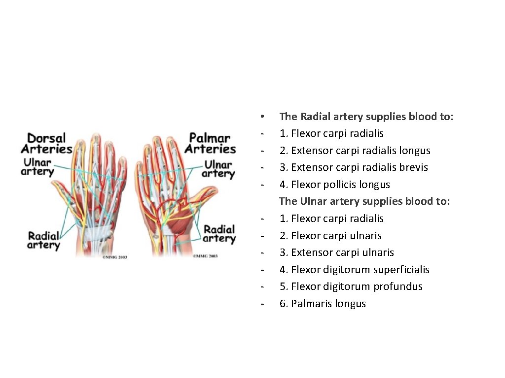
• - The Radial artery supplies blood to: 1. Flexor carpi radialis 2. Extensor carpi radialis longus 3. Extensor carpi radialis brevis 4. Flexor pollicis longus The Ulnar artery supplies blood to: 1. Flexor carpi radialis 2. Flexor carpi ulnaris 3. Extensor carpi ulnaris 4. Flexor digitorum superficialis 5. Flexor digitorum profundus 6. Palmaris longus
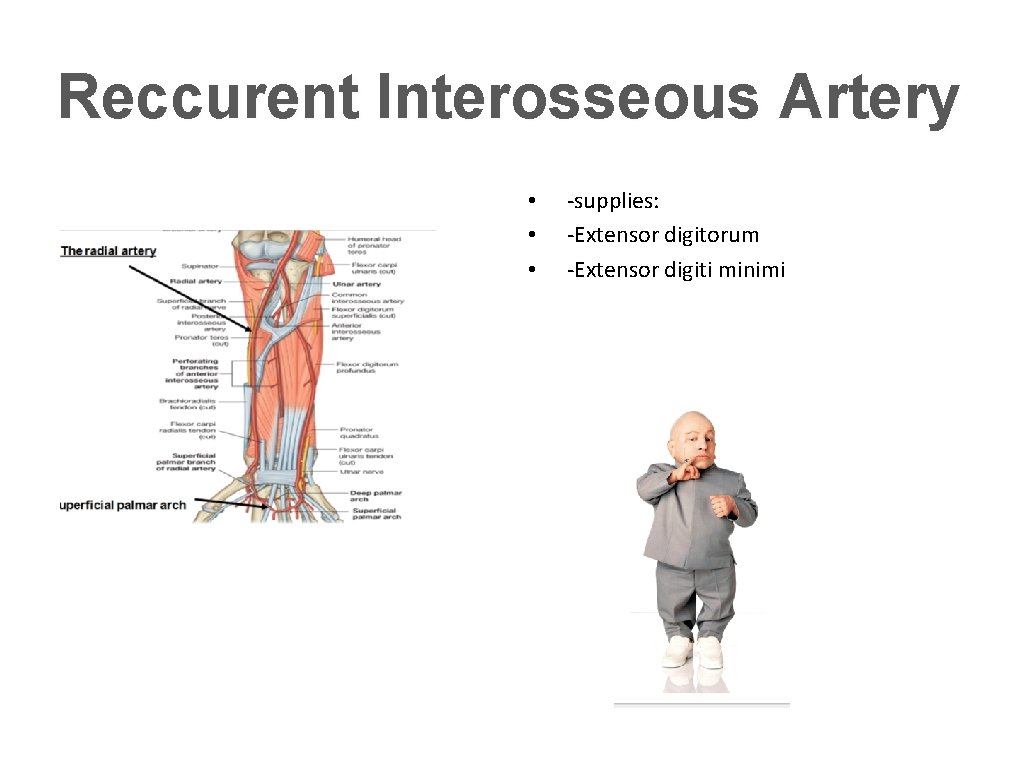
Reccurent Interosseous Artery • • • -supplies: -Extensor digitorum -Extensor digiti minimi
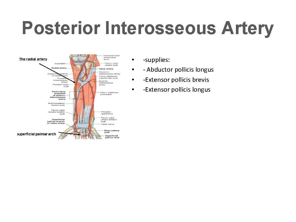
Posterior Interosseous Artery • • -supplies: - Abductor pollicis longus -Extensor pollicis brevis -Extensor pollicis longus
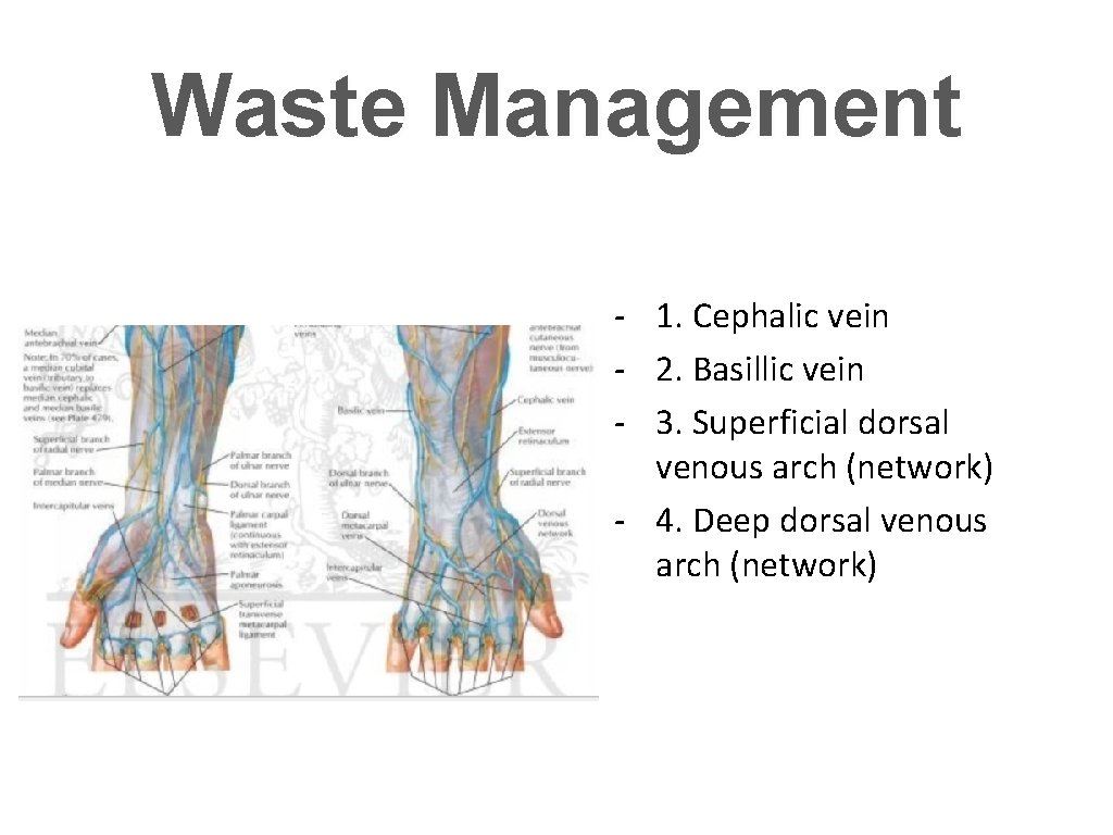
Waste Management - 1. Cephalic vein - 2. Basillic vein - 3. Superficial dorsal venous arch (network) - 4. Deep dorsal venous arch (network)
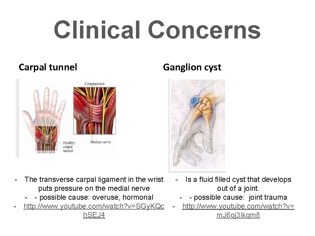
Clinical Concerns Carpal tunnel Ganglion cyst - The transverse carpal ligament in the wrist puts pressure on the medial nerve - - possible cause: overuse, hormonal - http: //www. youtube. com/watch? v=SGy. KQc h. SEJ 4 - Is a fluid filled cyst that develops out of a joint. - - possible cause: joint trauma - http: //www. youtube. com/watch? v= m. J 6 oj 3 lkqm 8
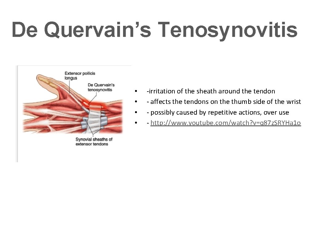
De Quervain’s Tenosynovitis • • -irritation of the sheath around the tendon - affects the tendons on the thumb side of the wrist - possibly caused by repetitive actions, over use - http: //www. youtube. com/watch? v=q 87 z. SRYHa 1 o

Spoiler Alert
- Slides: 40