
WOUND MANAGEMENT By Jenn Dailey RN BSN DIFFERENT
WOUND MANAGEMENT By: Jenn Dailey, RN, BSN
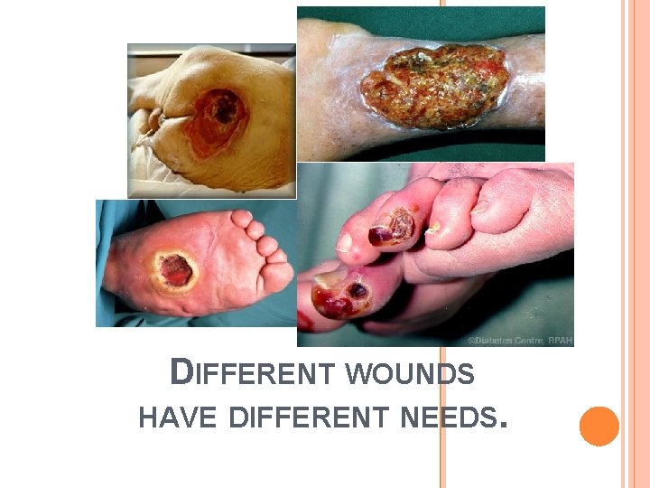
DIFFERENT WOUNDS HAVE DIFFERENT NEEDS.
Venous, Arterial and Diabetic Ulcers
For any lower extremity wound treatment, make sure you have the results from an ABI test. a) Ankle Brachial Index (ABI) • Test used to rule out significant arterial disease and to determine amount of compression that can be safely applied • Ratio of ankle systolic blood pressure over brachial systolic blood pressure. • Anything less than 0. 5 is ischemia and compression therapy is contraindicated. A referral to a vascular surgeon for evaluation is appropriate to regain circulation. • Not accurate in Diabetes
Compression • Promotes venous blood return, prevents blood pooling, decreases edema, improves efficiency of calf pump, enhances valve function, reverses capillary leak, reduces the pressure differential • Contraindicated in de-compensated CHF, peripheral arterial disease, ABI < 0. 5 • How much compression? • Modified (Low level) up to 23 mm. HG at ankle • ABI 0. 6 -0. 8 • High (Therapeutic level) up to 40 mm. HG at ankle • ABI > 0. 8
VENOUS ULCERS a) Result of malfunctioning venous valves causing pressure in the veins to increase which causes blood to pool when it is not as efficiently pumped back towards the heart. b) The increase in pressure stretches the vein walls, allowing proteins in the blood and blood cells to leach into the subcutaneous tissues resulting in edema and the eventual breakdown of tissues due to lack of oxygen and nutrients c) Symptoms: 1. Scaling and erythema, brownish/yellow patches under the skin 2. Dark red or purple skin coloring 3. Venous ulcer will present with shallow but large wounds with irregular margins. The base of the ulcer is typically red, can result in significant amount of exudate and will ooze venous blood when manipulated 4. Wound is relatively painless, pain would only be from infection or edema 5. Typically along the medial or lateral lower leg
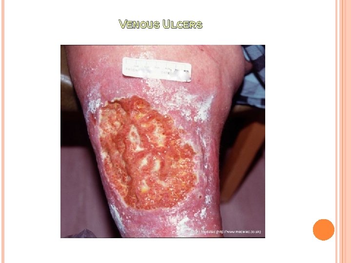
VENOUS ULCERS
VENOUS ULCERS Treatment of Venous Ulcers 1. Elevate Extremities 2. CORE i. Compression ii. Optimize local wound environment a) Keep wound from getting infected b) Alleviate edema c) Debridement if needed d) Wound should typically be moist e) Change dressings as infrequently as possible f) Skin grafts may be needed iii. Review contributing factors (obesity, DVT, pregnancy, incompetent valves, CHF, muscle weakness, decreased activity) iv. Establish a maintenance plan i. Examine feet and between toes daily for changes in color or development of sores ii. Avoid sitting or standing for extended periods of time iii. Avoid crossing legs iv. Quit smoking v. Elevate legs regularly vi. Exercise
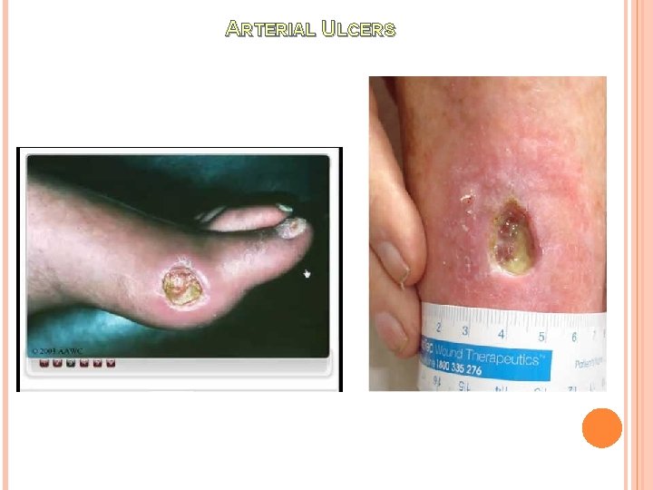
ARTERIAL ULCERS a) b) Result of poor perfusion to lower extremities. The overlying skin and tissues are deprived of oxygen and nutrients, killing the tissues and causing an ulcer. Also, the lack of blood supply can result in minor scrapes/cuts from healing and developing into ulcers Symptoms: Round in shape with well-defined, even wound margins Often found between or on the tips of toes, outer ankle, or where there is pressure from walking or footwear Wounds are characteristically deep, often extending down to underlying tendons, and will frequently display no signs of new tissue growth Base of wound typically does not bleed, and is yellow, brown, grey, or black in color Often the limb will feel cool to the touch and the extremity will have little to no distinguishable pulse Skin and nails on the extremity will appear atrophic, with hair loss, and skin will appear shiny, thin, dry, and taut in appearance Delayed capillary return These ulcers are generally very painful, especially when exercising, at rest or during the night 1. 2. 3. 4. 5. 6. 7. 8. i. Source of temporary relief is dangling affected leg(s) over the edge of the bed, allowing gravity to aid blood flow to the ulcer
ARTERIAL ULCERS
ARTERIAL ULCERS Treatment of Arterial Ulcers 1. Get it diagnosed – ABI, angiogram 2. If ischemia is not corrected, wound healing will not occur despite topical treatment. 3. Revascularization – Done by vascular surgeon 4. Angioplasty – Stent placement 5. Topical treatment 1. Stable gangrene (dry, hard, and wrinkled brown or black texture) – keep dry, no debridement, paint with alcohol, betadine or skin prep 2. Unstable gangrene (moist, gross swelling and blistering) – EMERGENT! Debridement required 6. Pain management 7. Eliminate tobacco use 8. Walk for 30 minutes 3 x/week 9. Weight control 10. Avoid cold, caffeine, and constrictive garments
Mixed Venous and Arterial Disease • ABI 0. 9 = more venous • ABI 0. 5 -0. 8 = mixed arterial and venous disease • ABI < 0. 5 = arterial • Treatment • ABI 0. 6 - 0. 8 initial trial of modified reduced compression of up to 23 mm. HG at ankle or less as tolerated by patient • ABI < 0. 5 refer to vascular MD (NEVER COMPRESS) • Tubular bandages • Single layer elastic bandages have low pressures
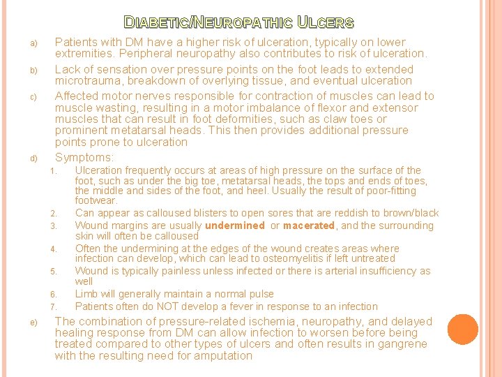
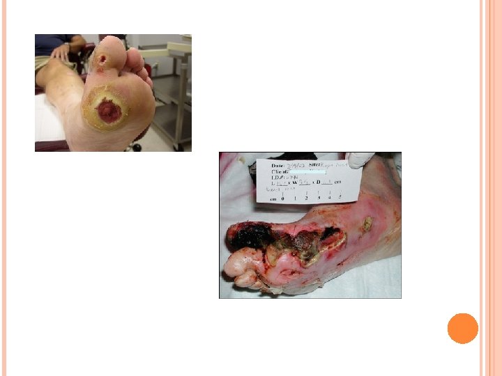
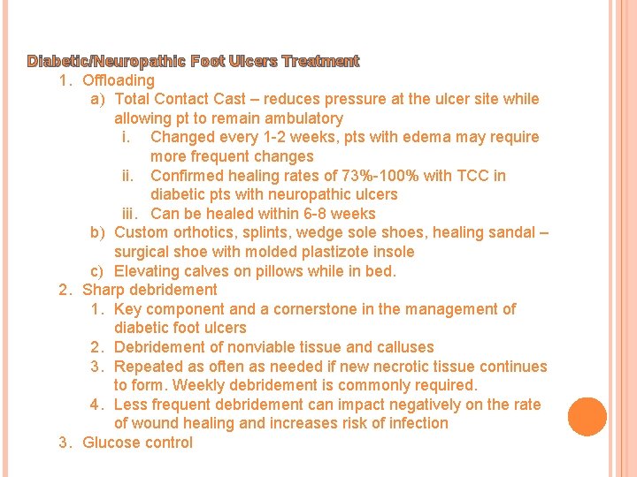
DIABETIC/NEUROPATHIC ULCERS a) b) c) d) Patients with DM have a higher risk of ulceration, typically on lower extremities. Peripheral neuropathy also contributes to risk of ulceration. Lack of sensation over pressure points on the foot leads to extended microtrauma, breakdown of overlying tissue, and eventual ulceration Affected motor nerves responsible for contraction of muscles can lead to muscle wasting, resulting in a motor imbalance of flexor and extensor muscles that can result in foot deformities, such as claw toes or prominent metatarsal heads. This then provides additional pressure points prone to ulceration Symptoms: 1. 2. 3. 4. 5. 6. 7. e) Ulceration frequently occurs at areas of high pressure on the surface of the foot, such as under the big toe, metatarsal heads, the tops and ends of toes, the middle and sides of the foot, and heel. Usually the result of poor-fitting footwear. Can appear as calloused blisters to open sores that are reddish to brown/black Wound margins are usually undermined or macerated, and the surrounding skin will often be calloused Often the undermining at the edges of the wound creates areas where infection can develop, which can lead to osteomyelitis if left untreated Wound is typically painless unless infected or there is arterial insufficiency as well Limb will generally maintain a normal pulse Patients often do NOT develop a fever in response to an infection The combination of pressure-related ischemia, neuropathy, and delayed healing response from DM can allow infection to worsen before being treated compared to other types of ulcers and often results in gangrene with the resulting need for amputation
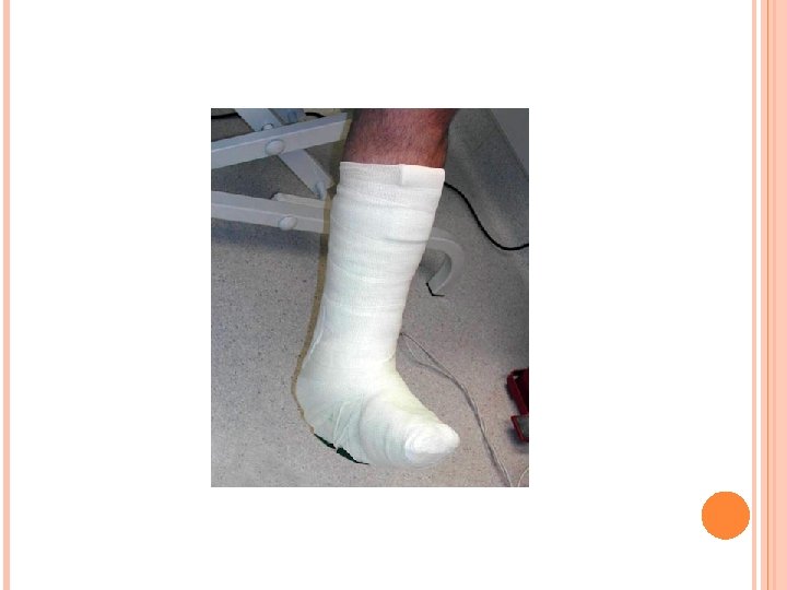
Diabetic/Neuropathic Foot Ulcers Treatment 1. Offloading a) Total Contact Cast – reduces pressure at the ulcer site while allowing pt to remain ambulatory i. Changed every 1 -2 weeks, pts with edema may require more frequent changes ii. Confirmed healing rates of 73%-100% with TCC in diabetic pts with neuropathic ulcers iii. Can be healed within 6 -8 weeks b) Custom orthotics, splints, wedge sole shoes, healing sandal – surgical shoe with molded plastizote insole c) Elevating calves on pillows while in bed. 2. Sharp debridement 1. Key component and a cornerstone in the management of diabetic foot ulcers 2. Debridement of nonviable tissue and calluses 3. Repeated as often as needed if new necrotic tissue continues to form. Weekly debridement is commonly required. 4. Less frequent debridement can impact negatively on the rate of wound healing and increases risk of infection 3. Glucose control
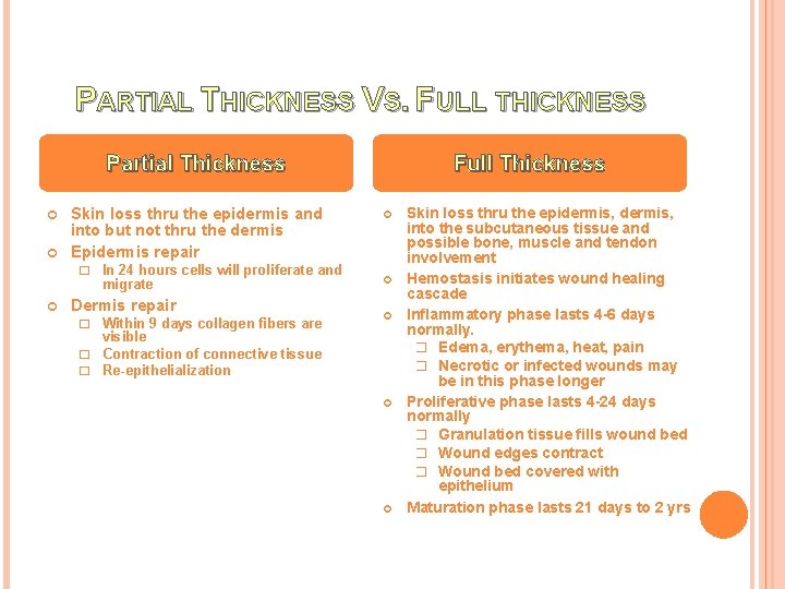
PARTIAL THICKNESS VS. FULL THICKNESS Partial Thickness Skin loss thru the epidermis and into but not thru the dermis Epidermis repair � In 24 hours cells will proliferate and migrate Dermis repair � Within 9 days collagen fibers are Full Thickness visible � Contraction of connective tissue � Re-epithelialization Skin loss thru the epidermis, into the subcutaneous tissue and possible bone, muscle and tendon involvement Hemostasis initiates wound healing cascade Inflammatory phase lasts 4 -6 days normally. � Edema, erythema, heat, pain � Necrotic or infected wounds may be in this phase longer Proliferative phase lasts 4 -24 days normally � Granulation tissue fills wound bed � Wound edges contract � Wound bed covered with epithelium Maturation phase lasts 21 days to 2 yrs
Necrotic Tissue Management
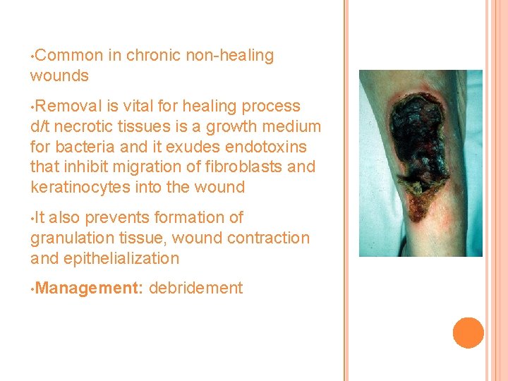
• Common in chronic non-healing wounds • Removal is vital for healing process d/t necrotic tissues is a growth medium for bacteria and it exudes endotoxins that inhibit migration of fibroblasts and keratinocytes into the wound • It also prevents formation of granulation tissue, wound contraction and epithelialization • Management: debridement
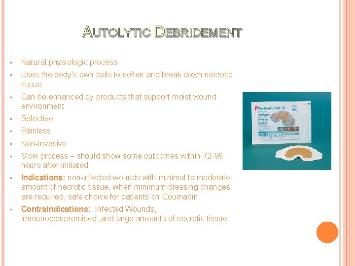
AUTOLYTIC DEBRIDEMENT • Natural physiologic process • Uses the body’s own cells to soften and break down necrotic tissue • Can be enhanced by products that support moist wound environment • Selective • Painless • Non-invasive • Slow process – should show some outcomes within 72 -96 hours after initiated • Indications: non-infected wounds with minimal to moderate amount of necrotic tissue, when minimum dressing changes are required, safe choice for patients on Coumadin • Contraindications: Infected Wounds, immunocompromised, and large amounts of necrotic tissue
Methods/Products Used for Autolytic Debridement • Any occlusive dressing that promotes moist wound bed • Hydrocolloid, transparent, foam dressing • If wound is dry use hydrogel • If wound exudative use an alginate or hydrofiber
WET TO DRY DRESSINGS ARE CONSIDERED TO BE MECHANICAL DEBRIDEMENT AND ARE NONSELECTIVE. THEY SHOULD NOT BE USED ON EPITHELIALIZING OR GRANULATING WOUNDS
Inflammation and Infection Control
SIGNS AND SYMPTOMS OF LOCAL INFECTIONS • • Redness Pain Heat Swelling Increased or purulent drainage Odor Discoloration of the tissue Delayed healing • • Friable granulation tissue (Crumbly, bleeds easily) Induration – big red flag! (Abnormal firmness of tissue) Increased serous exudate Increased pain
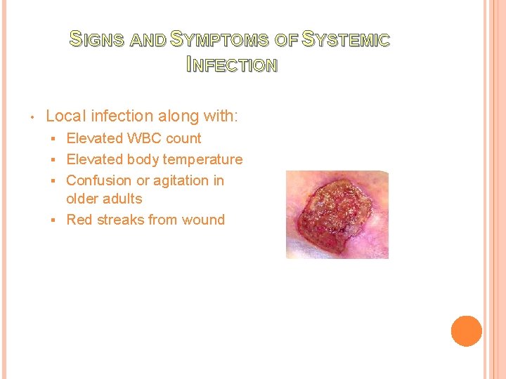
SIGNS AND SYMPTOMS OF SYSTEMIC INFECTION • Local infection along with: Elevated WBC count § Elevated body temperature § Confusion or agitation in older adults § Red streaks from wound §
BIOBURDEN a) Contamination: Presence of bacteria on the wound surface that are not actively multiplying. All wounds are contaminated and this condition does not elicit a host reaction and does not impair healing b) Colonization: Presence of replicating micro-organisms, which do not cause injury to the host. No s/s of injection observed. c) Critical Colonization: Presence of replicating micro-organisms, which are beginning to cause local tissue damage. 1 or 2 signs/symptoms present. d) Infection: Invasion of pathologic organisms into healthy surrounding tissue. 3 or more signs/symptoms present. i. If infection suspected, get orders for a wound culture, blood culture, CBC, and monitor vital signs.
Treatment of Infected Wounds 1. Topical Antiseptics - Only use for limited time periods or it can become cytotoxic (damaging to healthy cells) - Reassess use after 7 days for any improvement a) Betadine indications: gram-positive and gramnegative, MRSA, fungi/yeasts, viruses, and protozoa. External use only. b) Dakins Solution (Hydrochlorite solution/bleach) indications: gram-positive. Use BID. c) Acetic Acid (vinegar) indications: gram-positive and gram-negative, especially pseudomonas. Use TID. * Hydrogen peroxide’s cytotoxic properties outweigh its limited bactericidal effects as an antiseptic. Not recommended for wound care.
Treatment of Infected Wounds 2. Topical antibiotics - Used only when clinical infection is present and used only for a limited time period. a) Mupirocin indications: Gram-positive, MRSA. b) Polymyxin indications: Gram-negative, pseudomonas. c) Bacitracin indications: Most gram-negatives and positives. 3. Oral antibiotics - Not as effective in chronic wounds d/t impaired blood flow
Treatment of Infected Wounds 4. Topical antibacterial dressings a) Iodoflex – Indicated for use in cleaning wet ulcers and wounds such a venous stasis ulcers, pressure sores, and infected traumatic and surgical wounds. i. Iodosorb Gel and Iodoflex Pads assist in wound area protection and in reducing the microbial load in the wound environment. Apply Iodosorb Gel to at least 1/8 -inch thickness. ii. Changing Iodosorb: Should be changed approx. 3 x/week or when the Iodosorb has become saturated with wound exudate (change of color from brown to yellow/gray). Reduce applications as exudate diminishes. b) Aquacel AG – Kills bacteria, helps reduce the risk of airborne dissemination and cross-infection of wound pathogens, including MRSA, during dressing changes. Soaks up exudate, do not use in dry wounds.
Treatment of Infected Wounds 4. Topical Antibacterial Dressings cont. . c) Silver Sulfadiazine – Indicated for burns, skin grafts, incisions, leg ulcers and other clean lesions, abrasions, minor cuts and wounds. i. Apply to a thickness of at least 3 -5 mm to surfaces once or BID ii. Effective against a variety of bacteria and yeast. d) Silverlon – antibacterial, anti-fungal, hypoallergenic, cost -effective. i. Apply silver surface directly to wound and moisten with sterile water ii. Requires a covering dressing to assist in providing a moist environment iii. Changes depend on amount of exudate iv. NOT COMPATIBLE WITH PRODUCTS CONTAINING: saline, chlorine, petroleum, iodine, and hydrogen peroxide
Treatment of Infected Wounds 5. Anti-microbial Barrier Dressings a) Acticoat Moisture Control i. Waterproof, non-adhesive, highly absorbent antimicrobial dressing ii. Indications: Light to moderate drainage wounds, partial thickness, full thickness, pressure, diabetic, 1 st and 2 nd degree burns iii. Contraindications: Sensitivity to silver, MRI procedures, concurrent use of oil-based products, saline iv. Use for barrier to bacterial penetration, not treatment for infections v. Dressing change Q 7 days, silver-side face down b) Hydrofera Blue i. When moistened with NS or sterile water can be molded to a wound ii. Provides broad spectrum bacteriostatic protection iii. Can use on all wounds except 3 rd degree burns iv. Use secondary dressing to secure, leave it on for 13 days. Change when secondary dressing is saturated
Treatment of Infected Wounds 6. Impregnated Gauze a) Mesalte – For heavily discharging infected wounds, and deep cavity wounds. Cleanses the wound by absorbing exudate, bacteria and necrotic tissue. b) Iodoform Gauze c) Xeroform – Provides a deodorizing agent. Not recommended on heavily exudating wounds or sensitivity to 3% Xeroform d) Kerlix AMD - Attacks bacteria on and within the dressing fabric, helping keep infection out of the wound and limiting cross-contamination. i. Effective against MRSA, VRE, MDRAB, Staph aureus, pseudomonas, E. Coli, Candida Albicans ii. Available in 4 X 4 sponges, telfa dressings, drain sponges, and packing strips
Treatment of Infected Wounds 7. Gel Products a) Anasept Antimicrobial Skin and Wound Gel i. Topical hydrogel with rapid broad spectrum bactericidal, fungicidal and virucidal. ii. Effective against MRSA, VRE iii. Indications: stage I-IV pressure ulcers, partial and fullthickness wounds, diabetic and leg ulcers, post surgical wounds, 1 st and 2 nd degree burns iv. Apply a generous amount to entire wound bed, including undermining areas v. Apply a thin coat to peri-wound area and allow to dry vi. Cover with secondary dressing (avoid silver and heavy metal dressings) vii. Change dressing once a day. Maintain moist wound environment between dressing changes. b) Amerigel Wound dressing i. Antimicrobial, antifungal, anti-inflammatory hydrogel wound filler ii. Provides autolytic debridement, has antiseptic and skin protecting properties, maintains moist wound environment iii. Apply once daily

EXUDATE MANAGEMENT
Exudate from chronic wounds has higher concentrations of proteolytic enzymes which results in destruction of healthy tissues in and around the wound, inhibits fibroblast proliferation, and deprives the tissue cells of O 2 = retards healing
TREATMENT OF EXUDATE 1. Alginates a) Algisite, Curasorb, Kaltostat, Kalginate, Maxsorb Extra, Sorbsan, Tegaderm Alginate Dressings, Medihoney, Silverlon CA, Curasorb ZN b) Transforms into a moist gel consistency when they come in contact with exudate/moisture c) Moist gel consistency will maintain moist healing environment and facilitate autolytic debridement d) Indications: full or partial thickness wound with moderate to heavy exudate, bleeding wounds e) Contraindications: 3 rd degree burns, minimal exudate or dry wounds f) Change Q 1 -3 days when exudate saturates secondary dressing g) If change required more than 2 x/day, reassess appropriateness h) Tips: Do not moisten before applying, it can dehydrate wound bed and can adhere to granulation tissue and cause damage.
TREATMENT OF EXUDATE 2. Foam Dressings a) Allevyn, Curafoam, Flexzan, Hydrasorb, Lyofoam, Mitraflex, Polymem, Tielle, Mepilex, Biatain, 3 M Heel Foam, Comfeel Ulcer Dressing b) Capable of holding fluids and pulling them away from wound bed. c) Available in various shapes and sizes d) Indications: Moderate to heavy exudative wounds, granular wounds, softened necrotic tissue, shallow draining wounds, cavity foam fills dead space for packing e) Contraindications: Dry eschar, 3 rd degree burns, sinus tracts. f) Change Q 3 -7 days g) Tips: Dressings should be 1 inch larger than wound
TREATMENT OF EXUDATE 3. Specialty Absorptive Dressings a) Xtrasorb, Combiderm, Exu-dry, Softsorb, Tendersorb Wet-Pruf pads, Aquacel b) Hydrofiber dressing, multi-layered that consist of highly absorptive fiber layers c) Absorbs, provides moist wound healing, and autolytic debridement d) Indications: Heavy drainage, full or partial thickness e) Contraindications: Dry wounds, 3 rd degree burns f) Change Q 1 -3 days 4. Moisture Barrier Ointments a) Alo Vesta protective ointment, Baza Clear, Calmoseptine, Critic Aid, Selan, Soothe & Cool, A & D Ointment, Restore Moisture Barrier b) Comes in a cream, gel, ointment or paste c) Used to protect skin from excess moisture d) Ointments have a longer-lasting effect than creams because they are more occlusive e) Indications: Stage I pressure ulcers, peri-wound skin, perineal area
Other Dressing and Treatment Options
WOUND CLEANSING 1. Always cleanse wound before you assess 2. Solutions a) Cleanse healing, clean wounds with NS i. NS will not harm tissue and is cost-effective. ii. Once bottles are opened they must be used within 24 hrs. Available in spray bottles and spray cans, which don’t need to be used within 24 hrs. b) Wound Cleansers i. Cleanses wounds with debris, confirmed infection and suspected infection. ii. Facilitates removal of wound contaminants. c) Skin cleansers are formulated to remove fecal matter and should never be used on open wounds.
MISCELLANEOUS DRESSINGS 1. Collagens a) Fibercol, BGC, Matrix, Cellerate. RX, Stimulin, Promogran Matrix b) Freeze dried sheets of collagen, particles, pastes, or gels usually derived from cow hides c) Encourages deposition and organization of newly formed collagen fibers and granulation tissue in the wound bed d) Indications: Chronic, non-healing granulated wounds, partial/full thickness, tunneling wounds, wounds with minimal to heavy exudate, granulating or necrotic wounds. e) Contraindications: 3 rd degree burns, black wounds, sensitivity to bovine products f) Read product instructions for frequency. g) Requires secondary dressing 2. Composite Dressings a) Covaderm, Alldress, Telfa Island, Stratasorb, Tegaderm pad, Op. Site Postop b) Cover dressing, autolytic debridement c) Indications: Primary or secondary dressing, partial/full thickness, minimal exudate, granulation or necrotic tissue d) Frequency as needed, requires border of intact skin to anchor the dressing, shouldn’t be cut.
MISCELLANEOUS DRESSINGS 3. Contact Layers a) Tegapore, Dermanet, Mepitel, Telfa Clear b) Thin, non-adherent sheets placed directly on an open wound bed to protect the tissue from direct contact with other agents or dressings applied to the wound c) Porous to allow wound fluid to pass through for absorption by an overlying dressing d) Indications: Fragile wound tissue, pain at dressing change, secondary dressing adhering to wound bed, partial/full thickness, exudative wounds i. May be applied prior to wound vac therapy dressings. e) Contraindications: Shallow, dehydrated, or eschar covered wounds, thick exudate. f) Change once a week, does not need to be removed with every dressing change. g) Tips: May cut to fit, requires secondary dressing
MISCELLANEOUS DRESSINGS 4. Gauze Dressings - used for cleansing, packing, and covering. a) Woven Gauze Pad i. Fine mesh cotton gauze is used for packing, coarse mesh cotton gauze used for non-selective debriding. ii. Tip: Woven gauze should not be cut d/t loose fibers and strings may get lost in wound and delay healing b) Non-woven Gauze pad i. Provides improved wicking and greater absorbent capacity ii. Leaves fewer fibers behind when removed c) Impregnated gauze i. Mesalt – management of heavily discharging and discharging infected wounds in the inflammatory phase and deep cavity wounds such as pressure ulcers and surgical wounds. ii. Iodoform, Xeroform iii. Vaseline – clings and forms to the wound without sticking. Occlusive, indicated for use on fistulas, stomas, chest tube incisions, abrasions, burns, thoracic wounds or incisions
MISCELLANEOUS DRESSINGS d) Adaptic gauze – ideal for most draining wounds. Prevents dressing adherence while allowing exudate from moderate to heavy exudating wounds to easily pass through to the absorbent non-occlusive cellulous fiber layer. e) Wrapping gauze i. Kerlix – 100% woven gauze used to cushion and protect wound areas. Provides fast wicking action, superior aeration, and excellent absorbency. Ideal for bandaging heads, limbs and difficult to dress wounds. ii. Conform gauze – conforms to difficult body contours and clings to itself iii. Tubular elastic net bandage – applies gentle pressure to keep bandages securely in place. (Surgitube, Bandnet, Surgilast, Elastinet, Spandage) *NOTE: for wet-to-dry dressings, they need to be removed every 46 hrs and the gauze should be dry upon removal.
MISCELLANEOUS DRESSINGS 5. Hydrocolloids a) Bursamed HC, Duoderm, Derma. Film Hydrocolloid Dressing, Excel Hydrocolloid, Prima. Col, Replicare, Tegasorb, Restore, Comfeel b) Wafer dressing containing gel-forming agents with a water resistant outer layer c) Some contain alginate to increase absorptive capabilities d) Available in various thickness and in precut shapes for body areas such as sacrum, elbows and heels e) It is occlusive, provides moist wound healing environment, promotes autolytic debridement f) Indications: Non-infected wounds with scant to moderate drainage, necrotic or granular tissue, dry wound, to protect intact skin or newly healed wound, partial/full thickness g) Contraindications: 3 rd degree burns, infected wounds, heavy exudate, exposed tendon or bone, fragile surrounding skin, fungal or herpetic lesions, deep tunnels, tracts, and undermining h) Change dressing Q 3 -7 days depending on exudate, not appropriate for daily dressing changes i) Tips: apply wafer 1 -2 inches larger than wound, warm dressing prior to application, use a dressing with beveled edges to prevent dislodgement from shearing or friction j) Upon removal, dressing residue may adhere to wound bed, it may cause injury to wound bed, and may cause odor, which is common problem with moisture retentive dressings, may cause peri-wound maceration.
MISCELLANEOUS DRESSINGS 6. Hydrogels a) Donates moisture to wound, provides moist healing environment, promotes granulation and epithelialization, and autolytic debridement b) Helps with pain control. It has a cooling influence on the wound that causes and analgesic effect which lasts up to 6 hrs. c) Indications: Dry or slightly moist wounds, to soften eschar, cooling painful wounds, granulating wounds, slough, abrasions, minor burns, radiation skin damage d) Contraindications. Moderate to heavy exudative wounds, 3 rd degree burns e) Change dressing daily to Q 4 days f) Tips: may cause maceration, dehydrates easily if not covered
MISCELLANEOUS DRESSINGS 6. Hydrogels Cont. e) Available in: i. Amorphous hydrogel (Amerigel, Anasept, Antimicrobial Skin and Wound Gel, Aqua. Site, Curasol, Curafil Wound Gel, Dermasyn, Intra. Site Gel, Solosite, Nu. Gel, Normlgel, Regenecare, Duo. Derm Hydroactive Gel) • Free flowing gel, packaged in tubes, packets and spray bottles. ii. Impregnated Gauze Hydrogel (Carra. Gauze, Transigel, Curasol 4 X 4, Dermagran Gauze Dressing) • Hydrogel saturated gauze pad, non-woven sponges, ropes and strips. iii. Gel Sheets (Aquasite Hydrogel Sheet, Cool Magic Sheet Hydrogel, Kendall Curagel, Kendall Island, Elastogel, Vigilon, Nu. Gel, Tender Wet, Thera. Gauze) • Gel supported by a thin fiber mesh. Dressing can overlap intact skin and is available with or without adhesive borders and can be cut to fit the wound.
MISCELLANEOUS DRESSINGS 7. Transparent Films a) Tegaderm, Op. Site, Kendall Transparent-Polyskin b) Moisture vapor permeable (allows one-way passage of O 2 and moist vapor out of dressing but allows nothing in c) Waterproof and impermeable to bacteria and contaminants d) Provides moist healing environment, autolytic debridement, and protection from mechanical trauma and bacterial invasion e) Indications: flat, partial thickness, non-draining wounds, to protect intact skin, eschar to promote debridement, secondary dressing f) Contraindications: Moderate to heavy exudate, 3 rd degree burns, fragile skin, suspected infection, fungal infection or herpetic lesions g) Change dressing Q 1 -7 days h) Tips: skin must be clean and dry, may use alcohol and sealant prior to application, a buildup of exudate is indicative of autolytic debridement and a normal occurrence
TREATMENT THROUGH NUTRITION Mini Nutrition Screening Tool Nutritional deficiency is the term used when the reserves of nutrients stored in the body are insufficient to support healthy cellular function. When the individual is malnourished, any trauma may prove disastrous as they lack the reserves to respond to the metabolic demands of the wound. Key Nutrients to Wound Healing � � � Proteins: Vital to the healing process. Supports platelet function, fibroblast proliferation, and wound remodeling. Helps with formation of lymphocytes, collagen, and new vasculature. Carbohydrates: Provides the energy for recovery. Glucose is vital to wound healing as it supports cell growth, fibroblasts, and leukocytes. Also it plays a vital role in cell communication as a part of the immune response. Energy needs must be met to prevent the breakdown and utilization of protein for energy vs. healing. Fats: Helps meet the increased caloric demand that results from post-trauma hypermetabolism. Influences the localized metabolism of the wound. Helps modulate inflammation and the immune response. B Vitamins: Moderates local wound metabolism and tissue synthesis. Participates in collagen synthesis. Vitamin C: Helps stabilize collagen, which increases the tensile strength of the wound tissues. Supports free radical metabolism. Vitamin D: Regulates growth and differentiation of cells, including lymphocytes, melanocytes, and fibroblasts. Vitamin K: Required for blood clotting. Zinc: Supports protein synthesis and immune functions. Vital for cell growth and proliferation, liberates vitamin A from the liver, interacts with platelets in flood clotting; Mega doses of zinc may inhibit healing and cause copper deficiency. Iron: Delivers oxygen to tissues. Generation of energy from cells, transferrin, a blood protein, transfers iron to the tissues of the bed. Copper: Helps facilitate angiogenesis. Assists with cross linking of collagen fibers to rebuild tissue. Fluids serves as the solvent for minerals, vitamins, amino acids, and glucose, maintains fluid and electrolyte balance.

HELPFUL RESOURCES www. Wound. Care. Source. com www. Wound. Care. Advisor. com

Handouts Inservice for next month?
- Slides: 51