Working with Proteins Dr Aelya Yilmazer What to


- Slides: 10

Working with Proteins Dr. Açelya Yilmazer

What to Study about Peptides and Proteins? What is its sequence and composition? What is its three-dimensional structure? How does it find its native fold? How does it achieve its biochemical role? How is its function regulated? How does it interacts with other macromolecules? How is it related to other proteins? Where is it localized within the cell? What are its physico-chemical properties?

A Mixture of Proteins Can Be Separated • Separation relies on differences in physicochemical properties – – – Charge Size Affinity for a ligand Solubility Hydrophobicity Thermal stability • Chromatography is commonly used for preparative separation

Separation by Charge

Separation by Size

Separation by Affinity

Electrophoresis for Protein Analysis Separation in analytical scale is commonly done by electrophoresis – Electric field pulls proteins according to their charge – Gel matrix hinders mobility of proteins according to their size and shape

SDS PAGE: Molecular Weight • SDS – sodium dodecyl sulfate – a detergent • SDS micelles binds to, and unfold all the proteins – SDS gives all proteins an uniformly negative charge – The native shape of proteins does not matter – Rate of movement will only depend on size: small proteins will move faster

Protein Sequencing

Spectroscopic Detection of Aromatic Amino Acids • The aromatic amino acids absorb light in the UV region • Proteins typically have UV absorbance maxima around 275 -280 nm • Tryptophan and tyrosine are the strongest chromophores • Concentration can be determined by UV-visible spectrophotometry using Beers law: A = ·c·l
 Rasim yılmazer
 Prof. dr mehmet yilmazer
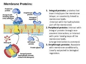
 Eritrosit periferal membran proteinleri
 Hot working and cold working difference
 Differentiate between hot working and cold working
 Smart work vs hard work
 Proses pengerjaan logam
 Hot hot
 Dna rna and proteins study guide answers
 Where are proteins found
 Section 8-1 carbohydrates fats and proteins answer key
