Wilms Tumor Wilms tumor or nephroblastoma is malignant

Wilms Tumor

• Wilms' tumor or nephroblastoma is malignant tumor of the kidneys that typically occurs in children. Dr. Max Wilms , the German surgeon (1867– 1918) first described this kind of tumor. • Wilms tumor is the fifth most common pediatric malignancy (7% of all childhood tumors).

incidence • Incidence is 1 in 10, 000, girls are slightly more affected • 70% of cases occur before the child is 5 years of age. • Most commonly unilateral, but in 5% 10% both kidneys are involved.

• Approx 500 cases are diagnosed in the U. S. annually. • The majority (75%) occurs in otherwise normal children; a minority (25%) is associated with other developmental abnormalities. • It is highly responsive to treatment, with about 90% of patients surviving at least five years.

Etiology • Unknown • Genetic abnormalities WT 1 gene; dominant oncogene (at chromosome 11 p 13), WT 2 gene (at chromosome 11 p 15)

It is believe that tumor begins to grow as a fetus develops in the womb, with some cells that are destined to form into the kidneys malfunctioning and forming a tumor. The exact etiology of the tumor are still being investigated.

• Tumor is exceedingly vascular, soft, mushy, or gelatinous in character. • Wilms tumor has capacity for rapid growth, usually grows to a large size.

• Tumor is usually uniform, well demarcated by a pseudocapsule of compressed renal tissue. • Tumor develops from primitive renal tissue and can have epithelial ( tubules and glomeruli), stromal (fat, skeletal muscles, cartilage) and blastemal elements (An immature material from which cells and tissues develop).

Wilms tumor may be associated with • Hemihypertrophy-one side of body is larger than other • Aniridia (complete loss of iris) and • Genitourinary anomalies.

, WT 2")
Pathophysiology • WT 1 gene; dominant oncogene (at chromosome 11 p 13), WT 2 gene (at chromosome 11 p 15) (a tissue-specific gene for renal blastemal cells and glomerular epithelium) • Leads to abnormal proliferation of the metanephric blastemal cells (primitive embryologic cells of the kidney). • promote changes that may lead to the formation of Wilms tumor.
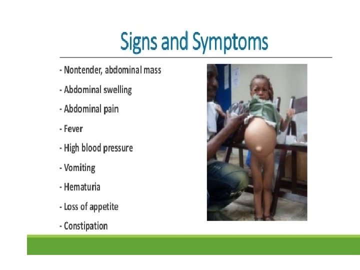

Clinical manifestations • Wilms tumor is diagnosed at a mean age of 3. 5 years. • The most common feature is an upper quadrant abdominal mass (firm, nontender)

• Abdominal pain occurs in 30%-40% of cases, related to rapid growth of tumor. • Urethral obstruction due to compression • Constipation, vomiting, abdominal ditress, anorexia, weight loss and dyspnea due to enlargement of tumor.

• Other signs and symptoms of Wilms tumor include hypertension, fever caused by tumor necrosis, hematuria, and anemia.

• The neoplasm metastasize either by direct extension or by bloodstream. They may invade perirenal tissues, lymph nodes, the liver, the diaphragm, abdominal muscles and the lungs. • Invasion of bone and brain are less common.

Diagnostic evaluation Laboratory studies: • CBC with differential for baseline data • Platelet count: Coagulation abnormalities • Urinalysis for hematuria and urine culture • Liver function tests • Renal function tests • Blood chemistry; sr. electrolytes, uric acid
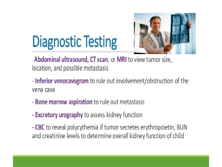

Imaging Studies • Ultrasonography – Initial diagnosis of a renal or abdominal mass, – Possible renal vein or inferior vena cava (IVC) thrombus (Doppler flow study may be helpful in the setting of vascular invasion. ) – Information regarding liver and other kidney

• CT scanning of the chest and abdomen – Differential diagnosis of a kidney tumor versus adrenal tumor (neuroblastoma) – Liver metastases – Status of opposite kidney – Lymph node assessment – Status of chest with respect to metastases

• IVP • Chest radiography - As a baseline for pulmonary metastases • Bone scan • Magnetic resonance imaging
 is characterized by all 3 histological")
Histologic Findings • Favorable histology (90% of cases) is characterized by all 3 histological elements, without any anaplastic features. The cure rate in these cases is close to 90%.
 is characterized by the presence of anaplasia.")
• Unfavorable histology (10% of cases) is characterized by the presence of anaplasia. • Anaplasia is defined as nuclear enlargement, and abnormal mitoses.

• Bone scan • Bone marrow aspirate • Biopsy


Staging and treatment • Staging is determined by combination of imaging studies and pathology findings. • Treatment strategy is determined by the stage.
 • For stage I Wilms' tumor, 1 or more")
Stage I (43% of patients) • For stage I Wilms' tumor, 1 or more of the following criteria must be met: • Tumor is limited to the kidney and is completely excised. • The surface of the renal capsule is intact. • The tumor is not ruptured or biopsied (open or needle) prior to removal. • No involvement of extrarenal or renal sinus lymphvascular spaces • No residual tumor apparent beyond the margins of excision. • Metastasis of tumor to lymph nodes not identified.

Treatment: • Nephrectomy +/- 18 weeks of chemotherapy depending on age of patient and weight of tumor. EG: less than 2 years old and less than 550 g only requires Nephrectomy with observation. • Outcome: 98% 4 -year survival; 85% 4 year survival if anaplastic
 For Stage II Wilms' tumor, 1 or more of")
Stage II (23% of patients) For Stage II Wilms' tumor, 1 or more of the following criteria must be met: • Tumor extends beyond the kidney but is completely excised. • No residual tumor apparent at or beyond the margins of excision. • Any of the following conditions may also exist: – Tumor involvement of the blood vessels of the renal and/or outside the renal parenchyma. – The tumor has been biopsied prior to removal or there is local spillage of tumor during surgery, confined to the flank. – Extensive tumor involvement of renal soft tissue.

Treatment: • Nephrectomy + abdominal radiation + 24 weeks of chemotherapy • Outcome: 96% 4 -year survival; 70% 4 year survival if anaplastic
 For Stage III Wilms' tumor, 1 or more of")
Stage III (23% of patients) For Stage III Wilms' tumor, 1 or more of the following criteria must be met: • Unresectable primary tumor. • Lymph node metastasis. • Tumor is present at surgical margins. • Tumor spillage involving peritoneal surfaces either before or during surgery.

Treatment: • Abdominal radiation + 24 weeks of chemotherapy + nephrectomy after tumor shrinkage • Outcome: 95% 4 -year survival; 56% 4 year survival if anaplastic
 • Stage IV Wilms' tumor is defined as the")
Stage IV (10% of patients) • Stage IV Wilms' tumor is defined as the presence of hematogenous metastases (lung, liver, bone, or brain), or lymph node metastases outside the abdomenopelvic region.

Treatment: • Nephrectomy + abdominal radiation + 24 weeks of chemotherapy + radiation of metastatic site as appropriate. • Outcome: 90% 4 -year survival; 17% 4 year survival if anaplastic
 • Stage V Wilms’ tumor is defined as bilateral")
Stage V (5% of patients) • Stage V Wilms’ tumor is defined as bilateral renal involvement at the time of initial diagnosis. • The 4 -year survival in 94% patients with stage I or stage II; 76% for with stage III. • Treatment: Individualized therapy based on tumor burden
 • Radiation therapy • Chemotherapy")
Management • Surgical ( partial/complete nephrectomy) • Radiation therapy • Chemotherapy

Chemotherapy • Actinomycin D; 0. 06 - 0. 12 mg/kg, IV • Doxorubicin ( adriamycin); 1. 25 – 1. 9 mg/ kg • Vincristine; 0. 125 – 0. 05 mg/kg

• Stage I actinomycin and vincristine for 1115 weeks • Stage II actinomycin and vincristine for 15 months • Stage III actinonycin, vincristine and adriamycin for 15 months,

• Stage IV actinomycin, vincristine, and adriamycin for 15 months. If response is slow then cyclophosphamide is added
 1. Excision of tumor 2. If not")
• Stage V (bilateral wilms tumor) 1. Excision of tumor 2. If not possible, confirmatory biopsies then treated with vincristine and actinomycin D for 3 -6 months. If no satisfactory resolution seen after 3 months, radiation is added to both kidneys

• A second look surgery is planned after 6 months of therapy, if not possible chemotherapy may be tried for an additional 9 months

• In totally inoperable cases with no metastatic disease bilateral nephrectomy and renal transplantation may be only viable operation.


- Slides: 47