
WHITE MATTER OF CEREBRUM White matter is a
WHITE MATTER OF CEREBRUM
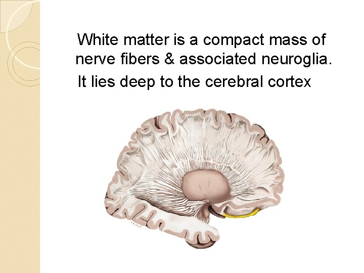
White matter is a compact mass of nerve fibers & associated neuroglia. It lies deep to the cerebral cortex

White matter is classified into 3 types: 1. Association fibers 2. Commissural fibers 3. Projection fibers

1. Association fibers connects the cortical areas of same cerebral hemisphere a) Short association fibers – which connects the adjacent gyri. b) Long association fibers – which connects the different lobes of the same cerebral hemisphere.
Long association fibers are……. a) Cingulum b) Uncinate fasciculus c) Superior longitudinal fasciculus d) Inferior longitudinal fasciculus e) Fronto occipital fasciculus
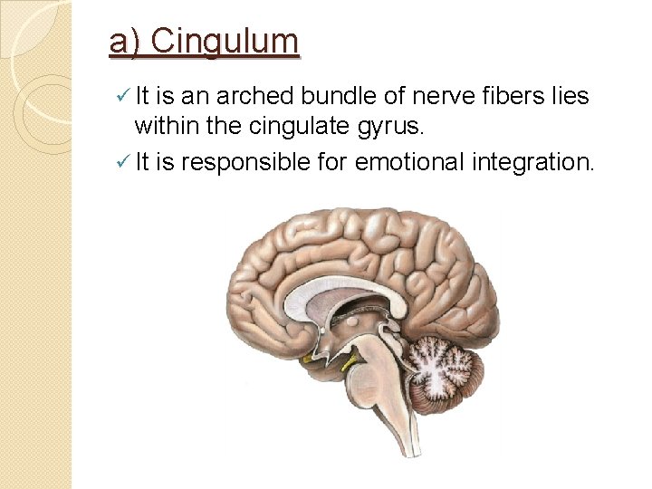
a) Cingulum ü It is an arched bundle of nerve fibers lies within the cingulate gyrus. ü It is responsible for emotional integration.
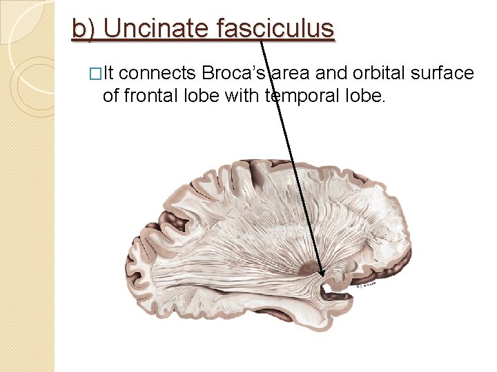
b) Uncinate fasciculus �It connects Broca’s area and orbital surface of frontal lobe with temporal lobe.

c) Superior longitudinal fasciculus ü It is the longest association bundle connects the frontal lobe to the occipital lobe and temporal lobes.
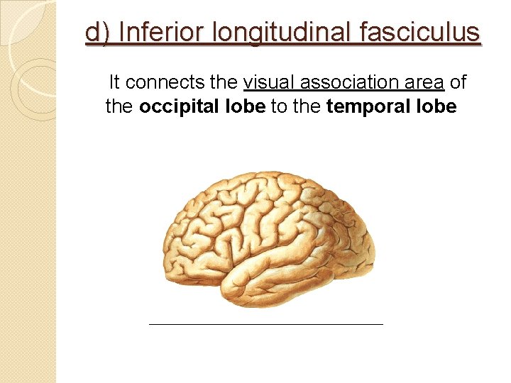
d) Inferior longitudinal fasciculus It connects the visual association area of the occipital lobe to the temporal lobe
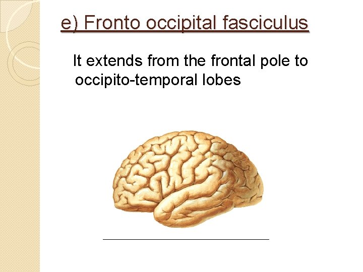
e) Fronto occipital fasciculus It extends from the frontal pole to occipito-temporal lobes
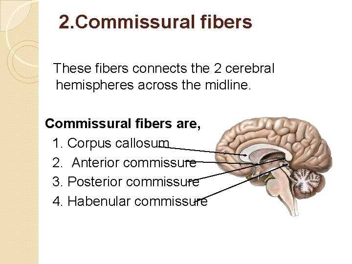
2. Commissural fibers These fibers connects the 2 cerebral hemispheres across the midline. Commissural fibers are, 1. Corpus callosum 2. Anterior commissure 3. Posterior commissure 4. Habenular commissure
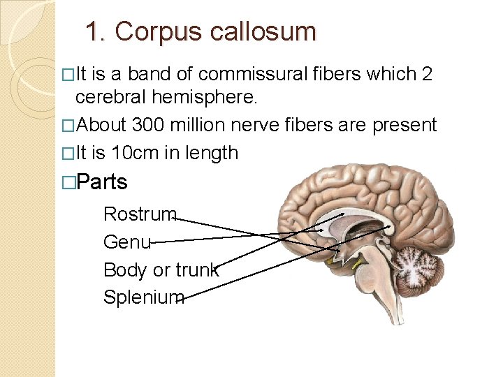
1. Corpus callosum �It is a band of commissural fibers which 2 cerebral hemisphere. �About 300 million nerve fibers are present �It is 10 cm in length �Parts Rostrum Genu Body or trunk Splenium
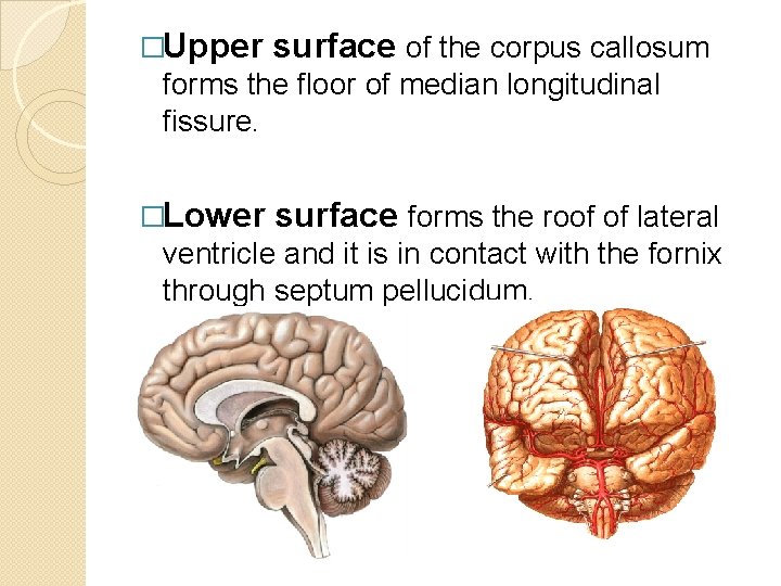
�Upper surface of the corpus callosum forms the floor of median longitudinal fissure. �Lower surface forms the roof of lateral ventricle and it is in contact with the fornix through septum pellucidum.
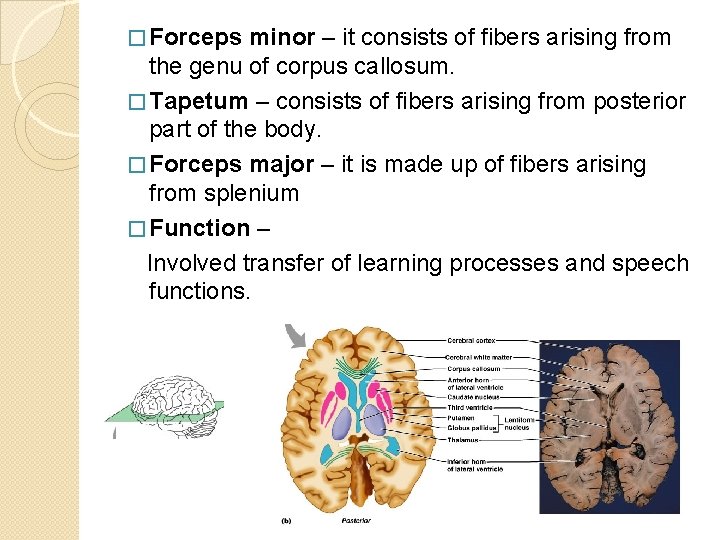
� Forceps minor – it consists of fibers arising from the genu of corpus callosum. � Tapetum – consists of fibers arising from posterior part of the body. � Forceps major – it is made up of fibers arising from splenium � Function – Involved transfer of learning processes and speech functions.

3. Projection fibers �These fibers connects cerebral cortex with subcortical grey matter, brain stem and spinal cord. �Example – corona radiata internal capsule
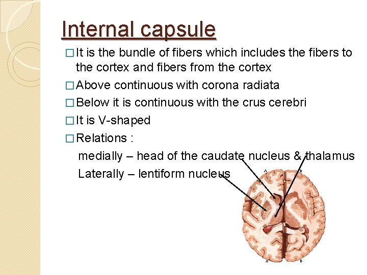
Internal capsule � It is the bundle of fibers which includes the fibers to the cortex and fibers from the cortex � Above continuous with corona radiata � Below it is continuous with the crus cerebri � It is V-shaped � Relations : medially – head of the caudate nucleus & thalamus Laterally – lentiform nucleus

Parts of internal capsule 1. 2. 3. 4. 5. Anterior limb Genu Posterior limb Sublentiform part Retrolentiform part
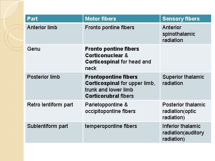
Part Motor fibers Sensory fibers Anterior limb Fronto pontine fibers Anterior spinothalamic radiation Genu Fronto pontine fibers Corticonuclear & Corticospinal for head and neck Posterior limb Frontopontine fibers Corticospinal for upper limb, trunk and lower limb Corticorubral fibers Superior thalamic radiation Retro lentiform part Parietoppontine & occipitopontine fibers Posterior thalamic radiation(optic radiation) Sublentiform part temperopontine fibers Inferior thalamic radiation(auditory radiation)
Clinical anatomy: Stroke – artery suppling the internal capsule may rupture and cause hemorrhage. This may result in contralateral hemiplegia.
- Slides: 19