White Matter of cerebral hemisphere The white matter


White Matter of cerebral hemisphere
 Association fibers (2) Commissural fibers (3)")
The white matter classified into three groups (1) Association fibers (2) Commissural fibers (3) Projection fibers

v. Association fibers § They connect parts of the cerebral cortex in the same hemisphere.
. F Uncus T O")
(Sup. Long. Bundle). F Uncus T O

A- Short association fibers; connect adjacent gyri with one another. B- Long association fibers: 1 - Superior longitudinal bundle; from frontal lobe and extends posteriorly then divided into 2 bands; a Superior band to the occipital lobe. b Inferior band to the temporal lobe. 2 - Inferior longitudinal bundle: from the occipital pole to the temporal pole. 3 - Uncinate bundle; from the frontal lobe and curved around the steam of lateral sulcus to the temporal lobe. 4 - Cingulum: from frontal lobe just below the rostrum of corpus callosum → extends backwards in the cingulate gyrus → curves in the isthmus → passes forwards in the parahippocampal gyrus → ends in the uncus. It is part of the limbic system

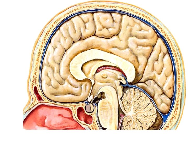
body of the fornix commissure of the fornix Anterior columns of the fornix mammillary bodies hippocampus The fornix the posterior columns of the fornix Fimbria of hypocampus sagittal section

septum pellucidum Corpus callosum Posterior column Body of fornix Anterior column Mammillary body Uncus Thalamus Fimbriae of hippocampus Parts & relations of fornix, sagittal section

5 - Fornix The fornix is the only efferent system of the hippocampus. It forms a part of the limbic system. - Structure; It is formed. 1 - Two Posterior columns (Crurae); efferent of hippocampus. They lie below and behind the posterior end of the thalamus. Each crus is the continuation fibers of the fimbria of the hippocampus. The 2 crurae are connected together by commissure of fornix. They come together in the median plane to form the body. 2 - Body; triangular in shape, with narrow anterior and broad posterior. - The upper surface is connected with the lower surface of the corpus callosum by the septum pellucidum. - The lower surface rests on upper surface of the 2 thalami and roof of the 3 rd ventricle. 3 - Two anterior columns; end in the mammillary body of the hypothalamus.

Medial longitudinal bundle • It is an associative coordinating tract present in the brain steam and extends from the upper end of the midbrain to the lower end of medulla oblongata. * Hearing reflex function • It connects the cochlear nuclei with: • a- Motor nuclei of 3, 4 & 6 cranial nerves together. • b- Motor nuclei of anterior horn cells of the spinal cord. • C- Spinal part of accessory nerve nuclei • - So, both eyes and neck move to the direction of the sound stimuli.

Medial longitudinal bundle v Speech function Connects Cranial nerves concerned in the speech; 1 - Facial nerve (7 th); supplies muscles of the lips. 2 - Cranial root of accessory nerve (11 th); supplies muscles of the larynx. 3 - Hypoglossal nerve (12 th); supplies the muscle of the tongue. - So, the muscles of the lips, larynx and tongue work together during speech. Connects motor nuclei of the cranial nerve 5 th, 7 th and 12 th together for the coordination of Mastication and swallowing Connects motor nuclei of the cranial nerve 3 rd, 4 th and 6 th and 7 th for the coordination of eye lids and eye movements.

v. Commissural Fibers • They connect parts of cerebral cortex of one hemisphere with the same part on opposite side. • They coordination of the activities of the right and left cerebral hemisphere

Spelnium The body septum pellucidum & lat. vent Cingulate gyrus Genu Rostrum lamina terminalis Fornix v Commissural Fibers v Corpus callosum

forceps minor is projection from genu forceps major is projection from splenium Tapetum fibers of the trunk and splenium Tapetum: fibers of the trunk and splenium which form the roof, lateral wall and floor of posterior horn of the lateral ventricle.
 Rostrum; connects fibers of frontal lobe. Downward")
* Parts of the corpus callosum a) Rostrum; connects fibers of frontal lobe. Downward tapering part of corpus callosum. It connects the genu with the lamina terminalis. It forms floor of anterior horn of lateral ventricle. b) Genu; connects the fibers of the frontal lobe It is the thick anterior end of the corpus callosum. It forms anterior boundary of anterior horn of lateral ventricle. On each side, the fibers curve forward and form the forceps minor.

Median log fissure Head caudate Septum pellucidum Body of Fornix Body corpus callosum TH 3 rd HT Coronal section Central Lat. ventricle Lentiform Tail caudate Inf horn
 Body (trunk); connects fibers of posterior part of the frontal lobe and parietal")
c) Body (trunk); connects fibers of posterior part of the frontal lobe and parietal lobe 1 - The inferior surface, a- Median plane, attached to fornix by septum pellucidum. b- On each side, forms roof central part of lateral ventricle. 2 - The superior surface; a- Median plane, it lies in the floor of the median longitudinal fissure and related to the falx cerebri. b- On each side, related to cingulate gyrus separated from it by callosal sulcus that contains anterior cerebral artery. d) Splenium; connects fibers of occipital and temporal lobes. It is the thick posterior end of the corpus callosum. Its posterior fibers curve posteriorly forming forceps major.

3 -Posterior commissure 2 -The anterior commissure Sagittal section Habenular commissure CG CC Pineal body

2 - Anterior commissure: It presents between lamina terminalis and anterior column of the fornix. It connects the olfactory bulbs and anterior parts of the temporal lobe. 3 - Posterior commissure: It presents above the tectum of the midbrain and below the pineal body (gland). It connects the superior colliculus, pretectal nuclei of the midbrain and medial longitudinal bundles. 4 - Habenular commissure; It presents at the root of the pineal body (gland). It connects the habenular nuclei of both sides.

Commissural fibres Sagittal section

Body of fornix Anterior commissure Lamina terminalis Sagittal section Tela choroida & choroid plexus Habenular commissure Pineal body Posterior commissure

The central part of the lateral ventricle Choroid plexus of the 3 rd ventricle Choroid Plexus of the Lateral Ventricle The inferior horn of the lateral ventricle Coronal section

Genu of corpus callosum septum pellucidum anterior columns of the fornix Head of caudate Tail of caudate the crus of splenium of the fornix corpus callosum Horizontal section of the brain

Horizontal section

§ Projection fibers They connecting the cerebral cortex with the lower centers and pass through Internal capsule

Th ank Qu you est ion s I/Azzam 2004
- Slides: 27