White blood cells White blood cellWBCLeucocytesstructural classification Normal

White blood cells
/Leucocytes(structural classification). Normal value: 4, 000 -11, 000 cells/ cu mm of")
White blood cell(WBC)/Leucocytes(structural classification). Normal value: 4, 000 -11, 000 cells/ cu mm of blood Granulocytes Neutrophil Eosinophil Basophil Agranulocytes Monocyte Lymphocyte Large AEJ Small 2

Neutrophil: ü Normal value: 40 -70% ü Normal size: 12 -14 μm ü Color of cytoplasm: Slight violet. ü Granules take both eosin and blue color. ü Size of the granules: Very fine sand like granules. ü Nucleus: Multi-lobed (1 -6 lobes). AEJ 3

Eosinophil: § Normal value: 1 -6% § Normal size: 12 -14 μm § Color of cytoplasm: Eosinophilic. § Size of the granules: Large, coarse , pinkish red granules. § Nucleus: Bilobed nucleus. (Spectacle shape) AEJ 4

Basophil: v Normal value: 0 -1% v Normal size: 12 -14 μm v Color of cytoplasm: Appear blue. v Size of the granules: Few , large and coarse, Overlying the nucleus. v Nucleus: Bilobed, not clearly seen. AEJ 5

Monocyte: • Normal value: 2 -10% • Normal size: 15 -22 μm. • Color of cytoplasm: Grey blue. • Granules: Absent. • Nucleus: Single, Usually Kidney /Horse shoe Shape. AEJ 6

Lymphocytes: Normal value: 20 -45% Normal size: Large: 12 -16 μm. Small: 7 -10 μm. Color of cytoplasm: Clear sky blue. Granules: Absent. Nucleus: Large : Single large occupies 3/4 th of the cell. Small: Single large occupies almost whole of the cell. AEJ 7

Functions of different WBC’s: NEUTROPHIL: First line of defence • Phagocytosis –Bacteria, antigen-antibody complexes, small particulate matter. • Release of chemicals involved in inflammationleucotrienes, prostaglandins, thomboxanes.

EOSINOPHIL: • Mild phagocytosis. • Provides mucosal immunity – respiratory, urinary, GI tract. • Anti allergic role –collects at the site and limit their intensity by degrading the effect of mediators, inhibit mast cell or basophil degranulation. • Destroy multicellular parasites – Peroxidase. • Clot lysis - Prefibrinolysin.

BASOPHIL: • Secretion Heparin • Role in allergic reaction- Release histamine, bradykinins • Mild phagocytosis • Involved in inflammation and healing process. Histamine, bradykinins Hyaluronic acid

Monocytes/ Macrophages: • Active Phagocytosis- Second line of defence. • Toxic chemical release. • Process and present antigens to helper T-cells. • Secrete cytokines. MAST CELLS: • Release Histamine and other chemicals involved in inflammation.

Macrophage consuming RBC

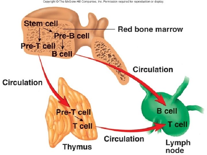
Pluripotent stem cells Lymphoid stem cells Bone marrow, Liver, Spleen Thymus T-Lymphocytes B-Lymphocytes & Natural killing cells Memory T- cell Cytotoxic T-cell Memory cell Plasma cell Suppressor T-cell Immunoglobulin's Helper T-cell

Difference b/w B-lymphocytes and T-lymphocytes B-Lymphocytes: • 20% of the total. T-Lymphocytes: • 80% of the total. • Produce Antibody. • Produce chemicalscytokines. • Processed in Bone Marrow. • Processed in Thymus. • Responsible for Humoral mediated immunity. • Responsible for Cell mediated immunity. • Protects from bacterial infection. • Protects against Viral infection, Tumor and Autoimmune diseases. • Shorter life span. • Longer life span.

LYMPHOCYTES Immunity B-LYMPHOCYTE: • Initiate antibody mediated immune response. • Major defence against bacterial infections, toxins.

T –LYMPHOCYTES • Cell mediated immunity General functions 1. Rejection of foreign grafts, destroy virus infected cells and cancer cells - Cytotoxic T cells. Binds to the antigens on plasma membrane of the target cell and directly destroy them. 2. Cooperates with B-lymphocytes in producing humoral immunity – Helper T cell Secrete cytokines that helps to activate other cells.

Two T lymphocyte cells attached to a cancer cell
 - Suppressor")
3. Suppress the production of antibodies against own tissue (prevent autoimmune disease) - Suppressor cell. 4. Major defence against virus, fungi and bacteria. 5. Participate in delayed hypersensitivity reaction.

NK CELLS: • Binds directly and non specifically on virus and cancer cells.
- Slides: 20