White Blood Cells Morphology and Counts Clinical Pathology

White Blood Cells Morphology and Counts Clinical Pathology

Granulocytes • Neutrophils • Basophils • Eosinophils • Produced predominately in the bone marrow. • Capable of mitotic division up to the myelocyte stage. • Responds to an increased demand • infection • Takes 3 -5 days to influence peripheral numbers.

Storage-Maturation Compartment • Cells mature into metamyelocytes→ band cells→ Segmented cells. • 80% of granulocytes are found in the bone marrow of healthy animals. • These cells are released from bone marrow with the oldest (segmented) to increased peripheral need. • In less than two days, the bone marrow can respond. • Dogs have the largest storage pool (5 days) whereas bovines have limited storage.

Functions of Leukocytes • Granulocytes: • Characterized by the presence of cytoplasmic granules • Function of these cells occurs in the tissues, not in the bloodstream. • These cells do not recirculate

Neutrophils • Primary functions: • Phagocytosis and killing of microbes. • Usually circulate for 10 hours before migrating into the tissues. • Ingest material and eventual bacterial killing.

Eosinophils • Inhibit chemical mediators such as histamine and serotonin which are released during allergic (hypersensitivity reactions). • Have phagocytic and bacteriocidal properties similar to neutrophils but not as effective. • Have parasiticidal properties. • Animals with heartworms may have hig numbers of eosinophils.
 associated with hypersensitivity reactions. • This release")
Basophils • Secrete mediators of inflammation (histamine) associated with hypersensitivity reactions. • This release occurs when antigens complex with Ig. E is located on the cellular surface.

Monocytes • Differentiate into the cells of the mononuclear/phagocyte system present in most tissues. • Become macrophages once they migrate into the tissues. • Capable of multiplying within the tissues. • Can survive for long periods of time

Monocyte Functions: • Phagocytosis and digestion of particulate material, bacteria, and dead cells. • Macrophages are less responsive to bacterial infections than neutrophils but are more effective against fungal infections. • Synthesis and release of substances invloved in inflammation and immune response. • Expressions of immune response by presenting antigens to T-lymphocytes. • Serve as a major source of colony stimulating factors and cytokines involved in hemtopoiesis. • Cause bone marrow to produce more granulocytes.

Lymphocytes • Distributed in lymphoid tissue to include lymph nodes, spleen, thymus, tonsils, bone marrow, and blood. • Capable of division. • Recirculate in the blood. • Functions: • B cells: turn into plasma cells which secrete immunoglobulins. Usually stayin the lymphoid tissue. • T cells: transform into effector cells that produce lymphokines which function in mediation of cellular immunity.

B cell

T cell

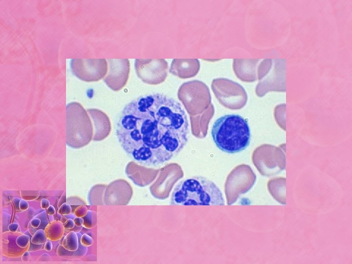
Normal morphology • Neutrophils: multiple nuclear lobules separated by constrictions. • Band granulocytes: band (horseshoe) shaped nuclei. • Eosinophil: Lobulated nucleus and cytoplasm containing reddish pink granules. • Basophil: Lobulated nucleus and purpleblue (Basophilic) granules.

Neutrophil compartments in Peripheral blood • Marginated neutrophil pool: during any moment of time, some neutrophils are loosely adhered to the vessel wall. These cells are not sampled when blood is drawn from non-stressed animals. • Circulating neutrophil pool: neutrophils moving with RBC’s and fluid. Are sampled pool of cells. • When an animal is stressed- marginated pool becomes circulating pool. Called stress leukogram.
")
Response to Inflammation • Inflammatory cells at sites of inflammation release substances (cytokines, interleukins) into the blood to attract neutrophils. • Segmented neutrophils are released from the mature storage compartment in the bone marrow. • Increase in peripheral numbers (measurable in 2 days). • If sudden demand for neutrophils depletes the storage compartment of segmented neutrophils, then band cells are released. • With continued depletion, cells in bone marrow begin to divide. • When source of inflammation is removed, demand for neutrophils decrease, and production slows down.
defined as the release")
Immature Neutrophils in Circulation • Immature neutrophils in circulation (left shift)defined as the release of immature neutrophils (usually bands) into the circulation to meet tissue demand. • The appearance of immature neutrophils is termed “Left shift”. • Regenerative left shift: the absolute number of neutrophils in circulation is increased. The bone marrow has increased the neutrophil release. • Degenerative left shift: the nummber of neutrophils has decreased. This is a poor diagnostic sign.

Cattle exception: • Adult cattle have a relatively low absolute number of neutrophils in circulation and have a small marrow storage pool. • A degenerative left shift is typical of the acute inflammatory response in cattle.

Some terminology for morphology • -penia: decreased number of cells in the blood (Neutropenia, lymphopenia). • -philia or –cytosis: increased number of cells in the blood (neutrophilia, lymphocytosis). • Left shift: increased numbers of immature neutrophils in the blood. • Leukemia: neoplastic cells in the blood or bone marrow. • Leukemoid response: marked leukocytosis (>50, 000/ul) usually a result of inflammatory disease. • Lymphoproliferative disorders: conditions in lymphocytes or plasma cells proliferate abnormally. • Myeloproliferative disorders: a group of bone marrow disorders, usually neoplastic, which stems from on of the bone marrow cell lines.

Neutrophilia • Not always due to infection. • Stress leukogram: endogenous or exogenous corticosteroids. • Characterized by neutrophilia without a left shift, monocytosis, lymphopenia, and eosionopenia. • Caused by a shift from the marginal to the circulating pool. • Physiological luekocytosis: transiet condition caused by excitement, epinephrine release and splenic contraction. • Neutrophilia without left shift and normal or increased lymphocyte number. • Occurs more commonly in the cat than the dog.

Inflammatory Leukogram • Increased bone marrow proliferation, shift from storage pool to blood. • Mild inflammation yields a leukocyte response similar to stress. • Purulent reaction: neutrophils with a left shift. • Intense response with left shift • Can confuse immature cells of myeloid series with neoplasia (leukemia) • Degenerative shift usually is due to extreme migration of cells into tissues and/or detrimental side effects of toxins.

Toxic Neutrophils • Characterized by ctyoplasm basophilia, Dohle bodies, toxic granulation, and/or foamy cytoplasm. • Cells have decreased functional abilities. • Animal with toxic, degenerative shift may be compromised by lack of adequate cell number and decrease ability of cells to function.

Dohle Bodies • Blue cytoplasmic inclusions. • Low numbers may be found in healthy cats. • Indicates toxicity in other species.

Neutrophil Hypersegmentation • Neutrophils with five or more nuclear lobes. • Normal aging change of neutrophils which normally occurs in the tissues, not bloodstream. • May occur with excessive levels of corticosteroids from administration or hyperadrenocorticism. • Chronic inflammation. • Artifactual change in the blood that sits for a period of time.

Pelger-Huet Anomaly • Hyposegemented neutrophils that function normally. • Hereditary disorder; failure of the nucleus in mature cells to undergo segmentation.

Lymphocytosis • Physiologic: due to epinephrine release. • Common in chronic inflammation and chronic antigenic stimulation. • Later stages of resolving infections. • Neoplastic lymphocytosis such as leukemia and lymphosarcoma

Lymphopenia • • • Common finding on CBC Associated with stress Immunosuppressive therapy Immunodeficiency syndrome Viral infections such as parvo.

Reactive Lymphocytes • Characteristic changes in the morphology of lymphocyte that has been stimulated to produce antibody (B cells) or lymphokines (T cells). • Morphological changes: increased cytoplasm, increased basophilia of cytoplasm, increased aggregation of chromatin, and may lack perinuclear zone.
- Slides: 28