
White blood cells CHNB Body Fluids By type

White blood cells CHNB

Body Fluids:
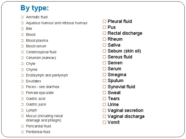
By type: � Amniotic fluid � Aqueous humour and vitreous humour � Bile � Blood plasma � Blood serum � Cerebrospinal fluid � Cerumen (earwax) � Chyle � Chyme � Endolymph and perilymph � Exudates � Feces - see diarrhea � Female ejaculate � Gastric acid � Gastric juice � Lymph � Mucus (including nasal drainage and phlegm) � Pericardial fluid � Peritoneal fluid � � � � � Pleural fluid Pus Rectal discharge Rheum Saliva Sebum (skin oil) Serous fluid Semen Serum Smegma Sputum Synovial fluid Sweat Tears Urine Vaginal secretion Vaginal discharge Vomit
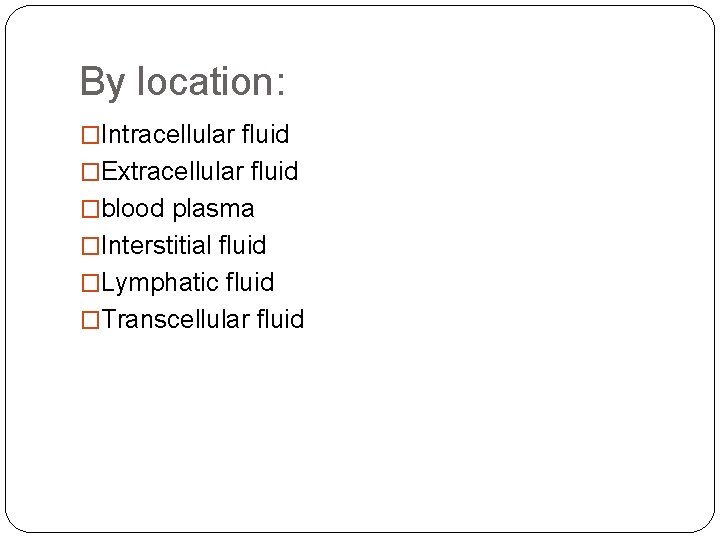
By location: �Intracellular fluid �Extracellular fluid �blood plasma �Interstitial fluid �Lymphatic fluid �Transcellular fluid
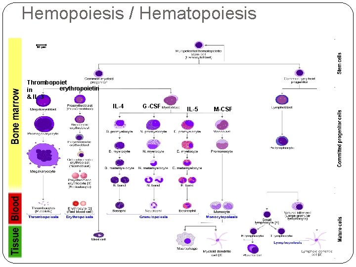
Hemopoiesis / Hematopoiesis Thrombopoiet erythropoietin in & IL-3 IL-4 G-CSF IL-5 M-CSF
Hemopoiesis / Hematopoiesis � All blood cells are derived from haematopoietic stem cells (HSCs). � HSCs are self-renewing cells: when they differentiate, at least some of their daughter cells remain as HSCs, so the pool of stem cells is not depleted (asymmetric division). � The other daughters of HSCs form myeloid and lymphoid progenitor cells.
Myeloid progenitor cells � The common myeloid progenitor cell (CFU-GEMM) is able to differentiate into erythrocytes, thrombocytes, lymphocytes, granulocytes, and monocytes. � CFU-GEMM is a colony forming unit that generates myeloid cells. � "GEMM" stands for granulocyte, erythrocyte, monocyte, megakaryocyte. � CFU-GEMM gives rise to �CFU-GM (leading to monoblasts and myeloblasts), �CFU-Meg (leading to megakaryoblasts), and �CFU-E (leading to proerythroblasts).
Common lymphoid progenitor �This is lymphopoiesis. �The lymphoid lineage is composed of � T-cells, �B-cells and �natural killer cells.
�Primitive erythroid cells can be found in the yolk sac between weeks 2 and 8 of gestation. From the second month of a pregnancy to the seventh month, the liver and spleen are the main hematopoietic sites. Some haematopoiesis occurs in the bone marrow post fifth month of gestation.
White blood cells / leukocytes / leucocytes � The number of leukocytes in the blood is often an indicator of disease. � The cells of the immune system that are involved in protecting the body against both infectious disease and foreign invaders. � Leukocytes are found throughout the body, including the blood and lymphatic system. � The normal white cell count is usually between 4, 000 to 11, 000/microliter of blood. � An increase in the number of leukocytes over the upper limits is called leukocytosis. � A decrease below the lower limit is called leukopenia. This indicates a weakened immune system. � Two broadest categories by structure are �granulocytes �agranulocytes
Granulocytes � Granulocytes are a category of white blood cells characterized by the presence of granules in their cytoplasm. � Granulocytes are produced via granulopoiesis in the bone marrow. � There are four different types of granulocytes: �Neutrophils �Basophils �Eosinophils �Mast cells � Except for the mast cells, their names are derived from their staining characteristics
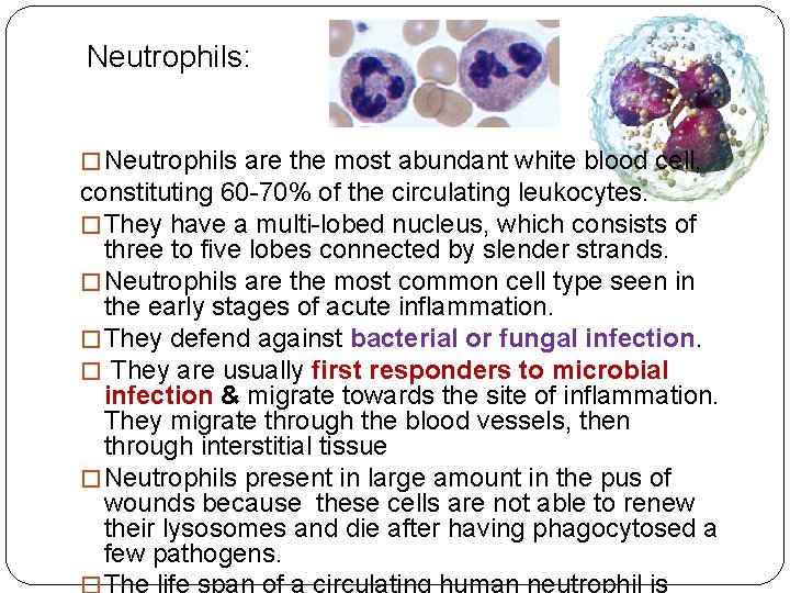
Neutrophils: � Neutrophils are the most abundant white blood cell, constituting 60 -70% of the circulating leukocytes. � They have a multi-lobed nucleus, which consists of three to five lobes connected by slender strands. � Neutrophils are the most common cell type seen in the early stages of acute inflammation. � They defend against bacterial or fungal infection. � They are usually first responders to microbial infection & migrate towards the site of inflammation. They migrate through the blood vessels, then through interstitial tissue � Neutrophils present in large amount in the pus of wounds because these cells are not able to renew their lysosomes and die after having phagocytosed a few pathogens.
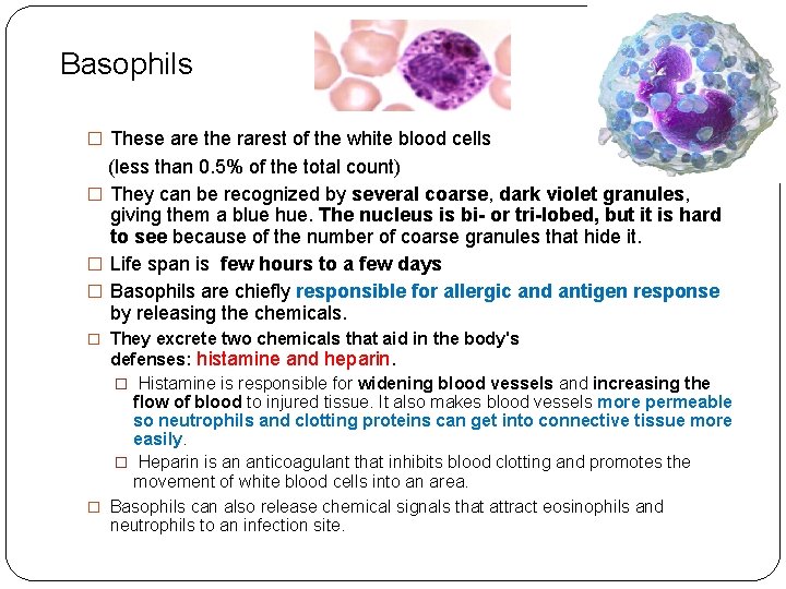
Basophils � These are the rarest of the white blood cells (less than 0. 5% of the total count) � They can be recognized by several coarse, dark violet granules, giving them a blue hue. The nucleus is bi- or tri-lobed, but it is hard to see because of the number of coarse granules that hide it. � Life span is few hours to a few days � Basophils are chiefly responsible for allergic and antigen response by releasing the chemicals. � They excrete two chemicals that aid in the body's defenses: histamine and heparin. � Histamine is responsible for widening blood vessels and increasing the flow of blood to injured tissue. It also makes blood vessels more permeable so neutrophils and clotting proteins can get into connective tissue more easily. � Heparin is an anticoagulant that inhibits blood clotting and promotes the movement of white blood cells into an area. � Basophils can also release chemical signals that attract eosinophils and neutrophils to an infection site.
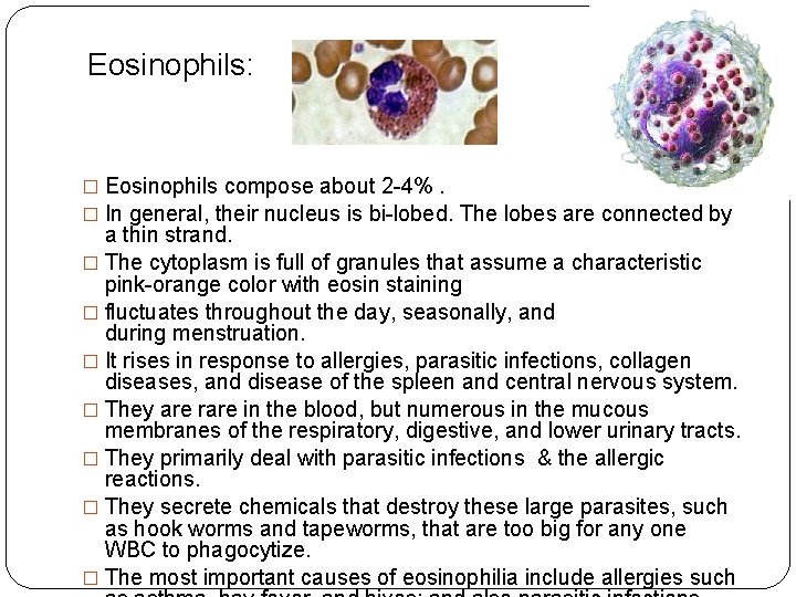
Eosinophils: � Eosinophils compose about 2 -4%. � In general, their nucleus is bi-lobed. The lobes are connected by a thin strand. � The cytoplasm is full of granules that assume a characteristic pink-orange color with eosin staining � fluctuates throughout the day, seasonally, and during menstruation. � It rises in response to allergies, parasitic infections, collagen diseases, and disease of the spleen and central nervous system. � They are rare in the blood, but numerous in the mucous membranes of the respiratory, digestive, and lower urinary tracts. � They primarily deal with parasitic infections & the allergic reactions. � They secrete chemicals that destroy these large parasites, such as hook worms and tapeworms, that are too big for any one WBC to phagocytize. � The most important causes of eosinophilia include allergies such
Agranulocytes: � They are characterized by the absence of granules in their cytoplasm. � These are about 35% of all white blood cells. � The 2 types of agranulocytes in the blood circulation are � lymphocytes and � monocytes. � A third type of agranulocyte, the macrophage, is formed in the tissue when monocytes leave the circulation and differentiate into macrophages.
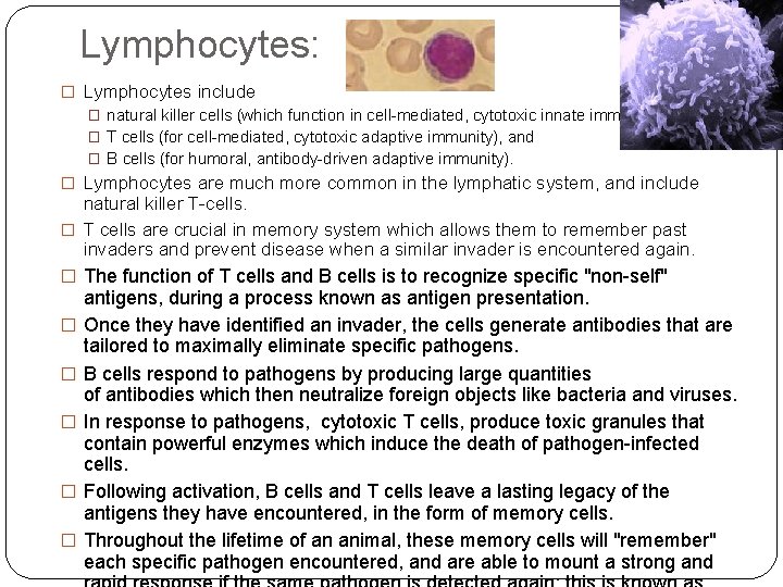
Lymphocytes: � Lymphocytes include � natural killer cells (which function in cell-mediated, cytotoxic innate immunity), � T cells (for cell-mediated, cytotoxic adaptive immunity), and � B cells (for humoral, antibody-driven adaptive immunity). � Lymphocytes are much more common in the lymphatic system, and include natural killer T-cells. � T cells are crucial in memory system which allows them to remember past invaders and prevent disease when a similar invader is encountered again. � The function of T cells and B cells is to recognize specific "non-self" � � � antigens, during a process known as antigen presentation. Once they have identified an invader, the cells generate antibodies that are tailored to maximally eliminate specific pathogens. B cells respond to pathogens by producing large quantities of antibodies which then neutralize foreign objects like bacteria and viruses. In response to pathogens, cytotoxic T cells, produce toxic granules that contain powerful enzymes which induce the death of pathogen-infected cells. Following activation, B cells and T cells leave a lasting legacy of the antigens they have encountered, in the form of memory cells. Throughout the lifetime of an animal, these memory cells will "remember" each specific pathogen encountered, and are able to mount a strong and
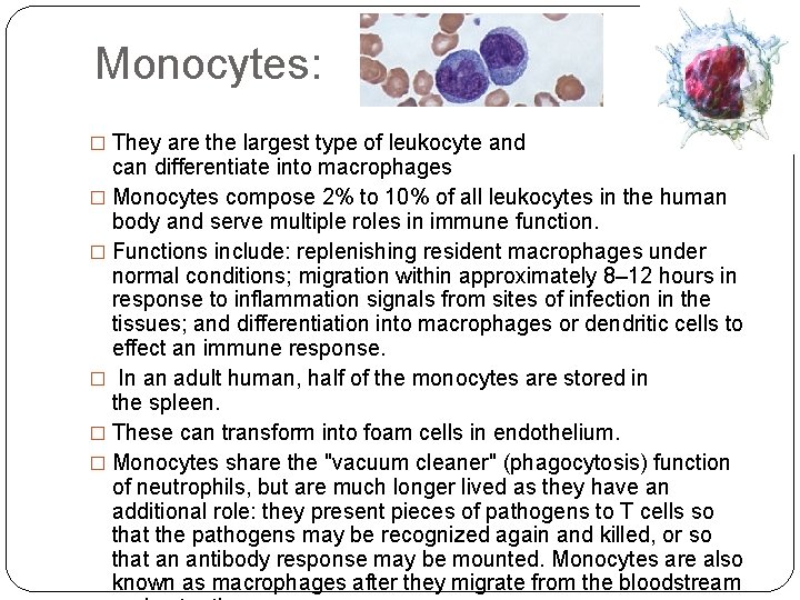
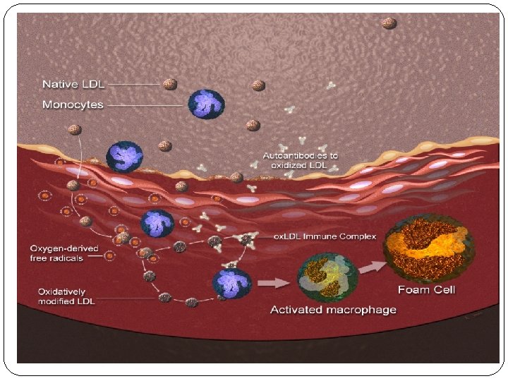
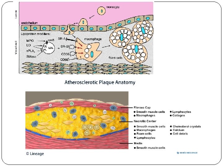
Monocytes: � They are the largest type of leukocyte and can differentiate into macrophages � Monocytes compose 2% to 10% of all leukocytes in the human body and serve multiple roles in immune function. � Functions include: replenishing resident macrophages under normal conditions; migration within approximately 8– 12 hours in response to inflammation signals from sites of infection in the tissues; and differentiation into macrophages or dendritic cells to effect an immune response. � In an adult human, half of the monocytes are stored in the spleen. � These can transform into foam cells in endothelium. � Monocytes share the "vacuum cleaner" (phagocytosis) function of neutrophils, but are much longer lived as they have an additional role: they present pieces of pathogens to T cells so that the pathogens may be recognized again and killed, or so that an antibody response may be mounted. Monocytes are also known as macrophages after they migrate from the bloodstream
Formation of Hemoglobin (Hb): � It is the iron-containing oxygen- transport metalloprotein in the red blood cells (erythrocytes) of almost all vertebrates. � Hemoglobin (Hb) is synthesized in a complex series of steps. � The heme part is synthesized in a series of steps in the mitochondria and the cytosol of immature red blood cells, while the globin protein parts are synthesized by ribosomes in the cytosol. � Production of Hb continues in the cell throughout its early development from the proerythroblast to the reticulocyte in the bone marrow. At this point, the nucleus is lost in mammalian red blood cells. � Even after the loss of the nucleus in mammals, residual ribosomal RNA allows further synthesis of Hb until the reticulocyte loses its RNA soon after entering the vasculature.

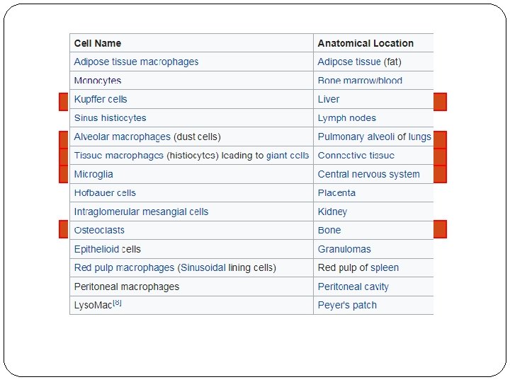
Reticuloendothelial system (RES): � mononuclear phagocyte system or � mononuclear phagocytic system (MPS) (also known as the reticuloendothelial system or � Macrophage system. � "Reticuloendothelial system" is an older term for the mononuclear phagocyte system. � It is a part of the immune system that consists of the phagocytic cells - Monocytes, Macrophages located in reticular connective tissue i. e. in lymph nodes and the spleen. � The spleen is the largest unit of the mononuclear phagocyte system.
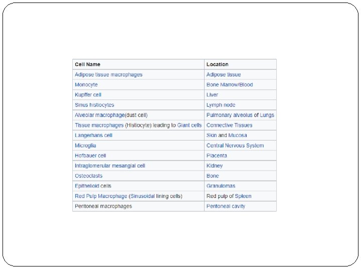
Cell types & Location: � The monocyte is formed in the bone marrow and transported by the blood; it migrates into the tissues, where it transforms into a histiocyte or a macrophage. � Macrophages are diffusely scattered in the connective tissue and in liver (Kupffer cells), spleen and lymph nodes (sinus histiocytes), lungs (alveolar macrophages), and central nervous system (microglia). � The half-life of blood monocytes is about 1 day, whereas the life span of tissue macrophages is several months or years. � The mononuclear phagocyte system is part of both humoral and cell-mediated immunity. � The mononuclear phagocyte system has an important role in defense against microorganisms, including mycobacteria, fungi, bacteria, protozoa, and viruses. � Macrophages remove senescent erythrocytes, leukocytes, and megakaryocytes by phagocytosis and digestion.
Functions: �Formation of new red blood cells (RBCs) and white blood cells (WBCs). �Destruction of old RBCs and WBCs �Formation of plasma proteins. �Formation of bile pigments. �Storage of iron. In the liver, Kupffer cells store excess iron from catabolism of heme from the breakdown of red blood cells. In bone marrow and spleen, iron is stored in MPS cells mostly as ferritin; in iron overload states, most of the iron is stored as hemosiderin. �Clearance of Heparin via Heparinases
- Slides: 27