
WHIT MATTER OF THE CERBRUM BY PROF SAEED
WHIT MATTER OF THE CERBRUM BY PROF. SAEED ABUEL MAKAREM
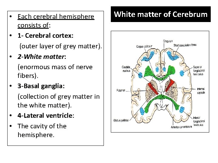
• Each cerebral hemisphere consists of: • 1 - Cerebral cortex: (outer layer of grey matter). • 2 -White matter: (enormous mass of nerve fibers). • 3 -Basal ganglia: (collection of grey matter in the white matter). • 4 -Lateral ventricle: • The cavity of the hemisphere. White matter of Cerebrum
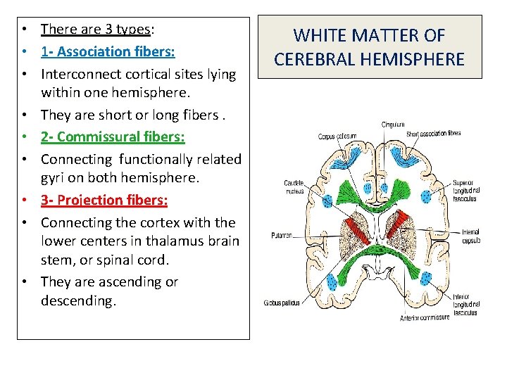
• • • There are 3 types: 1 - Association fibers: Interconnect cortical sites lying within one hemisphere. They are short or long fibers. 2 - Commissural fibers: Connecting functionally related gyri on both hemisphere. 3 - Projection fibers: Connecting the cortex with the lower centers in thalamus brain stem, or spinal cord. They are ascending or descending. WHITE MATTER OF CEREBRAL HEMISPHERE
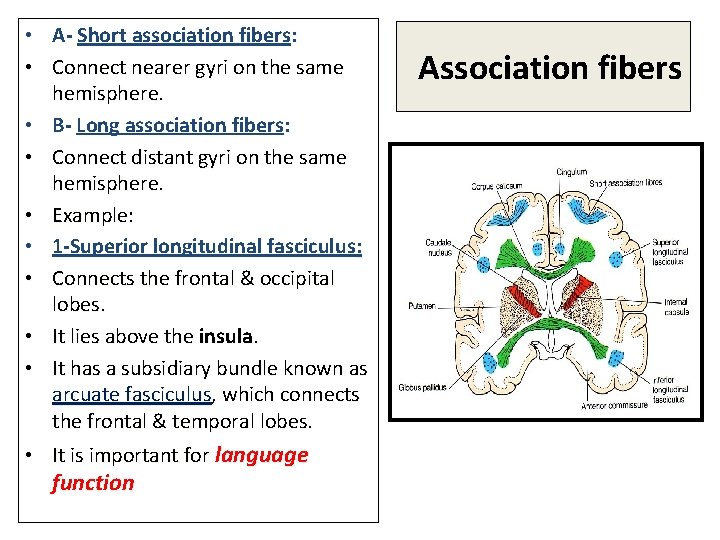
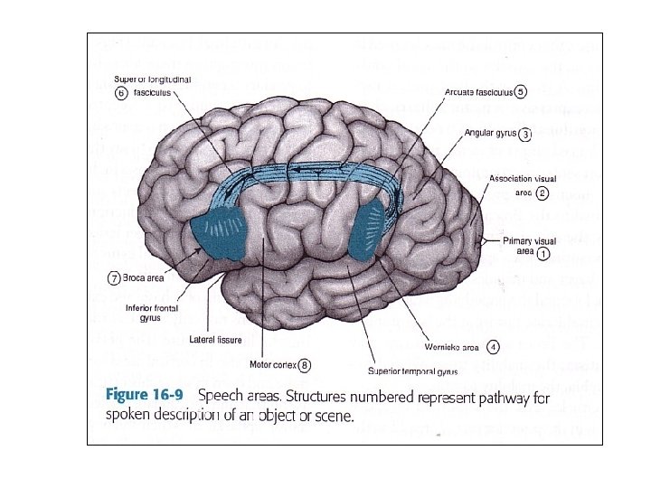
• A- Short association fibers: • Connect nearer gyri on the same hemisphere. • B- Long association fibers: • Connect distant gyri on the same hemisphere. • Example: • 1 -Superior longitudinal fasciculus: • Connects the frontal & occipital lobes. • It lies above the insula. • It has a subsidiary bundle known as arcuate fasciculus, which connects the frontal & temporal lobes. • It is important for language function Association fibers
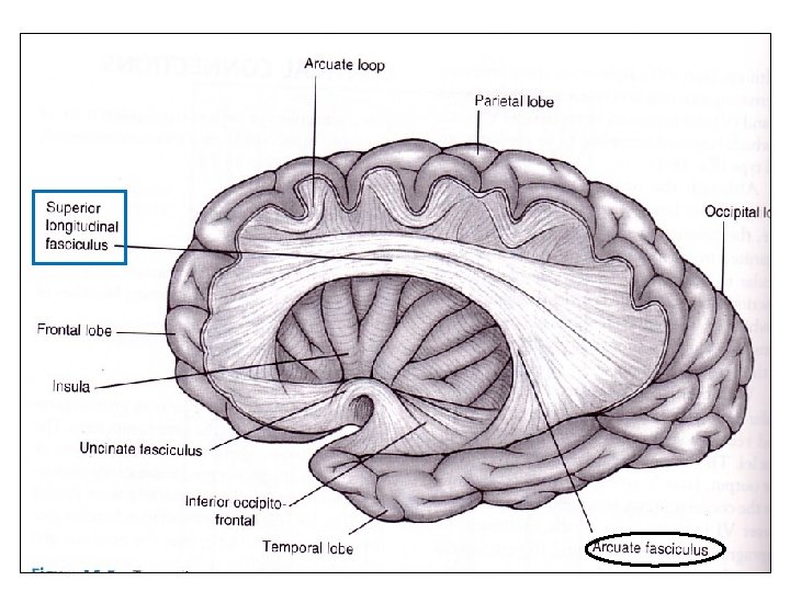
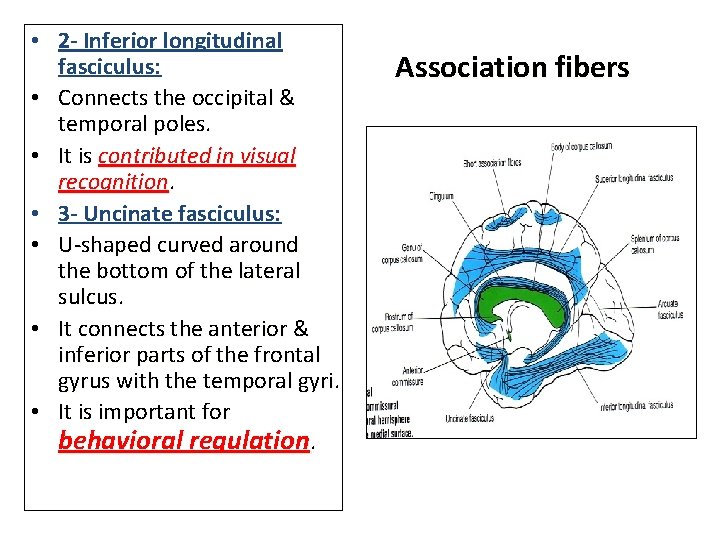
• 2 - Inferior longitudinal fasciculus: • Connects the occipital & temporal poles. • It is contributed in visual recognition. • 3 - Uncinate fasciculus: • U-shaped curved around the bottom of the lateral sulcus. • It connects the anterior & inferior parts of the frontal gyrus with the temporal gyri. • It is important for behavioral regulation. Association fibers

• 4 - Cingulum: • Begins in the anterior perforated substance in the frontal lobe. • Runs in cingulate gyrus. • Curves inferiorly to run in the Parahippocampal gyrus. • It is a part of the limbic system • It terminates in the Uncus. Association fibers

COMMISSURAL FIBERS • The major commissural fibers are: • 1 -Corps callosum. • 2 -Anterior commissure. • 3 -Hippocampal commissure ( Commissure of the fornix).
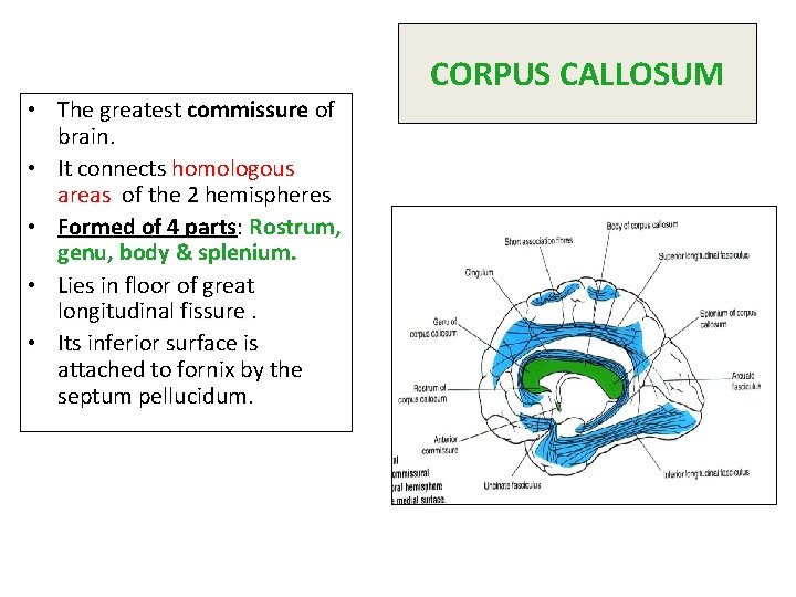
• The greatest commissure of brain. • It connects homologous areas of the 2 hemispheres • Formed of 4 parts: Rostrum, genu, body & splenium. • Lies in floor of great longitudinal fissure. • Its inferior surface is attached to fornix by the septum pellucidum. CORPUS CALLOSUM
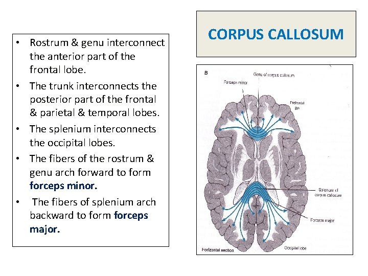
• Rostrum & genu interconnect the anterior part of the frontal lobe. • The trunk interconnects the posterior part of the frontal & parietal & temporal lobes. • The splenium interconnects the occipital lobes. • The fibers of the rostrum & genu arch forward to form forceps minor. • The fibers of splenium arch backward to form forceps major. CORPUS CALLOSUM


ANTERIOR COMISSURE Rounded commissure. Lies behind the lamina terminals. • Runs transversely in front of the anterior column of fornix. • Interconnects the middle & inferior temporal gyri. • Also, it connects the olfactory regions of the 2 hemisphere. • •

HIPPOCAMPAL COMMISSURE • Composed of transverse fibers. • Interconnects the posterior columns of the fornix.
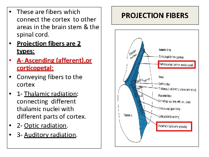
• These are fibers which connect the cortex to other areas in the brain stem & the spinal cord. • Projection fibers are 2 types: • A- Ascending (afferent), or corticopetal: • Conveying fibers to the cortex • 1 - Thalamic radiation: connecting different thalamic nuclei with different parts of cortex. • 2 - Optic radiation. • 3 - Auditory radiation. PROJECTION FIBERS
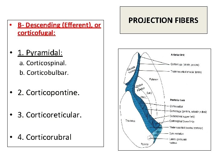
• B- Descending (Efferent), or corticofugal: • 1. Pyramidal: a. Corticospinal. b. Corticobulbar. • 2. Corticopontine. • 3. Corticoreticular. • 4. Corticorubral PROJECTION FIBERS
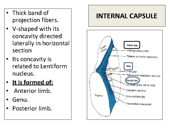
• Thick band of projection fibers. • V-shaped with its concavity directed laterally in horizontal section • Its concavity is related to Lentiform nucleus. • It is formed of: • Anterior limb. • Genu. • Posterior limb. INTERNAL CAPSULE
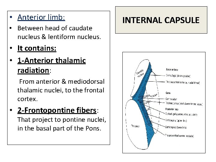
• Anterior limb: • Between head of caudate nucleus & lentiform nucleus. • It contains: • 1 -Anterior thalamic radiation: From anterior & mediodorsal thalamic nuclei, to the frontal cortex. • 2 -Frontopontine fibers: That project to pontine nuclei, in the basal part of the Pons. INTERNAL CAPSULE

• Genu of internal capsule: • Is the angle of the V which points medially, between head of caudate nucleus and thalamus. • It contains: • 1 - Corticonuclear (Corticobulbar). • 2 - Anterior part of sensory (superior thalamic) radiation, from ventral anterior & ventral lateral nuclei to motor regions of the frontal lobe. INTERNAL CAPSULE
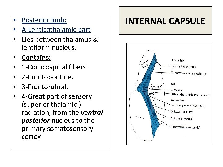
• • Posterior limb: A-Lenticothalamic part Lies between thalamus & lentiform nucleus. Contains: 1 -Corticospinal fibers. 2 -Frontopontine. 3 -Frontorubral. 4 -Great part of sensory (superior thalamic ) radiation, from the ventral posterior nucleus to the primary somatosensory cortex. INTERNAL CAPSULE
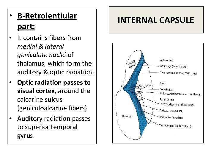
• B-Retrolentiular part: • It contains fibers from medial & lateral geniculate nuclei of thalamus, which form the auditory & optic radiation. • Optic radiation passes to visual cortex, around the calcarine sulcus (geniculoalcarine fibers). • Auditory radiation passes to superior temporal gyrus. INTERNAL CAPSULE
- Slides: 21