Which Fabrics Best Block UV Rays Kris Sabatini

Which Fabrics Best Block UV Rays? Kris Sabatini
 rays are light waves that have shorter wavelengths than")
Ultraviolet Rays - Ultraviolent (UV) rays are light waves that have shorter wavelengths than visible light. They range from 400 nm to 10 nm. - Given off from the sun but most are absorbed by the ozone layer. - The waves that reach Earth can lead to many problems in humans caused by DNA damage: skin burn, sun poisoning, skin irritation, redness, photoaging, nausea, and possibly skin cancer. - The FDA Protection methods include sun screen, hats, and radiation-blocking clothing when outside for long periods of time.

Fabrics • Cotton - Common naturally grown fabric - Past Experiments show it to be effective at blocking UV rays • Polyester - Manmade fabric made from fossil-fuels - Contains trace amounts of zinc-oxide, a UV inhibiter • Nylon - Manmade fabric made from plastic fibers - Past experiments show it to be ineffective at blocking UV rays • Wool - Natural fabric made the hair of sheep - Very densely woven together making harder for UV rays to penetrate

Yeast • Most studied cell in the world • Easy to grow and culture • Similar cell cycle, biochemistry and genetics to other eukaryotic cells • Saccharomyces cerevisiae

Objective/Purpose - To determine the fabric among four common types that is most effective at blocking UV rays

Hypothesis - The fabrics will not vary significantly in blocking UV light, yielding statistically similar yeast survivorship.

Materials • 60 YEPD agar plates(1% yeast extract, 2% peptone, 2% dextrose, 1. 5% agar) • Sterile dilution fluid [SDF] (10 m. M KH 2 PO 4, 10 m. M K 2 HPO 4, 1 m. M Mg. SO 4, . 1 m. M Ca. Cl 2, 100 m. M Na. Cl) • Klett spectrophotometer • Sterile pipette tips and Micropipettors • Vortex • Sidearm flask • Spreader bar • Ethanol • Micro burner • Saccharomyces cerevisiae (yeast) • UV Hood • Rubber Gloves • Test tubes • Test Tube Rack • SDF Test Tubes • Autoclave • 4 white fabrics, nylon, cotton, polyester and wool, cut into squares of 80 x 80 mm

Procedure 1. Sterilized Fabrics by autoclave. 2. Saccharomyces cerevisiae was grown overnight in sterilized YEPD media. 3. A sample of the overnight culture was added to YEPD in a sterile sidearm flask. 4. The culture was incubated at 30°C until a density of 50 Klett spectrophotometer units was reached. This represents a cell density of approximately and 107 cells per m. L. 5. The cultures were diluted in sterile dilution fluid to a concentration of approximately 103 cells per m. L. 6. 0. 1 m. L aliquots were spread onto the YEPD agar plates. 7. Sterilized hands by washing, putting on rubber gloves, and rubbing with ethanol. 8. Sterilized UV hood by rubbing with ethanol. 9. Placed agar plates in UV hood.
 Explanation of Step 4 and 5 105")
100 u. L 107 cells/m. L (yeast) Explanation of Step 4 and 5 105 cells/m. L wit 103 cells/m. L 102 cells

10. Uncovered agar plates. 11. Placed sterile fabrics over uncovered yeast plates. 12. Exposed to UV rays (for 0, 1, and 4 minutes). 13. Removed fabrics for agar plates. 14. Recovered the agar plates with its lid. 15. Removed from UV hood. 16. Repeated for each exposure time period. 17. Incubated for 48 hours at 30 C. 18. The resulting colonies were counted and recorded.
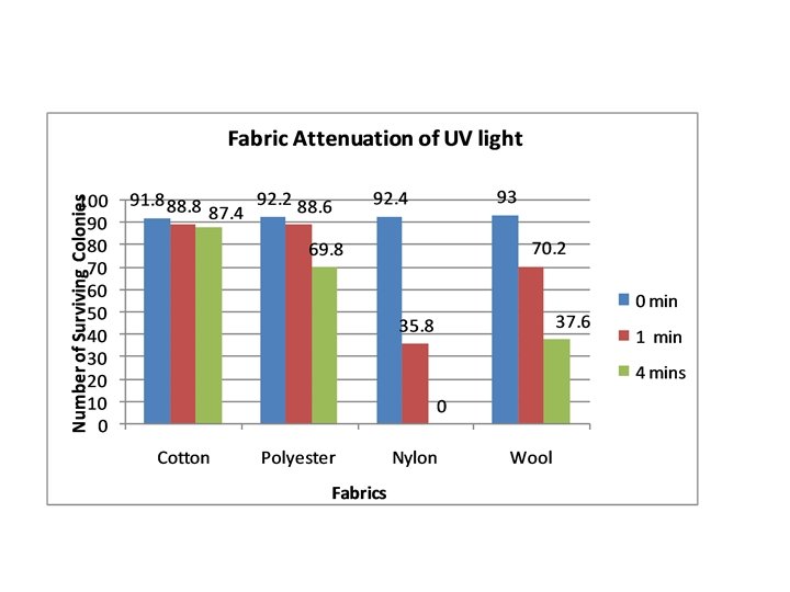
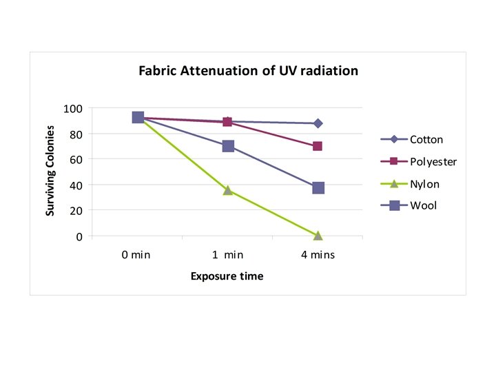

ANOVA Analyze Exposure Time 0 Minutes 1 Minute 4 Minutes P-Value What the P-Value means. 0. 99509 8. 8 E-09 1. 34 E-10 The relative small number means that no variation occurred, since the variable had not yet been introduced no variation was expected. The number means variation occurred, does not supported hypothesis.

Conclusion • The null Hypothesis was rejected. • In the experiment, cotton was the most effective fabric at inhibiting ultraviolent rays. • The experiment also supports past studies that show nylon to be relatively ineffective at blocking UV rays. • Polyester appeared to be the best synthetic at inhibiting the UV rays.

Limitations -Due to slight differences in positioning in the culture hood, the cultures may have received slight differences in amount of ultra-violent rays. - Fabrics may have touched the growing cultures during UV exposure (Would not have unsterilized the cultures but may have inhibited the growth)

Further Testing Different Fabrics Different UV exposure length Different model Varying growth times Photometer to measure UV penetration

Sources • www. FDA. com • http: //www. uvguide. co. uk/
- Slides: 17