Which blood vessel is which Learning Objectives What

Which blood vessel is which?

Learning Objectives: • What are the structures of arteries, arterioles and veins? • How is the structure of each of the blood vessels related to its function? • What is the structure of capillaries and how is it related to their function?

Blood Vessels • Arteries: Carry blood away from the heart • Arterioles: Control blood flow from arteries to capillaries • Capillaries: Link arterioles to veins • Veins: Carry blood towards the heart
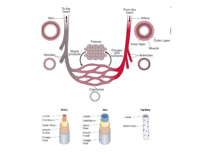

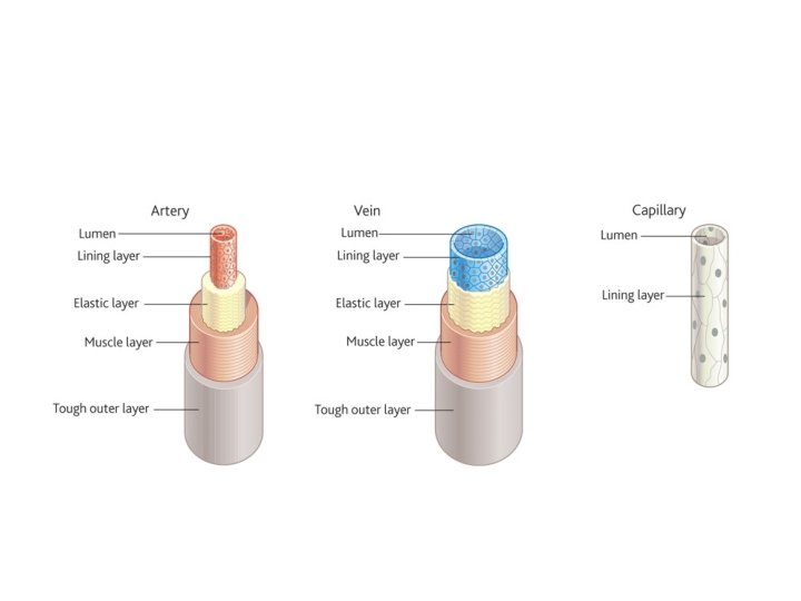
Structure of Blood Vessels • Tough outer layer • Muscle layer – can contract and relax control the flow of blood • Elastic layer – can stretch and recoil to maintain blood pressure • Endothelium – smooth layer to prevent friction • Lumen – the space in the middle

Structure of Arteries • Thick muscle layer – control the flow of blood • Thick elastic layer – smooth surges from the heart • No valves

Structure of Arterioles • Thicker muscle layer than arteries • Thinner elastic layer than arteries • No valves

Structure of Veins • Thin muscle layer • Thin elastic layer • Valves

Structure of Capillaries • No muscle • No elastic • No valves • Thin layer of endothelial cells only

Capillary Structure to Function • Thin layer of cells – short diffusion distance. • Numerous and highly branched – large SA for diffusion. • Narrow diameter – keep all cells close by. • Narrow lumen – bring RBC close to the cells = short diffusion distance. • Spaces between cells – allow WBC to escape. • Have a little fibrous tissue around them to prevent certain cells and proteins leaking out

https: //www. youtube. com/watch? v=Q 530 H 1 Wxt. Ow Tissue Fluid

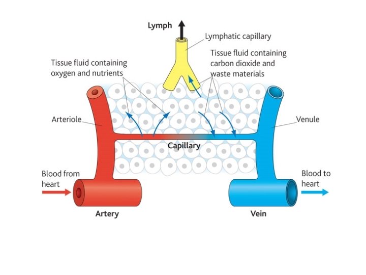
Tissue Fluid • What is the role of tissue fluid? It is the fluid which allows the exchange of substances between the blood and cells • What substances are found in tissue fluid? glucose, amino acids, fatty acids, salts and oxygen = all delivered to the cells. carbon dioxide and other waste substances = removed from the cells.

Tissue Fluid Formation

Hydrostatic Pressure • As the capillaries are narrower than the arterioles, a pressure builds up which forces tissue fluid out of the blood plasma = hydrostatic pressure. • This pressure is resisted by: • Pressure of the tissue fluid on the capillaries (from the outside) • The lower water potential of the blood (caused by plasma proteins – too large to leave the blood) • Overall, pressure pushes tissue fluid and small molecules out of the capillary, leaving cells and large proteins behind = ultrafiltration.

Return of tissue fluid • Most tissue fluid is returned to the blood plasma via the capillaries. • Hydrostatic pressure at the venule end of the capillary is higher outside the capillary and tissue fluid is forced back in. • Osmotic forces (resulting from the proteins in the plasma) pull water back into capillaries. • Remaining tissue fluid enters the lymph vessels – drain back into the veins close to the heart.

Lymph System

Lymph • Lymph is moved by: • Hydrostatic pressure • Contraction of body muscles (aided by valves in the lymph vessels)

Exam Questions • Describe the advantage of having elastic tissue in the wall of an artery. (2) • Explain how tissue fluid is formed and how it may be returned to the circulatory system. (6)

1. elastic tissue allows recoil (reject if wording implies a muscle e. g. contract / relax)(ignore expand); 2. maintains blood pressure / constant / smooth blood flow (not increases blood pressure);
 pressure of blood high at arterial end; 2. fluid / water /")
1. (hydrostatic) pressure of blood high at arterial end; 2. fluid / water / soluble molecules pass out (reject plasma); 3. proteins / large molecules remain; 4. this lowers the water potential / water potential becomes more negative; 5. water moves back into venous end of capillary (reject tissue fluid) by osmosis / diffusion; 6. lymph system collects any excess tissue fluid which returns to blood / circulatory system / link with vena cava / returns tissue fluid to vein;
- Slides: 22