
Whats New in Neurology Recent Discoveries Holly E



- Slides: 31
What’s New in Neurology? Recent Discoveries Holly E. Hinson, MD, MCR, FAAN Courtney Takahashi, MD MCR Associate Professor of Neurology Assistant Professor of and Emergency Medicine Neurology and Neurosurgery Oregon Health & Science Boston University Medical University Center Slide 1
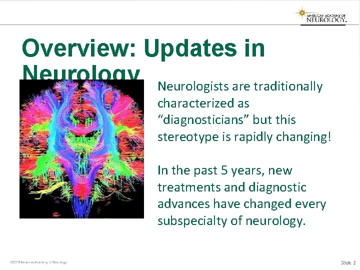
Overview: Updates in Neurology Neurologists are traditionally characterized as “diagnosticians” but this stereotype is rapidly changing! In the past 5 years, new treatments and diagnostic advances have changed every subspecialty of neurology. Slide 2
Updates: Vascular Neurology Slide 3
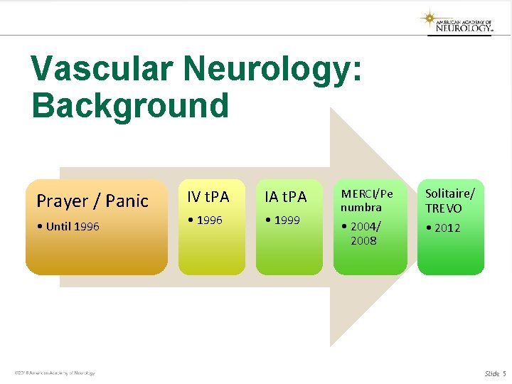
Vascular Neurology: Background • Before 1995: Stroke patients are managed for secondary prevention only • 1996: t. PA approved for the treatment of acute stroke, shown to improve functional outcomes (modified rankin scores) at 3 months • 2004: Thrombectomy devices approved • 2012: Stent-trievers introduced Slide 4
Vascular Neurology: Background Prayer / Panic • Until 1996 IV t. PA IA t. PA • 1996 • 1999 MERCI/Pe numbra • 2004/ 2008 Solitaire/ TREVO • 2012 Slide 5
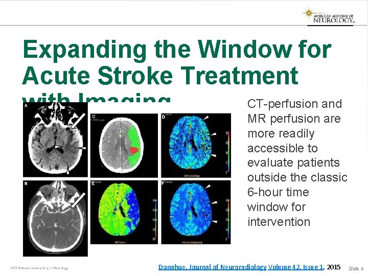
Expanding the Window for Acute Stroke Treatment CT-perfusion and with Imaging MR perfusion are more readily accessible to evaluate patients outside the classic 6 -hour time window for intervention Donahue. Journal of Neuroradiology Volume 42, Issue 1, 2015 Slide 6
Hyperacute MRI May Change t. PA Time Windows NEJM published data that suggest MRI mismatch may be able to supplement and possibly replace time windows based on last known well times. Slide 7
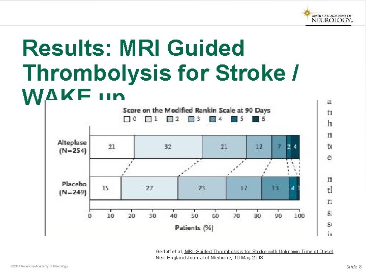
Results: MRI Guided Thrombolysis for Stroke / WAKE up Gerloff et al, MRI-Guided Thrombolysis for Stroke with Unknown Time of Onset, New England Journal of Medicine, 16 May 2018 Slide 8
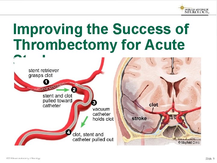
Improving the Success of Thrombectomy for Acute Stroke Slide 9
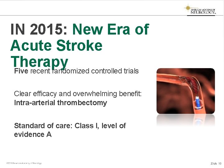
IN 2015: New Era of Acute Stroke Therapy Five recent randomized controlled trials Clear efficacy and overwhelming benefit: Intra-arterial thrombectomy Standard of care: Class I, level of evidence A Slide 10
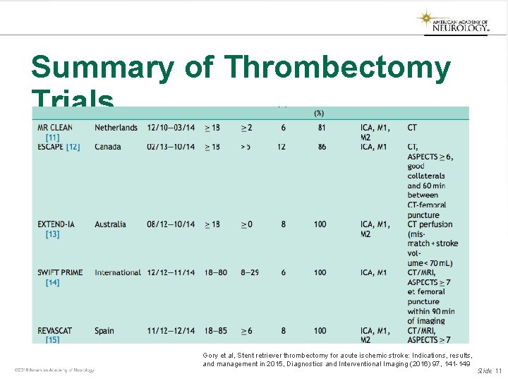
Summary of Thrombectomy Trials Gory et al, Stent retriever thrombectomy for acute ischemic stroke: Indications, results, and management in 2015, Diagnostics and Interventional Imaging (2016) 97, 141 -149 Slide 11
Updates: Traumatic Brain Injury Slide 12
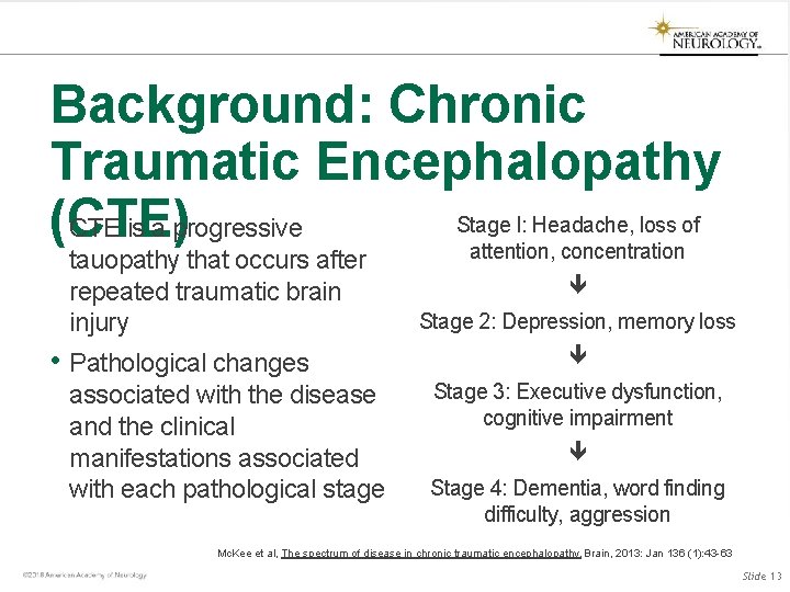
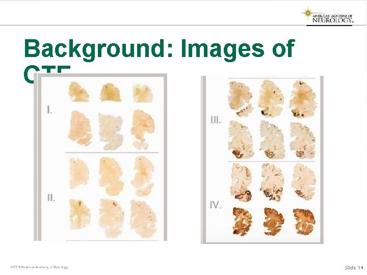
Background: Chronic Traumatic Encephalopathy Stage I: Headache, loss of • (CTE) CTE is a progressive attention, concentration tauopathy that occurs after repeated traumatic brain injury • Pathological changes associated with the disease and the clinical manifestations associated with each pathological stage Stage 2: Depression, memory loss Stage 3: Executive dysfunction, cognitive impairment Stage 4: Dementia, word finding difficulty, aggression Mc. Kee et al, The spectrum of disease in chronic traumatic encephalopathy, Brain, 2013: Jan 136 (1): 43 -63 Slide 13
Background: Images of CTE Slide 14
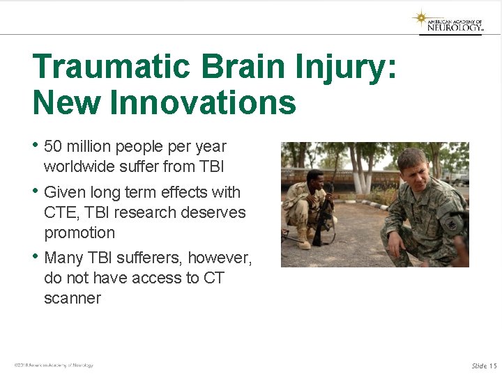
Traumatic Brain Injury: New Innovations • 50 million people per year worldwide suffer from TBI • Given long term effects with CTE, TBI research deserves promotion • Many TBI sufferers, however, do not have access to CT scanner Slide 15
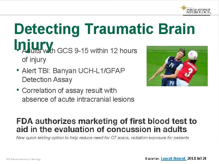
Detecting Traumatic Brain Injury • Adults with GCS 9 -15 within 12 hours of injury • Alert TBI: Banyan UCH-L 1/GFAP Detection Assay • Correlation of assay result with absence of acute intracranial lesions Bazarian. Lancet Neurol. 2018 Jul 24
ALERT-TBI: Results • Can blood-based biomarkers (GFAP, UCH-L 1) predict head injury after TBI? • Primary study outcomes: test sensitivity and negative predictive value (NPV) • 1977 patients enrolled; sensitivity 0. 976 and NPV 0. 996. Less than 1% of patients had a positive CT scan when the test was negative. Bazarian et al, Serum GFAP and UCH-L 1 for prediction of absence of intracranial injuries on head CT (ALERTTBI): a multicentre observational study, The Lancet Neurology, Vol 17, issue 9, September 2018, 782 -789 17
Updates: Neuromuscular Disease 18
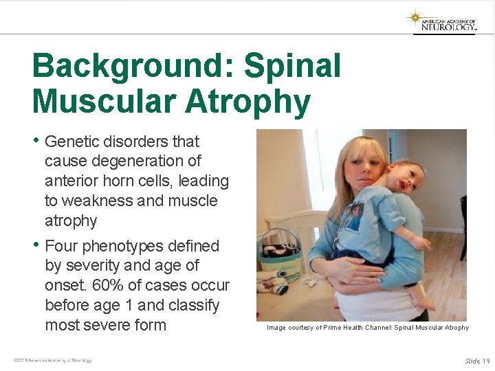
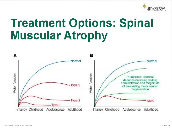
Background: Spinal Muscular Atrophy • Genetic disorders that cause degeneration of anterior horn cells, leading to weakness and muscle atrophy • Four phenotypes defined by severity and age of onset. 60% of cases occur before age 1 and classify most severe form Image courtesy of Prime Health Channel: Spinal Muscular Atrophy Slide 19
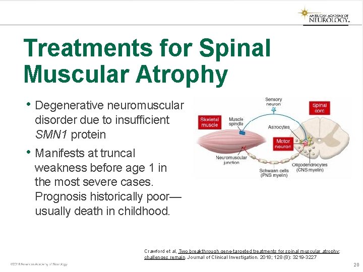
Treatments for Spinal Muscular Atrophy • Degenerative neuromuscular disorder due to insufficient SMN 1 protein • Manifests at truncal weakness before age 1 in the most severe cases. Prognosis historically poor— usually death in childhood. Crawford et al, Two breakthrough gene-targeted treatments for spinal muscular atrophy: challenges remain. Journal of Clinical Investigation. 2018; 128 (8): 3219 -3227 20
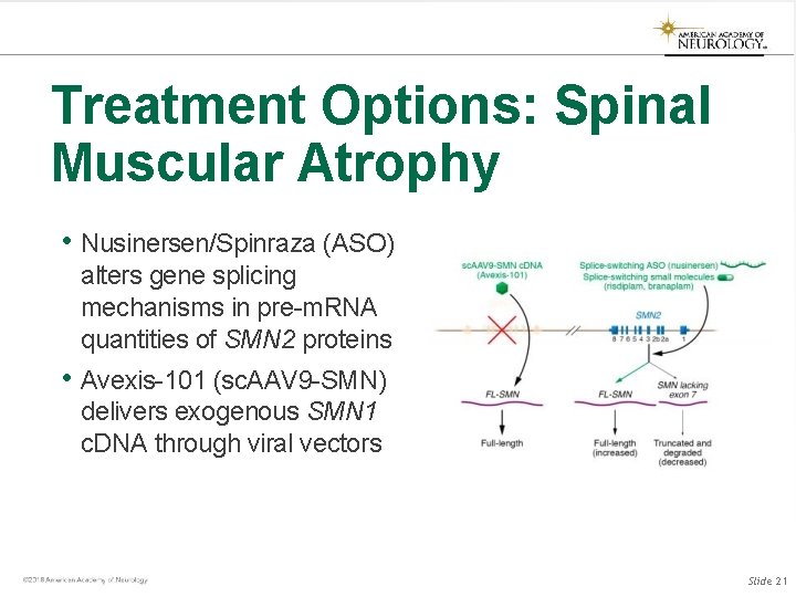
Treatment Options: Spinal Muscular Atrophy • Nusinersen/Spinraza (ASO) alters gene splicing mechanisms in pre-m. RNA quantities of SMN 2 proteins • Avexis-101 (sc. AAV 9 -SMN) delivers exogenous SMN 1 c. DNA through viral vectors Slide 21
Treatment Options: Spinal Muscular Atrophy Slide 22
Background: Myasthenia Gravis • Myasthenia gravis is an autoimmune disease that prevents transmission between motor neuron and muscle • Traditional treatments include symptom control with pyridostigmine and immunosuppression with steroids, azathioprine, and mycophenolate mofetil Slide 23
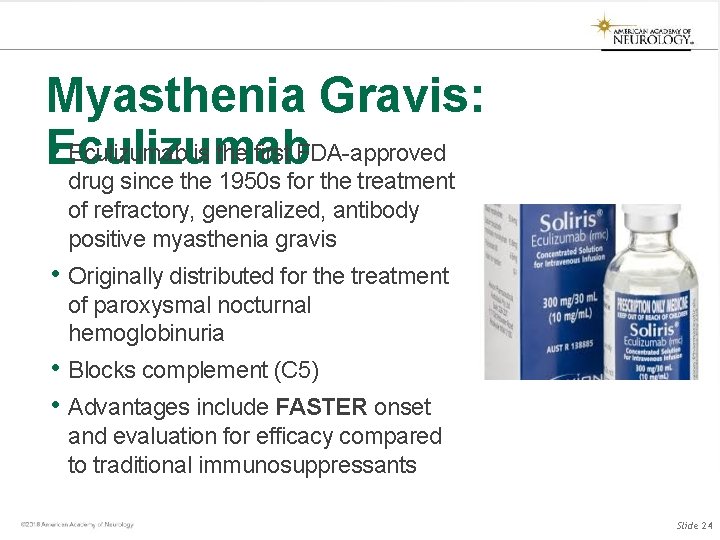
Myasthenia Gravis: • Eculizumab is the first FDA-approved Eculizumab drug since the 1950 s for the treatment of refractory, generalized, antibody positive myasthenia gravis • Originally distributed for the treatment of paroxysmal nocturnal hemoglobinuria • Blocks complement (C 5) • Advantages include FASTER onset and evaluation for efficacy compared to traditional immunosuppressants Slide 24
Updates: Movement Disorders Slide 25
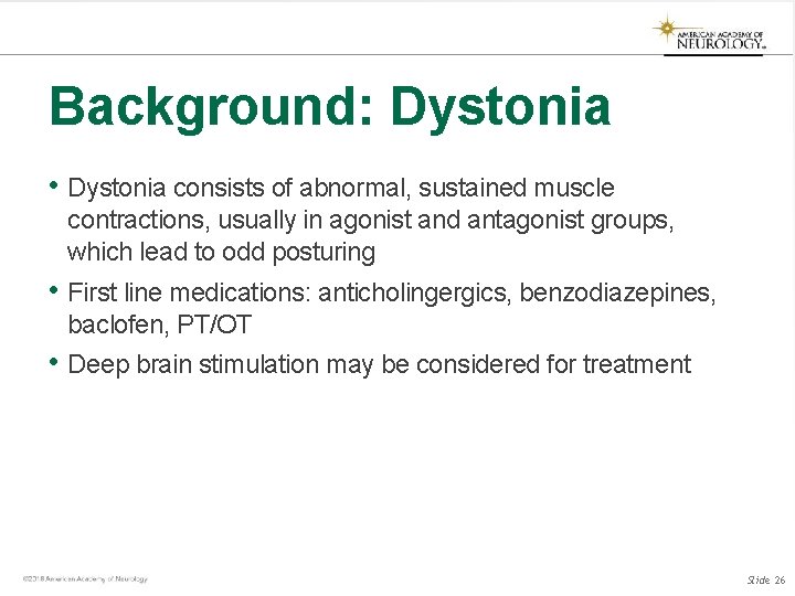
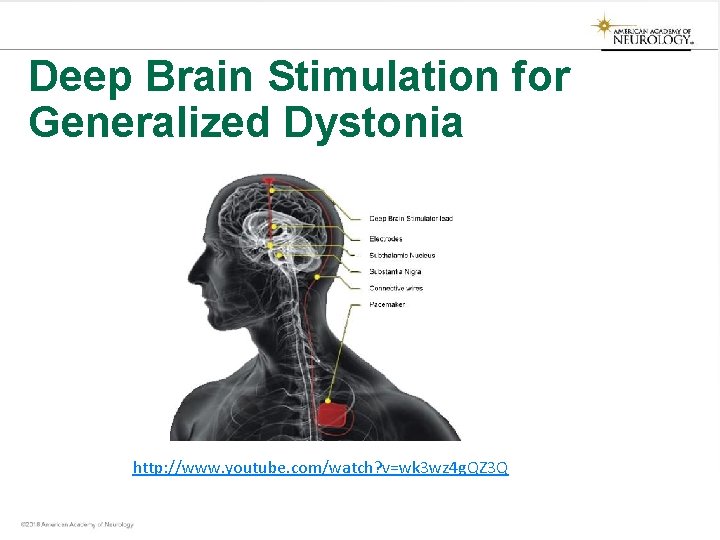
Background: Dystonia • Dystonia consists of abnormal, sustained muscle contractions, usually in agonist and antagonist groups, which lead to odd posturing • First line medications: anticholingergics, benzodiazepines, baclofen, PT/OT • Deep brain stimulation may be considered for treatment Slide 26
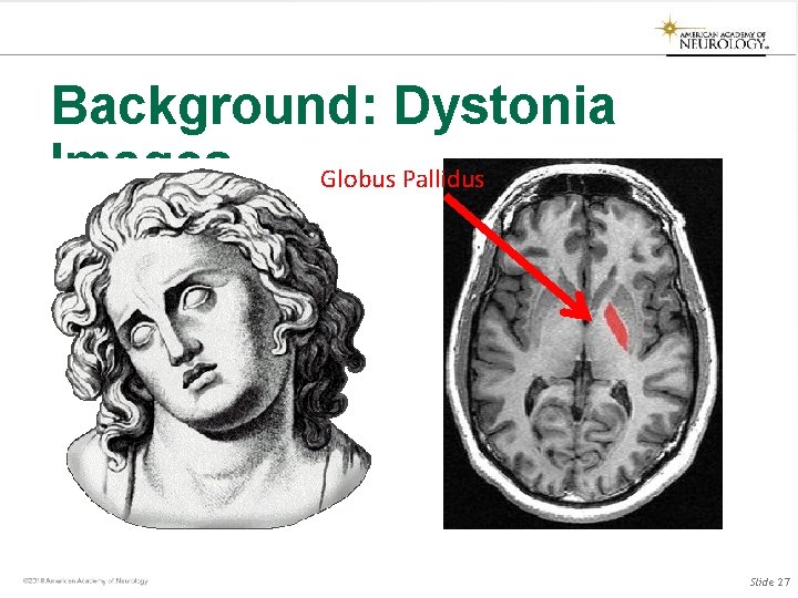
Background: Dystonia Images Globus Pallidus Slide 27
Deep Brain Stimulation for Generalized Dystonia http: //www. youtube. com/watch? v=wk 3 wz 4 g. QZ 3 Q
Thank You
Acknowledgement to Dr. Mohamad Abdalkader from Boston University Medical Center for assistance with these slides