
What you need to know about CBC and
What you need to know about CBC and coagulation profile Ghada El Gohary MB Bch, MSC, MD Prof Of Internal Medicine/BMT Ain Shams University Cairo , Egypt Consultant Adult hematology and stem cell transplantation KSUH

Slide credits to many contributors
Objectives • Practical approach to CBC • How to approach coagulation defect
• CBC is one of the commonest investigation we use and you need to understand it. • Many items listed , some of them tell the same information in different way. • Most of the CBC now done in automated way
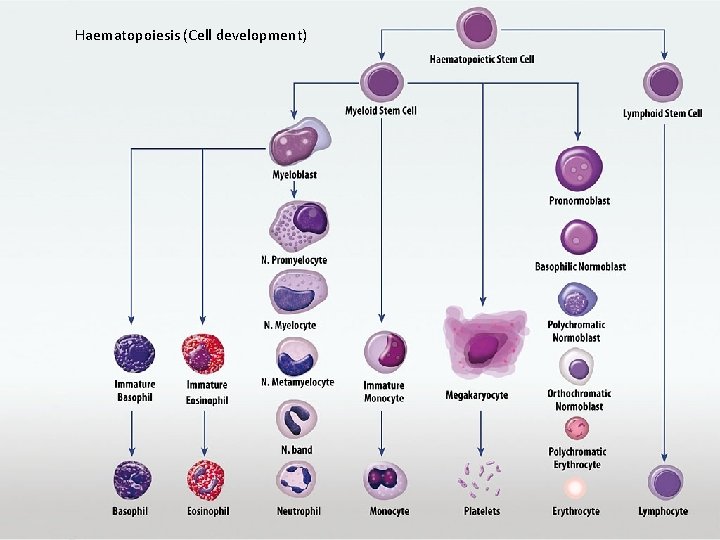
Haematopoiesis (Cell development)

Blood consists of (Red Blood Cells, White blood cells, Platelets, Plasma) • • WBC and diff RBC HB Htc MCV MCHC Plat and MPV • • ESR Blood film RDW Retics
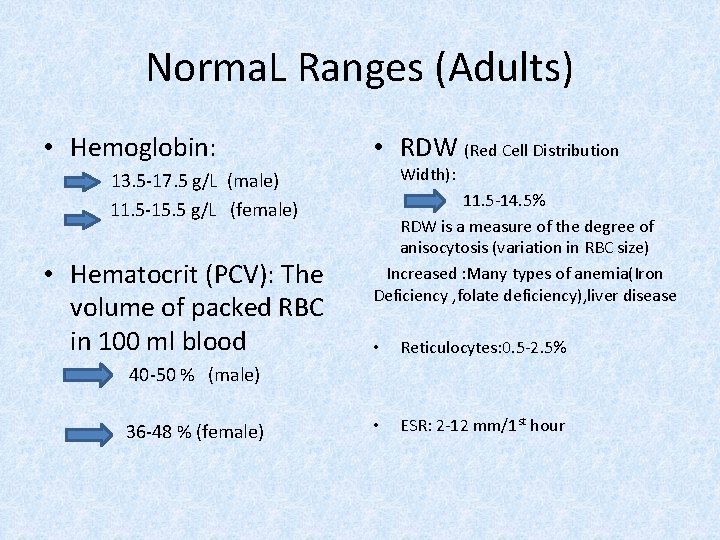
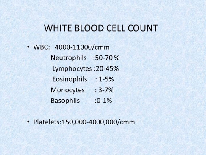
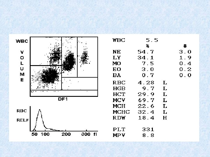
Norma. L Ranges (Adults) • Hemoglobin: 13. 5 -17. 5 g/L (male) 11. 5 -15. 5 g/L (female) • Hematocrit (PCV): The volume of packed RBC in 100 ml blood • RDW (Red Cell Distribution Width): 11. 5 -14. 5% RDW is a measure of the degree of anisocytosis (variation in RBC size) Increased : Many types of anemia(Iron Deficiency , folate deficiency), liver disease • Reticulocytes: 0. 5 -2. 5% • ESR: 2 -12 mm/1 st hour 40 -50 % (male) 36 -48 % (female)
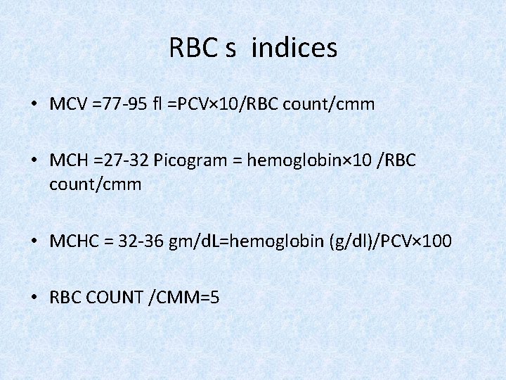
RBC s indices • MCV =77 -95 fl =PCV× 10/RBC count/cmm • MCH =27 -32 Picogram = hemoglobin× 10 /RBC count/cmm • MCHC = 32 -36 gm/d. L=hemoglobin (g/dl)/PCV× 100 • RBC COUNT /CMM=5
Interpret results in clinical context • All haematology results need to be interpreted in the context of a thorough history and physical examination, as well as previous results.
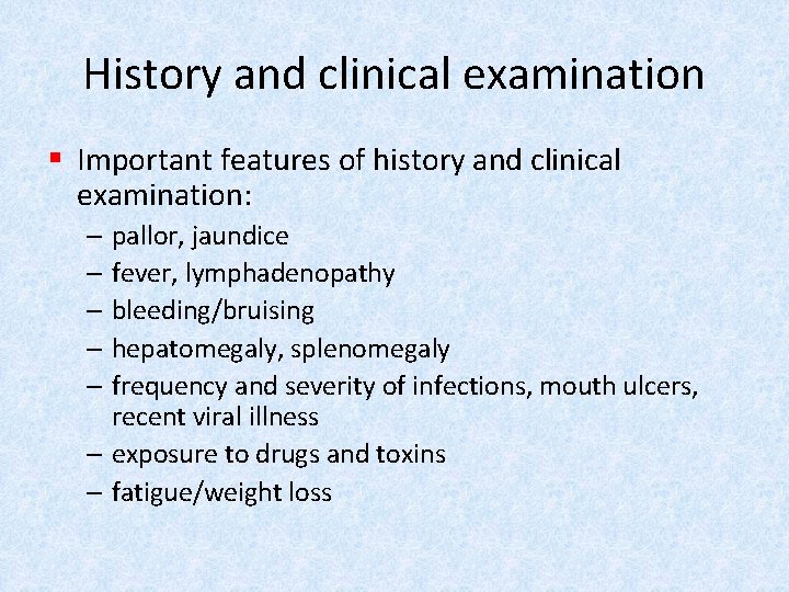
History and clinical examination § Important features of history and clinical examination: – pallor, jaundice – fever, lymphadenopathy – bleeding/bruising – hepatomegaly, splenomegaly – frequency and severity of infections, mouth ulcers, recent viral illness – exposure to drugs and toxins – fatigue/weight loss
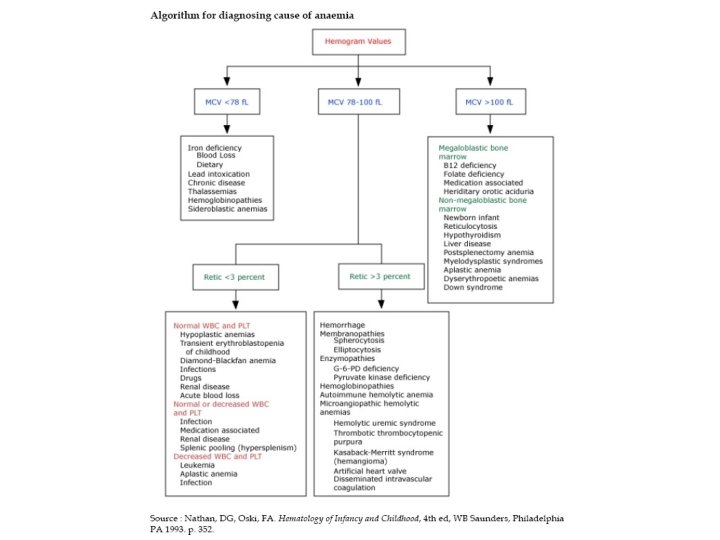
Low haemoglobin § Useful to use MCV to classify the anaemia: – Microcytic, MCV < 80 fl – Normocytic, MCV 80 – 100 fl – Macrocytic, MCV > 100 fl
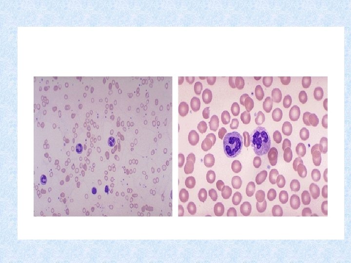
Microcytic Anaemia § The three most common causes for microcytic anaemia are: – Iron deficiency – Thalassaemia – Anaemia of Chronic disease
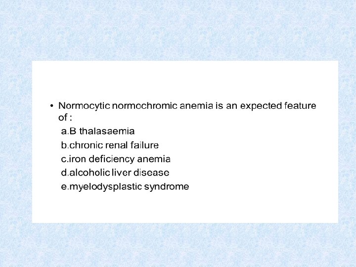
Normocytic anaemia § The causes of normocytic anaemia include: – Bleeding – Early nutritional anaemia (iron, B 12, folate deficiencies) – Anaemia of renal insufficiency – Anaemia of chronic disease/chronic inflammation – Haemolysis – Primary bone marrow disorder
Macrocytic anaemia § Common causes: – Alcohol – Liver disease – B 12 or folate deficiency – Thyroid disease – Some drugs (especially hydroxyurea)
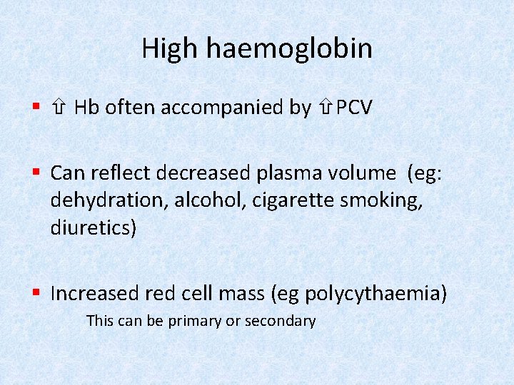
High haemoglobin § Hb often accompanied by PCV § Can reflect decreased plasma volume (eg: dehydration, alcohol, cigarette smoking, diuretics) § Increased red cell mass (eg polycythaemia) This can be primary or secondary
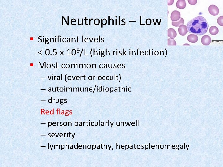
Neutrophils – Low § Significant levels < 0. 5 x 10⁹/L (high risk infection) § Most common causes – viral (overt or occult) – autoimmune/idiopathic – drugs Red flags – person particularly unwell – severity – lymphadenopathy, hepatosplenomegaly
Neutrophils – High § Most common causes – – – infection/inflammation Necrosis/malignancy any stressor/heavy exercise Drugs CML § Red flags – person particularly unwell – Severity – presence of left shift or blast
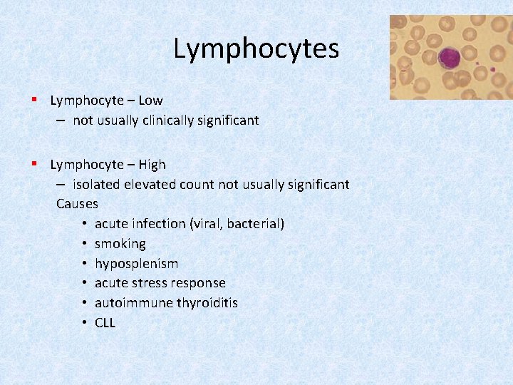
Lymphocytes § Lymphocyte – Low – not usually clinically significant § Lymphocyte – High – isolated elevated count not usually significant Causes • acute infection (viral, bacterial) • smoking • hyposplenism • acute stress response • autoimmune thyroiditis • CLL
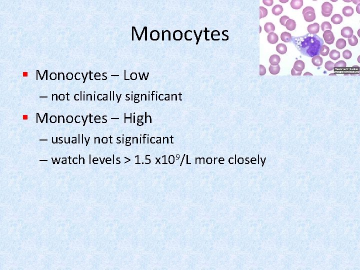
Monocytes § Monocytes – Low – not clinically significant § Monocytes – High – usually not significant – watch levels > 1. 5 x 10⁹/L more closely
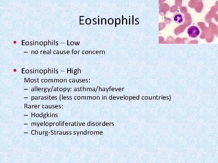
Eosinophils § Eosinophils – Low – no real cause for concern § Eosinophils – High Most common causes: – allergy/atopy: asthma/hayfever – parasites (less common in developed countries) Rarer causes: – Hodgkins – myeloproliferative disorders – Churg-Strauss syndrome
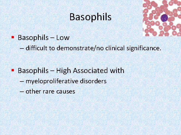
Basophils § Basophils – Low – difficult to demonstrate/no clinical significance. § Basophils – High Associated with – myeloproliferative disorders – other rare causes
§ Platelets – Low Significant levels < 100 x 109/L § Most common causes – viral infection – idiopathic thrombocytopenic purpura – liver disease – drugs – hypersplenism – autoimmune disease – Pregnancy – Artificial confirm on blood film § Red flags – bruising – petechiae – signs of bleeding
Platelets – High § Significant levels > 500 x 109/L § Most likely causes – reactive conditions eg infection, inflammation – pregnancy – iron deficiency – post splenectomy – essential thrombocythaemia
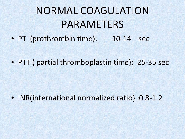
NORMAL COAGULATION PARAMETERS • PT (prothrombin time): 10 -14 sec • PTT ( partial thromboplastin time): 25 -35 sec • INR(international normalized ratio) : 0. 8 -1. 2
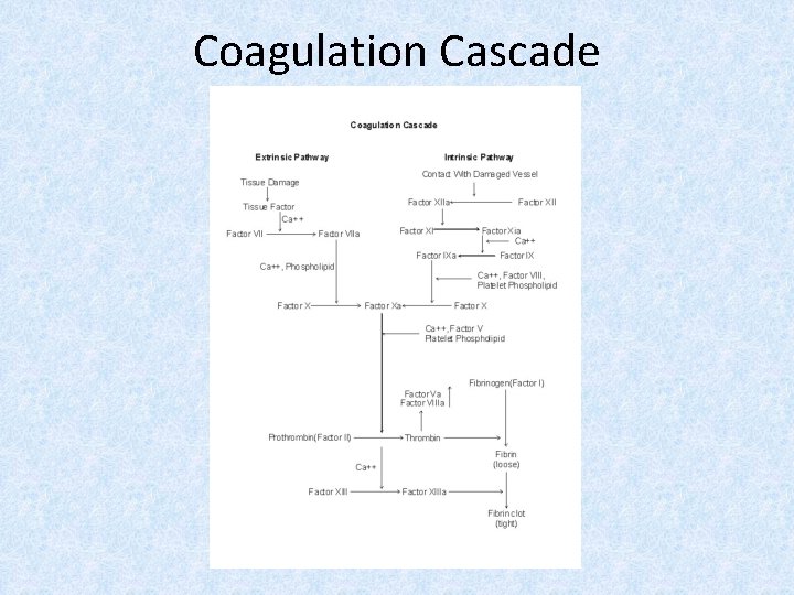
Coagulation Cascade
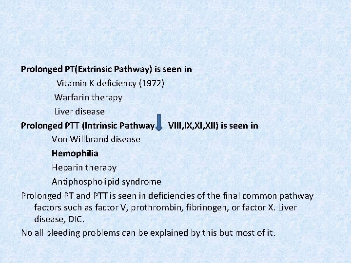
Prolonged PT(Extrinsic Pathway) is seen in Vitamin K deficiency (1972) Warfarin therapy Liver disease Prolonged PTT (Intrinsic Pathway VIII, IX, XII) is seen in Von Willbrand disease Hemophilia Heparin therapy Antiphospholipid syndrome Prolonged PT and PTT is seen in deficiencies of the final common pathway factors such as factor V, prothrombin, fibrinogen, or factor X. Liver disease, DIC. No all bleeding problems can be explained by this but most of it.
• Hemophilia A: deficiency of factor VIII (XR) • Hemophilia B : deficiency of factor IX (XR) • Hemophilia C : deficiency of factor XI (AR)
• Warfarin is oral anticoagulant which is a vitamin K antagonist that inhibits Vit K dependant coagulation factors : II, VII, IX, X
ANTICOAGULANTS: IIa (thrombin) • Heparin (great efficacy) • LMWH (dalteparin, enoxaparin) • Direct thrombin inhibitors (argatroban, bivalirudin, dabigatran)
ANTICOAGULANTS: Factor Xa • • LMW (greatest efficacy) Heparin Direct Xa inhibitors (apixaban, rivaroxaban) Fondaparinux
• Which of the following anemias is associated with splenomegaly a. chronic renal failure b. aplastic anemia c. hereditary spherocytosis d. sickle cell anemia
• All of the following produce microcytic anemia EXCEPT: a. sideroblastic anemia b. Thalassemia c. Prenicious anemia d. Lead poisoning
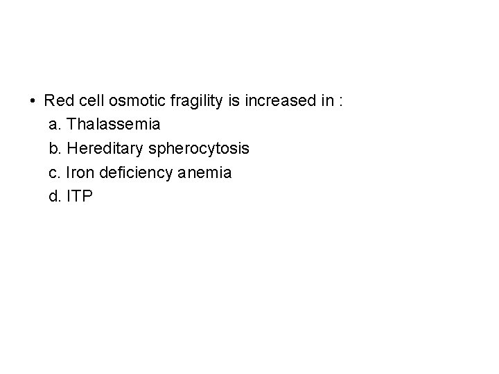
• Red cell osmotic fragility is increased in : a. Thalassemia b. Hereditary spherocytosis c. Iron deficiency anemia d. ITP
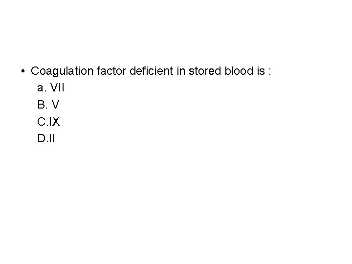
• Coagulation factor deficient in stored blood is : a. VII B. V C. IX D. II
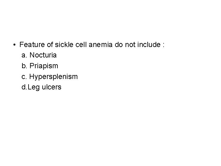
• Feature of sickle cell anemia do not include : a. Nocturia b. Priapism c. Hypersplenism d. Leg ulcers


Thank you
- Slides: 43