What is the optimal method of fetal surveillance

 and biophysical scoring are")
 Amniotic fluid volume Biophysical profile")


 • CTG should not be used as the only form of surveillance")


 • Biophysical profile should not be used for fetal surveillance in")

- Slides: 34
• What is the optimal method of fetal surveillance in a SGA infant ? • What is the frequency of fetal surveillance in a SGA infant ? • What is/are the optimal test/s to time delivery ?
• Biophysical tests, including amniotic fluid volume, cardiotocography (CTG) and biophysical scoring are poor at diagnosing a small or growth restricted fetus. • A systematic review of the accuracy of umbilical artery Doppler in a high–risk population to diagnose a SGA neonate has shown moderate.
• • • Umbilical artery Doppler Cardiotocography (CTG) Amniotic fluid volume Biophysical profile Middle cerebral artery Ductus venusus (DV) & Umbilical vein Doppler
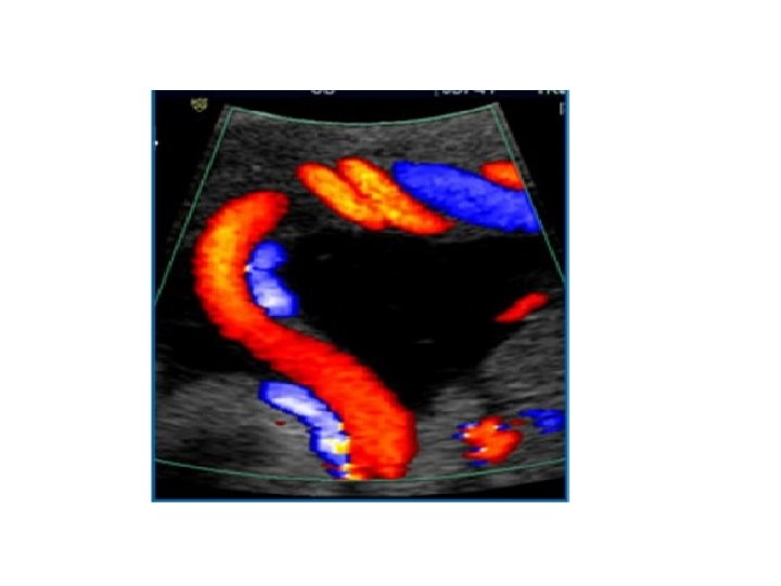
Umbilical artery Doppler • In high-risk population , the use of umbilical artery Doppler has been shown to reduce prenatal morbidity & mortality
Umbilical artery Doppler • Umbilical artery Doppler should be performed in all fetuses with an estimated fetal weight or an abdominal circumference < 10 th percentile (I- A)
Umbilical artery Doppler • Umbilical artery Doppler should be the primary surveillance tool in the SGA fetus.
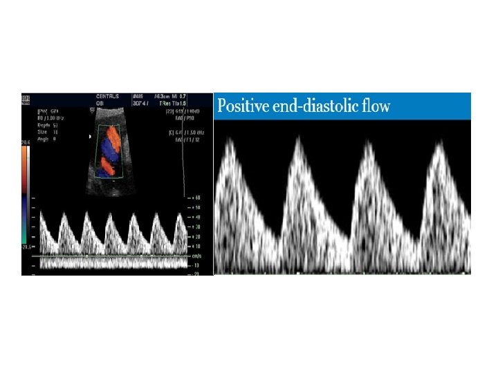
Frequency of Normal Umbilical artery Doppler flow indices in SGA fetus: • Defined by customized fetal weight standards 81 % of SGA fetuses have a normal umbilical artery Doppler
Management of Normal Umbilical artery Doppler flow indices in SGA fetus: • Outpatient management is safe in this group • When umbilical artery Doppler flow indices are normal it is reasonable to repeat surveillance every 14 days. • More frequent Doppler surveillance may be appropriate in a severely SGA infant.
• However Compare to AGA, SGA fetuses with a normal umbilical artery Doppler are still at increased risk of neonatal morbidity & adverse neurodevelopmental outcome
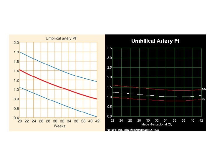
Which Umbilical artery Doppler waveform index ? • The large systematic review of test accuracy couldn't comment on which waveform index to use. • Although PI has been widely adopted in the UK , an analysis using receiver operator curves found that IR had the best discriminatory ability to predict a range of adverse perinatal out come
Routine umbilical artery Doppler • In a low risk or unselected population, systematic review found no conclusive evidence that routine umbilical artery Doppler benefits mother or baby. • As , such, umbilical artery Doppler is not recommended for screening an unselected population.
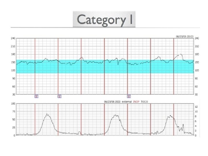
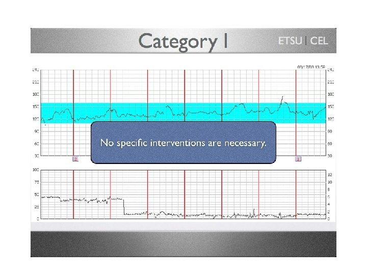
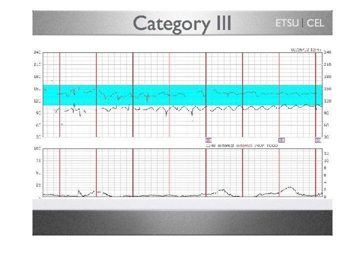
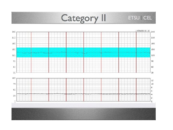
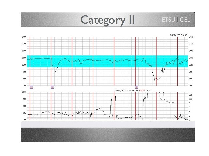
Cardiotocography (CTG) • CTG should not be used as the only form of surveillance in SGA fetuses. • Interpretation of the CTG should be based on short term fetal heart rate variation from computerized analysis (A)
The most useful CTG predictor: • FHR variation is the most useful predictor of fetal wellbeing in SGA fetuses. • A short term variation 3 ms (within 24 h of delivery ) has been associated with a higher rate of metabolic acidemia & early neonatal death
• Comparison of c. CTG with traditional CTG showed a reduction in perinatal mortality with c. CTG but no significant difference in perinatal mortality excluding congenital anomalies
Amniotic fluid volume • Ultrasound assessment of amniotic fluid volume should not to be used as the only form of surveillance in SGA fetus
• Amniotic fluid volume is usually estimated by the single deepest vertical pocket (SDVP) or amniotic fluid index(AFI), although both correlate poorly with actual amniotic fluid volume
• Interpretation of amniotic fluid volume should be based on single deepest vertical pocket (SDVP).
• The incidence of an AFI ≤ 5 cm in a low risk population is 1. 5%. • Compared to cases with a normal AFI, the risk of perinatal mortality and morbidity was not increased in cases with isolated oligohydramnios (RR 0. 7, 95% CI 0. 2– 2. 7) nor in those with associated conditions, including SGA fetuses (RR 1. 6, 95% CI 0. 9– 2. 6).
Oligohydramnios is associated with labour outcome: • a systematic review of 18 studies involving 10551 women, found an AFI ≤ 5 cm was associated with an increased risk of caesarean section for fetal distress (RR 2. 2, 95% CI 1. 5– 3. 4) and an Apgar score < 7 at 5 minutes (RR 5. 2, 95% CI 2. 4– 11. 3) but not acidaemia. • limited information is available about the accuracy of oligohydramnios to independently predict perinatal mortality and substantive perinatal morbidity in non– anomalous SGA fetuses monitored with umbilical artery Doppler
Biophysical profile • Consist of a NST + 4 ultrasound component : fetal movement, fetal muscle tone , amniotic fluid volume, fetal breathing movement. • A BPP is an appropriate second line (back-up) testing strategy when the NST component is non-reactive or none – interpretable
Biophysical profile (BPP) • Biophysical profile should not be used for fetal surveillance in preterm SGA fetuses.
• What is the optimal method of fetal surveillance in a SGA infant ? • What is the frequency of fetal surveillance in a SGA infant ? • What is/are the optimal test/s to time delivery ?