
What is thalassemia Genetic blood disorder resulting in
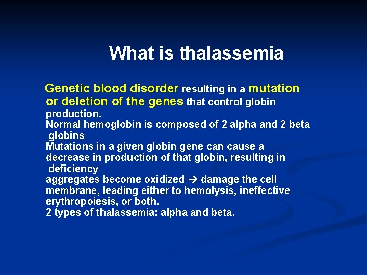
What is thalassemia Genetic blood disorder resulting in a mutation or deletion of the genes that control globin production. Normal hemoglobin is composed of 2 alpha and 2 beta globins Mutations in a given globin gene can cause a decrease in production of that globin, resulting in deficiency aggregates become oxidized damage the cell membrane, leading either to hemolysis, ineffective erythropoiesis, or both. 2 types of thalassemia: alpha and beta.
Types Of Thalassemia Two types: Alpha thalassemia occurs when a gene or genes related to the alpha globin protein are missing or mutated. Beta thalassemia occurs when gene defects affect production of the beta globin protein
Demographics The thalassemia gene may be maintained in the human population, in part because of the greater immunity of heterozygous individuals against malaria and is found in parts of the world where malaria is common These include Southeast Asia, China, India, Africa, and parts of the Mediterranean.
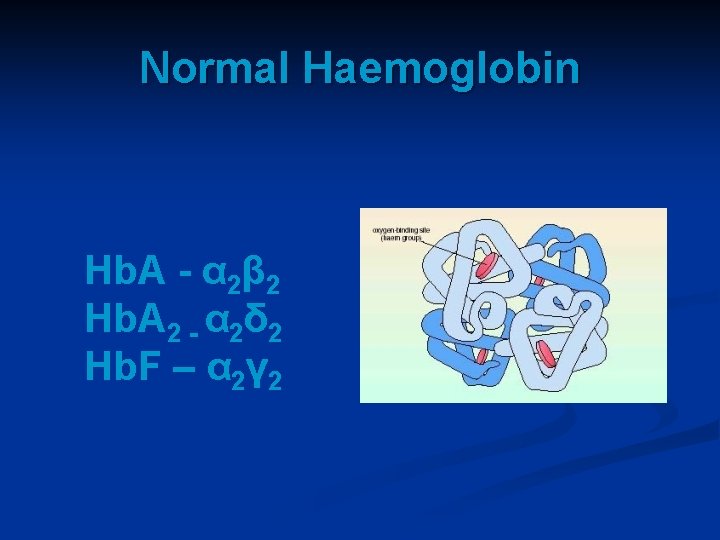
Normal Haemoglobin Hb. A - α 2β 2 Hb. A 2 - α 2δ 2 Hb. F – α 2γ 2
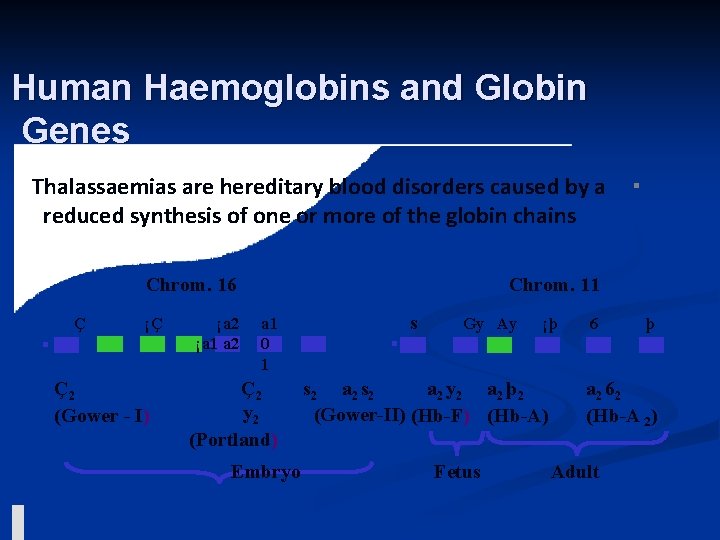
Human Haemoglobins and Globin Genes Thalassaemias are hereditary blood disorders caused by a reduced synthesis of one or more of the globin chains Chrom. 16 Ç ¡Ç Ç2 (Gower - I) ¡a 2 ¡a 1 a 2 ▪ Chrom. 11 a 1 0 1 Ç2 y 2 (Portland) Embryo s Gy Ay ¡þ s 2 a 2 y 2 a 2 þ 2 (Gower-II) (Hb-F) (Hb-A) Fetus 6 þ a 2 62 (Hb-A 2) Adult
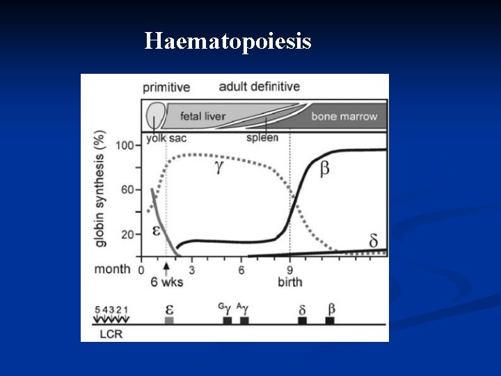
Haematopoiesis
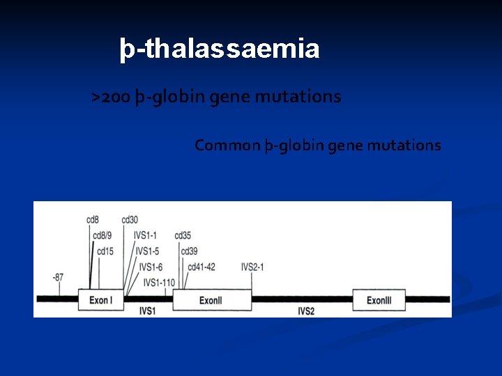
þ-thalassaemia >200 þ-globin gene mutations Common þ-globin gene mutations
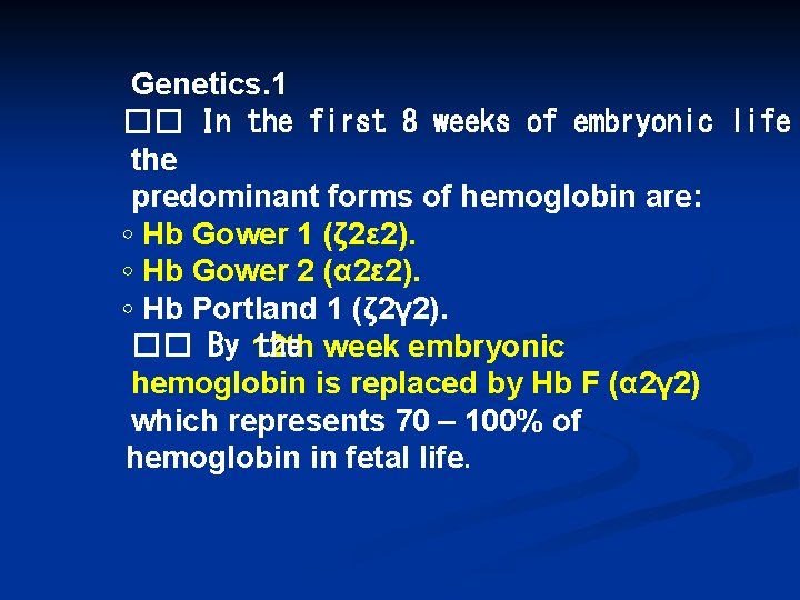
Genetics. 1 �� In the first 8 weeks of embryonic life the predominant forms of hemoglobin are: ◦ Hb Gower 1 (ζ 2ε 2). ◦ Hb Gower 2 (α 2ε 2). ◦ Hb Portland 1 (ζ 2γ 2). �� By 12 th the week embryonic hemoglobin is replaced by Hb F (α 2γ 2) which represents 70 – 100% of hemoglobin in fetal life.

Genetics (2). Adult hemoglobin Hb A (α 2β 2) detectable from 6/12 after birth. �� Hemoglobin Hb. A 2 (α 2Δ 2) is present in utero but only very minor in normal adults. �� In normal adults 96 – 98% of hemoglobin is Hb. A, Hb A 2 (2 – 3%) and Hb. F (<1%) constitute a minor component of the total hemoglobin.
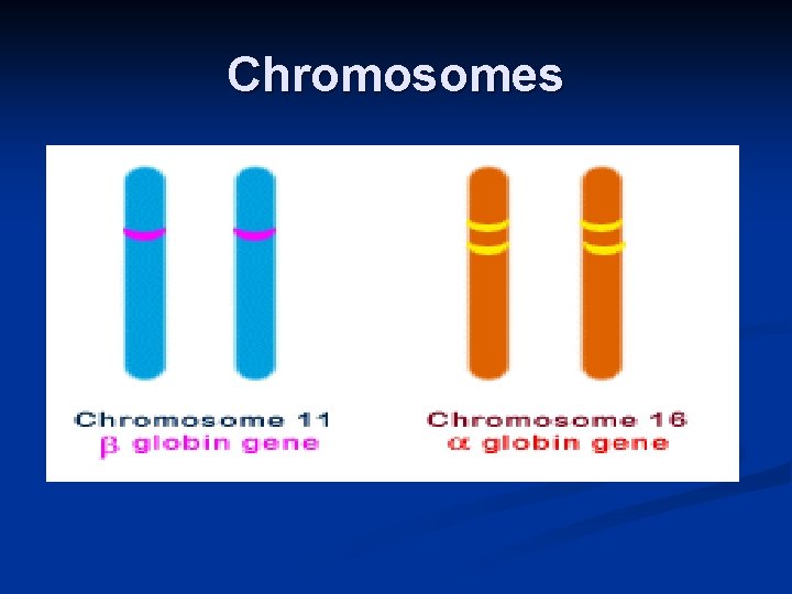
Chromosomes
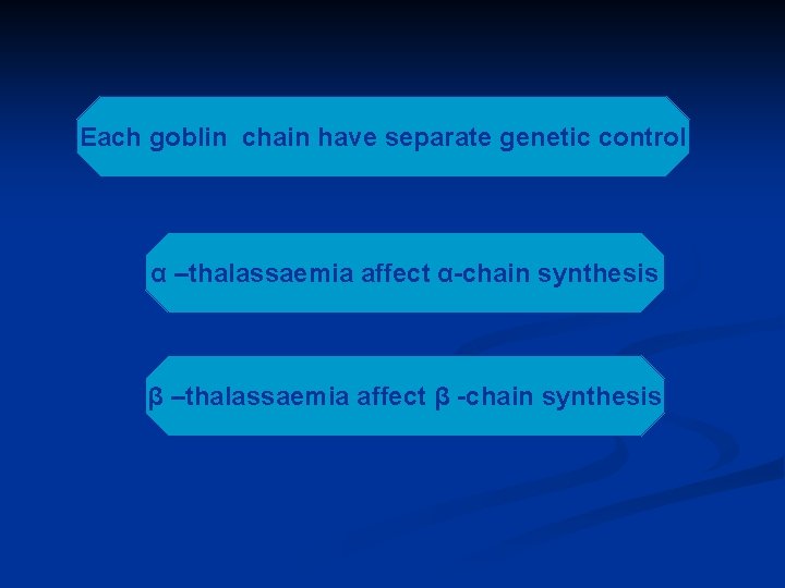
Each goblin chain have separate genetic control α –thalassaemia affect α-chain synthesis β –thalassaemia affect β -chain synthesis
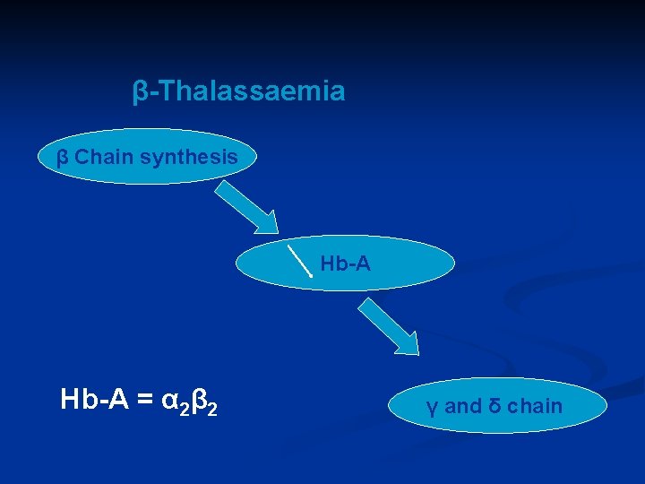
β-Thalassaemia β Chain synthesis Hb-A = α 2β 2 γ and δ chain
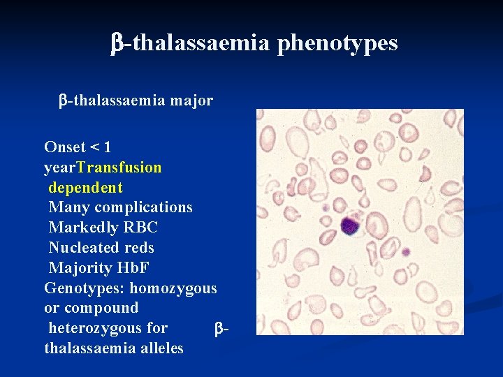
-thalassaemia phenotypes -thalassaemia major Onset < 1 year. Transfusion dependent Many complications Markedly RBC Nucleated reds Majority Hb. F Genotypes: homozygous or compound heterozygous for thalassaemia alleles
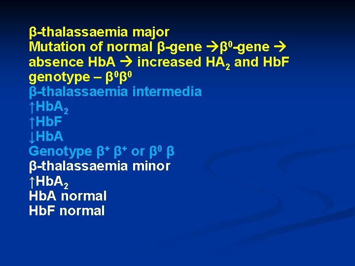
β-thalassaemia major Mutation of normal β-gene β 0 -gene absence Hb. A increased HA 2 and Hb. F genotype – β 0β 0 β-thalassaemia intermedia ↑Hb. A 2 ↑Hb. F ↓Hb. A Genotype β+ β+ or β 0 β β-thalassaemia minor ↑Hb. A 2 Hb. A normal Hb. F normal
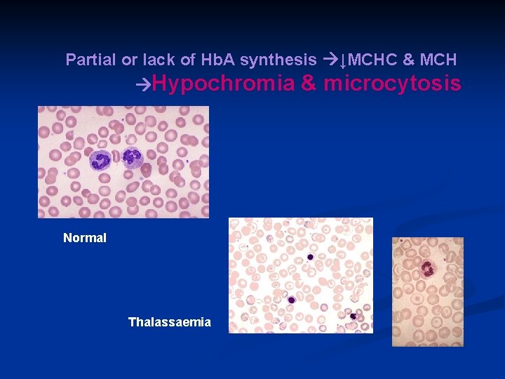
Partial or lack of Hb. A synthesis ↓MCHC & MCH Hypochromia Normal Thalassaemia & microcytosis
α-Thassaemia An absence or deficiency of αchain synthesis due to delation of α-genes. Silent Carriers (heterozygotes +/-) 3 functional alpha globin genes No symptoms, but thalassemia could potentially appear in offspring
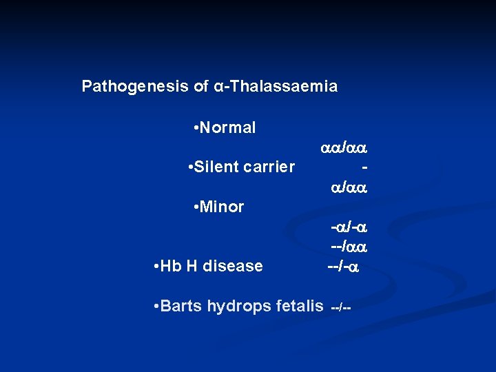
Pathogenesis of α-Thalassaemia • Normal. • Silent carrier / / • Minor • Hb H disease - /- --/- • Barts hydrops fetalis --/--
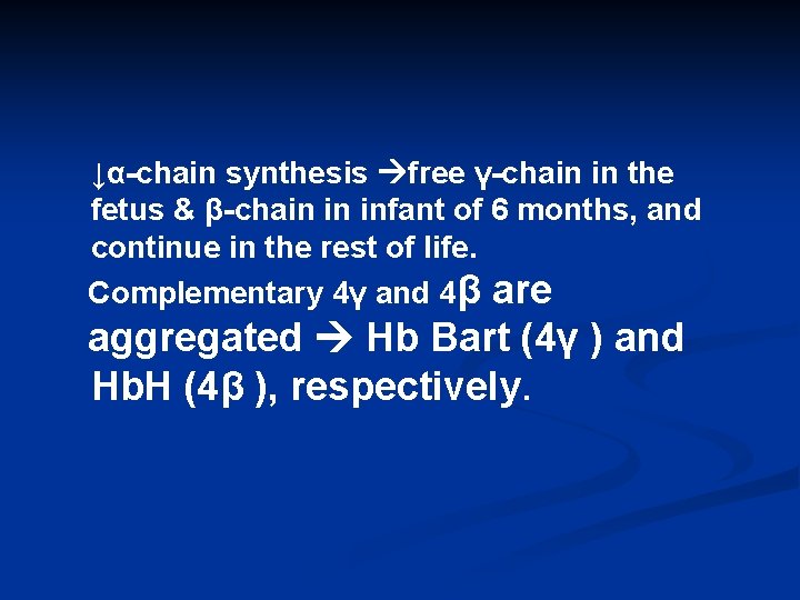
↓α-chain synthesis free γ-chain in the fetus & β-chain in infant of 6 months, and continue in the rest of life. Complementary 4γ and 4β are aggregated Hb Bart (4γ ) and Hb. H (4β ), respectively.
![Example 1 Hamoud alhanzal has 1 mutation (IVS 1. 5 [G>C]) and the genotype](http://slidetodoc.com/presentation_image_h2/3ccbe9be3bc57cded3e4cf1f81de07c0/image-26.jpg "Example 1 Hamoud alhanzal has 1 mutation (IVS 1. 5 [G>C]) and the genotype")
Example 1 Hamoud alhanzal has 1 mutation (IVS 1. 5 [G>C]) and the genotype is heterozygote. 2 - Amina alhamoud has 1 mutation (IVS 1. 5 [G>C]) and the genotype is heterozygote. 3 - The embryo has 2 mutations (IVS 1. 5 [G>C]) / (IVS 1. 5 [G>C]) and the genotype is homozygote. Conclusion: The embryo has 2 mutations in the β –globin gene. Therefore, the embryo has β –thalassemia
![Dxample 2 M. Al-Kahr has 1 mutation (Codon 39 [C>T]) and the genotype is](http://slidetodoc.com/presentation_image_h2/3ccbe9be3bc57cded3e4cf1f81de07c0/image-27.jpg "Dxample 2 M. Al-Kahr has 1 mutation (Codon 39 [C>T]) and the genotype is")
Dxample 2 M. Al-Kahr has 1 mutation (Codon 39 [C>T]) and the genotype is heterozygote. 2 - K. Al-Kahr has 1 mutation (Codon 39 [C>T]) and the genotype is heterozygote. 3 - The embryo has 2 mutations (Codon 39 [C>T]/Codon 39 [C>T]) and the genotype is homozygote. Conclusion: The embryo has 2 mutations in the β –globin gene. Therefore, the embryo has β – thalassemia.
![Example 3: 1 - Ali Khalaf has 1 mutation (Codon 39 [C>T]) and the](http://slidetodoc.com/presentation_image_h2/3ccbe9be3bc57cded3e4cf1f81de07c0/image-28.jpg "Example 3: 1 - Ali Khalaf has 1 mutation (Codon 39 [C>T]) and the")
Example 3: 1 - Ali Khalaf has 1 mutation (Codon 39 [C>T]) and the genotype is heterozygote. 2 - Amina Jadouah has 1 mutation (Codon 39 [C>T]) and the genotype is heterozygote. 3 - The embryo has no mutations and the genotype is normal. Conclusion: The embryo has no mutations in the β –globin gene. Therefore, the embryo is normal
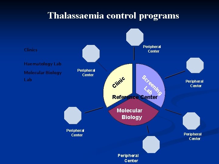
Thalassaemia control programs Peripheral Center Clinics Haematology Lab Molecular Biology Lab Peripheral Center Sc re in e l La nin C b g Reference Center ic Peripheral Center Molecular Biology Peripheral Center
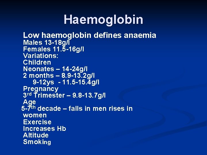
Haemoglobin Low haemoglobin defines anaemia Males 13 -18 g/l Females 11. 5 -16 g/l Variations: Children Neonates – 14 -24 g/l 2 months – 8. 9 -13. 2 g/l 9 -12 ys - 11. 5 -15. 4 g/l Pregnancy 3 rd Trimester – 9. 8 -13. 7 g/l Age 5 -7 th decade – falls in men rises in women Exercise Increases Hb Altitude Smoking
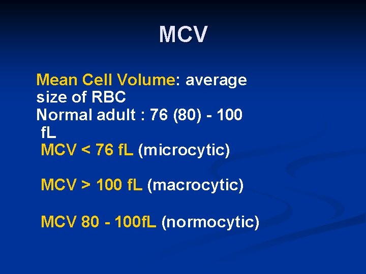
MCV Mean Cell Volume: average size of RBC Normal adult : 76 (80) - 100 f. L MCV < 76 f. L (microcytic) MCV > 100 f. L (macrocytic) MCV 80 - 100 f. L (normocytic)
Reticulocyte count: In the investigation of anaemia Reduced: Failure of • erythropoiesis Increased: Appropriate BM • erythroid response
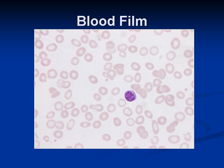
Blood Film
BLOOD SMEAR DIAGNOSIS Previous profiles show basophilic stippling is not the only abnormality present on a blood smear Merck manual states that a blood smear is almost diagnostic for beta thalassemia major due to many nucleated erythroblasts, target cells, small pale RBC and punctuate and diffuse basophilia. Basophilic stippling alone is not enough to diagnose and differentiate between the types of thalassemias
Red Cell Distribution Width (RDW) The degree of variation in size of RBC: N <14 Increased RDW corresponds with anisocytosis: Iron deficiency (increased RDW is the earliest lab feature: anisocytosis precedes the anaemia) Megaloblastic anaemia (can be very high >20) Anaemia with bone marrow erythroid response (i. e. reticulocytosis) RDW useful in DDx of microcytic anaemias. Most cases of iron deficiency: raised RDW Most cases thalassaemia trait: normal RDW
Haemoglobin Electrophoresis Different types of haemoglobins contain different surface charge which determine the elecrtrophoretic mobility and gives a specific bands on the electrophoretic papers when haemolysate is undergone eletrophoresis. Hb. F, Hb. A 2, Hb. C, Hb. E, Hb. D, Hb. S, Hb. H and Hb Bart can be measured accurately by electrophoresis
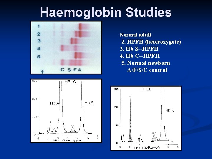
Haemoglobin Studies Normal adult 2. HPFH (heterozygote) 3. Hb S--HPFH 4. Hb C--HPFH 5. Normal newborn A/F/S/C control
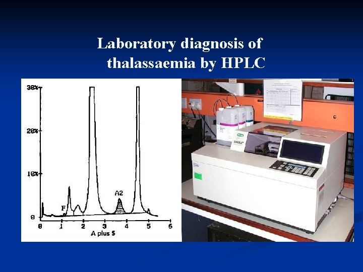
Laboratory diagnosis of thalassaemia by HPLC
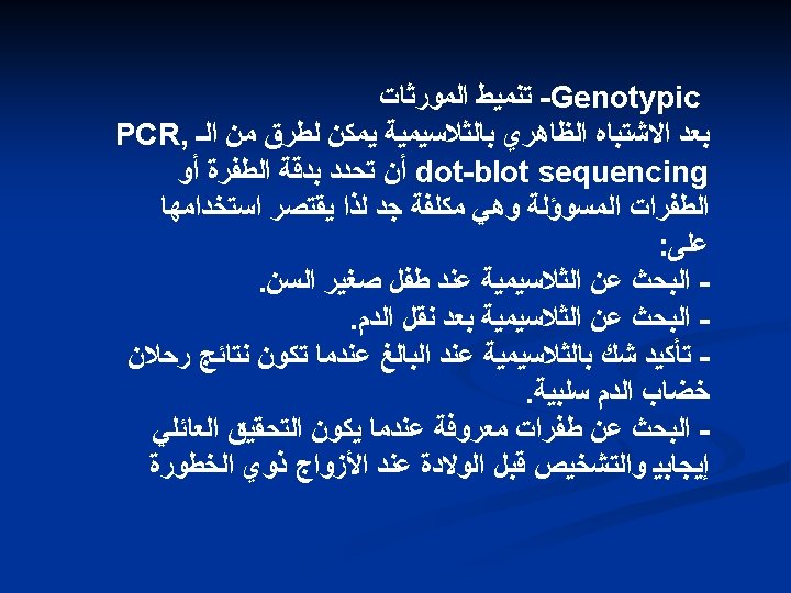
Molecular diagnosis. To date many molecular defects have been defined in thalassemias by. The common polymerase chain reaction (PCR) procedures
n. Prenatal diagnosis. Early prenatal diagnosis can be done using first fetal blood sampling, and later chorion villus biopsy and direct analysis of the globin genes. n. The error rate in experienced centers is now well under 1%.
Prevention efforts Pre marital screening to make sure that the couple are not both carriers. Provision of counseling and health education for the thalassemics, their families and the public. Provision of prenatal testing for thalassemia. Reduction of marriages between relatives.
Counselling for hemoglobin disorders. Couples at risk for hemoglobin disorders may be identified retrospectively after the birth of an affected child, or prospectively by analyzing childless spouses. Prospective identification allows parents to have a disease -free family. Education of the public by mass media, posters, lectures, etc. was carried out in Sardinia, Italy. Special meetings were held with physicians and especially with pediatricians and obstetricians, family planning associations, nurses and social workers.
Problems commonly faced by thalassemia major patients in developing countries Reduced availability of blood for transfusion. Reduced availability of Desferal pumps, less than third of the patients have access to pumps. High cost of treatment. Limited services that blood banks are able to give. Unavailability of counseling services. Lack of experience and appropriate training among the health providers to handle thalassemia cases.
More Permanent Options §Bone Marrow Transplants §Replacing patient’s marrow with donor marrow §First performed on thalassemia patient in 1981 §Difficult, because donor must be exact match for recipient §Even a sibling would only have a 1 in 4 chance of being a donor §Cord Blood Transplants §Rich in stem cells §Also needs to be an exact match

THANKS
- Slides: 47