What is a Brain Introduction to neuronal circuits

What is a Brain? Introduction to neuronal circuits, neurons, synapses, and non-invasive imaging

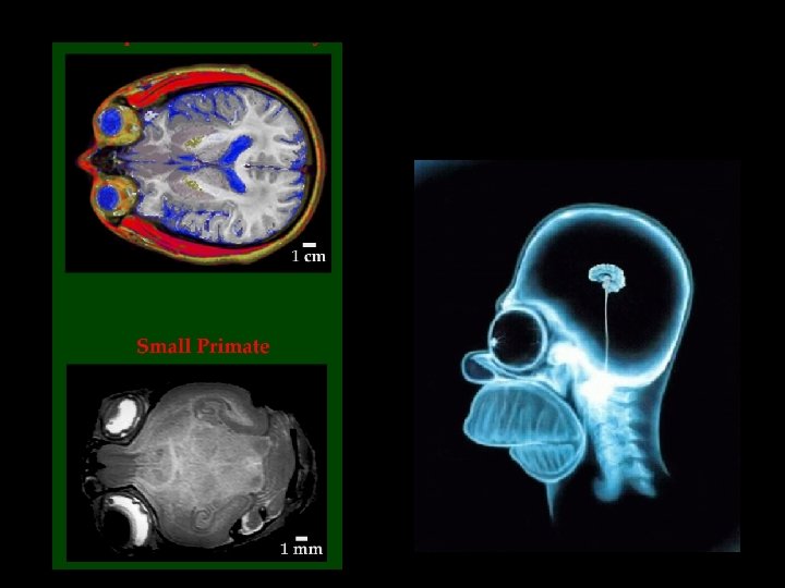
Organization of the Brain MRI imaging Hemispheres PET imaging Regions Connections Neurons Synapses 2 -deoxyglucose labelling Multi-cell recordings Single-cell recordings Electrical and chemical events

Front Back Brain The Central Nervous System: Spinal cord

A split-brain patient fixates on the dot in the middle of a screen. Then a picture of a spoon is flashed to the right of the dot. Language, Math, Logic Spatial abilities, Face recognition, Visual imagery, Music The visual information about the spoon crosses in the optic chiasm and ends up in the LEFT HEMISPHERE. The person correctly identifies the spoon verbally.

Now the picture of a spoon is flashed to the left of the dot. Language, Math, Logic Spatial abilities, Face recognition, Visual imagery, Music Now the visual information travels to the RIGHT HEMISPHERE. Now if the subject is asked to identify the picture, he reports seeing nothing. But, when this same subject is asked to pick out an object using only the LEFT hand, he correctly picks out the spoon. This is because touch information from the left hand crosses over to the right hemisphere - the side that "saw" the spoon. However, if he is again asked to identify the object verablly, even when it is in his hand, he cannot do so because the right hemisphere cannot "talk. " So, the right hemisphere is not stupid, it just has little ability for language - it is "non-verbal. "
 and dopamine")
Function is often localized to specific brain regions Front Back acetylcholine (nicotine) and dopamine

A typical pathway: sensation of pain and the reaction to pain

Spinal reflexes, such as the knee-jerk, involve just two neurons. sensory neuron motor neuron the sensory neuron acts like a strain gauge wrapped around a special muscle fiber.

Electricity is also a language of the nervous system

2 -deoxyglucose can label thousands or millions of active cells at once glucose cell activity glucose phosphates metabolic products, ATP cell 2 -deoxyglucose (radiolabelled) activity 2 -deoxyglucose phosphates No metabolic products; label remains in cell
![Positron emission tomography (PET). The probe is [18 F]fluoro-2 -deoxyglucose. The 18 F nucleus](http://slidetodoc.com/presentation_image_h2/ae66051d2168aede44c505a9cb0d94ae/image-11.jpg "Positron emission tomography (PET). The probe is [18 F]fluoro-2 -deoxyglucose. The 18 F nucleus")
Positron emission tomography (PET). The probe is [18 F]fluoro-2 -deoxyglucose. The 18 F nucleus decays, eventually yielding a positron which annihilates with an electron to produce a pair of g rays (photons). These travel in opposite directions. The two coincident photons intersect an array of detectors. The point of origin is on the line between the two detectors; and “tomography” is the set of algorithms that compute the point of origin from many independent events.
 image. This is the rear view of")
A functional magnetic resonance imaging (f. MRI) image. This is the rear view of a human brain activation associated with performance of a task that utilizes motor, visual and planning cognitive processes. The subject was moving a joystick to follow a target around a video display.

Nuclear Magnetic Resonance Nuclei of interest 1 H Proton Carbon Sodium Phosphorus Xenon H 2 O, fat, NAA 13 C 23 Na 31 P ATP, ADP, Pi 129 Xe These nuclei possess spin angular momentum (mh/2 p) & thus a magnetic moment (m) m= I, I-1, …-I 2 I+1 values of m m=g. Ih/2 p g gyromagnetic ratio


Action Potentials and Single Channels

The “Na+ pump” splits ATP to make a Na+ and K+ concentration gradient What are the consequences of the charge imbalance? A transporter protein moves a few ions for each conformational change

Na pump = = current source = R very large E very large

V pump channel

V pump ATP channel

V + pump ATP DV = IR - channel The Na pump drives the membrane potential more negative (the cell “hyperpolarizes”)

Major Roles for Ion Channels actually, DE electrical transmission in axons: electric field open closed Monday: chemical transmission at synapses: [neurotransmitter] closed open

Little Alberts Fig 12 -30

action potential noun Date: 1926 : a momentary change in electrical potential (as between the inside of a nerve cell and the extracellular medium) that occurs when a cell or tissue has been activated by a stimulus.

The frequency of impulses represents signaling among cells in the nervous system. -from sense organs to the brain -within the brain -from the brain to muscles -even in a muscle or in the heart -even in the pancreas

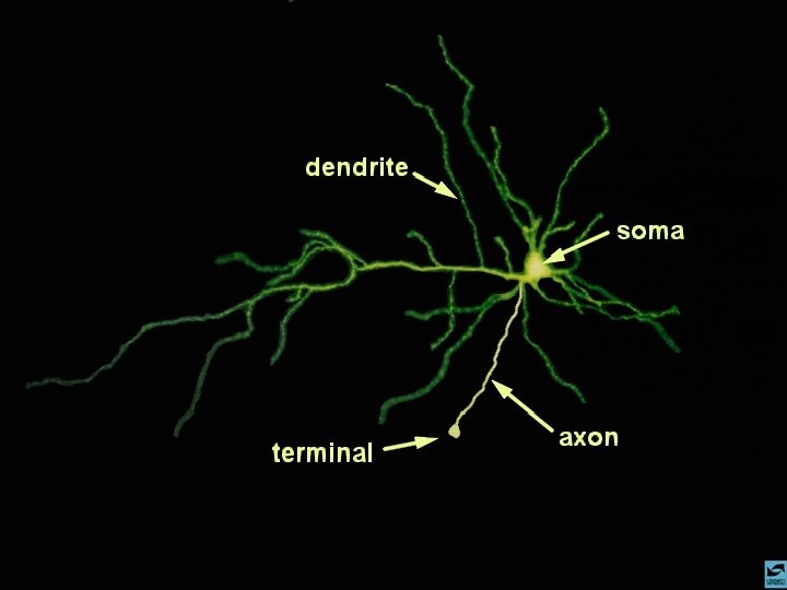
The synapse is a point of information processing An adult human brain contains ~ 1011 neurons , and each of these might receive 103 synapses apiece, for a total of 1014 synapses. Most of these synapses form during the first 2 yr of life. Thus 1014 synapses/108 s = 106 synapses/s form in a fetus and infant!

Synapses
, mitochondria (watermelons) 1. Movements do not")
Cerebral cortex Axons Dendrites Synapses Synaptic vesicles (volleyballs), mitochondria (watermelons) 1. Movements do not occur in 1 ms; vesicles are already “docked”. 2. Neurotransmitter diffuses across the synaptic cleft in a few ms 3. “Slight” errors cause mental illness? Unproven! (But single-codon mutations do cause some neuroscience diseases)

presynaptic terminal cell body kinesin 50 nm ~ 20 distinct proteins cytosol Neurotransmitter and ATP vesicle transport; pumping protons; pumping neurotransmitter; docking; fusion; recycling.

H+ ATP-driven proton pump cytosol proton-coupled neurotransmitter pump cytosol Neurotransmitter and ATP (1, 000 to 10, 000 molecules of each) ~ isotonic!

Electricity, then chemistry triggers synaptic vesicle fusion docked vesicle neurotransmitter nerve impulse voltage-gated Ca 2+ channel

Electricity, then chemistry triggers synaptic vesicle fusion docked vesicle Ca 2+ nerve impulse voltage-gated Ca 2+ channel neurotransmitter

Electricity, then chemistry triggers synaptic vesicle fusion fused vesicle Ca 2+ neurotransmitter

The heavy chain of botulinum toxin is an enzyme that cleaves synaptic vesicle fusion proteins

What is a Receptor? Receptors and Ion Channels as Examples of Proteins

receptor a molecule on the cell surface or in the cell interior that has an affinity for a specific molecule (the ligand). Latin, “to tie” Most drug receptors are proteins. Greek, “first”

Chemistry is a language of the nervous system, for instance at synapses

Na+-coupled cell membrane serotonin transporter Na+-coupled cell membrane dopamine transporter cytosol synaptic cleft postsynaptic G protein-coupled serotonin receptor postsynaptic G protein-coupled dopamine receptor cytosol

Drugs act on the G protein pathway. How fast? 100 ms to 10 s Neurotransmitter or hormone binds to receptor activates G protein How far? Probably less 1 mm Effector: enzyme or channel outside inside b g a GTP a GDP + Pi b g
- Slides: 41