What Imaging is Required During TAVR Fluoroscopy is


- Slides: 33
What Imaging is Required During TAVR: Fluoroscopy is usually all that is required Ganesh Manoharan Consultant Cardiologist Regional Cardiology Department Royal Victoria Hospital Belfast, UK
Disclosures I am a consultant for: St. Jude Medical Medtronic Cardio. Vascular GDS Inc
My Echo TEAM What possible reason could I have not to have them in the cath lab during TAVR? Answer: I DONT NEED THEM FOR TAVR
Commonly used ‘Justification’ for TEE Finalise valve sizing Accurate valve positioning Assess degree of paravalvular regurgitation Diagnose pericardial Effusion Assess degree of valvular regurgitation Mitral valve involvement Know where the wire is Exclude coronary obstruction
What is essential during TAVR? Excellence
What is essential during TAVR? YOU WILL NEED AN EXCELLENT: TEAM IMAGING VISION INSTITUTION
Have a ‘complete’ plan before patient is on the table • Perform TEE before day of procedure if valve sizing is in doubt • Perform CT angio of anatomy • Know dimensions of the ‘working’ area – Root, STJ, coronary ostia height etc • Know other valve status before procedure
‘Imaging’ for TAVR Fluoroscopy Haemodynamics Transthoracic Echo
Transthoracic Echo • Do it before procedure, in the cath lab, on the table • Do it after the procedure, in the cath lab, on the table • Do it during if haemodynamics change unexplained • Thats it!
Fluoroscopy • Fixed system – Monoplane is adequate • State of the art – Excellent digital reconstruction – Large monitors • Slave monitor – Allows simultaneous visualisation of fixed land mark image • Ability to store/save fluoro images
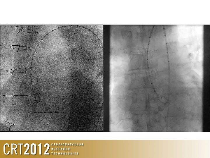
The Aortic Working View • Important for initial pre-assessment measurements • Essential to ensure implantation proceeds correctly • Should be obtained during diagnostic workup • Should be reaffirmed prior to crossing the aortic valve during TAVI procedure
Essentials of Aortic Angiography • Use imager angles that are ergonomic • Don’t ignore the anaesthetist • That does not require too much movement between RAO and implant view • Access to patient is still possible • Use graduated pigtail • Helps with spacial orientation and depth • Helps with post implant analysis • Use contrast volume and pressure that will delineate all 3 sinuses • Typically, 10 -15 mls at 900 Psi
‘Rules’ of using angiography for TAVR • Align the 3 sinuses in 1 plane – Convert 3 D anatomy to 2 D • Position the pigtail deep into the base of the non-coronary sinus – Helps with implantation • Adequate volume of contrast, under adequate pressure
Be prepared to make subtle changes to fluoro angle to ‘align’ valve to anulus 3 D Start at LAO 5 -10 2 D End at LAO 15, CAU 10
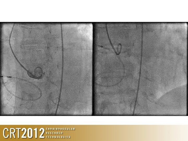
Positioning of stiff wire • After crossing AV, exchange to pigtail, ensure pigtail is free • Advance stiff wire with pigtail
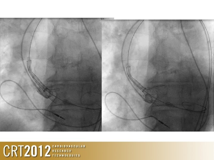
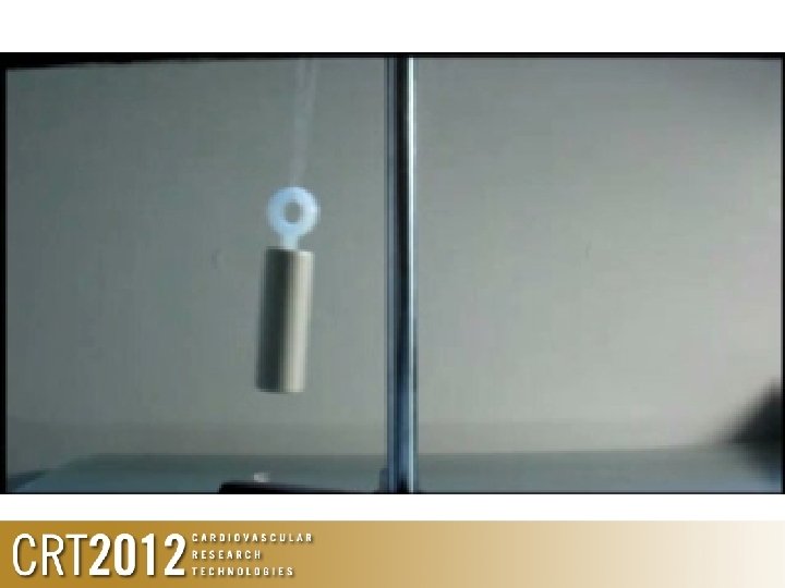
Positioning of valve
What about Edwards valve? 2 D • • ‘Rules’ Usually aim for 50: 50 If LV severely hypertrophied, go more ventricular If LV dilated/impaired and STJ small, go more aortic Ensure adequate pacing, with significant drop in cardiac output during valve deployment
Assessment of paravalvular leak • Echo is great at telling you the obvious • Its poor with the borderline. . . and is confirming what you are already thinking about – If the haemodynamics before the valve is better than after the valve (especially diastolic), then YOU HAVE A LEAK! • Haemodynamics and pressure waveform are best, and when combined with angio gives all you need to know
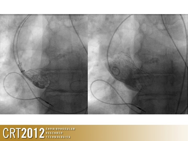
Valve too low
Valve too low • If the valve is so low that its affective MV, then, YOU WILL KNOW ABOUT IT – Significant angio leak – Significant haemodynamic change
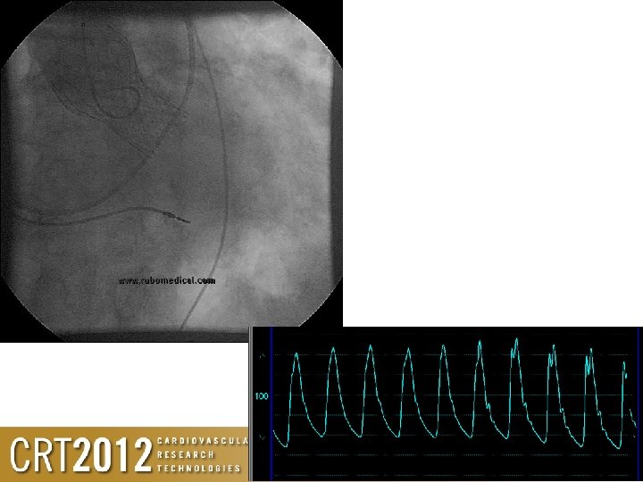
What’s happening here?
Poor haemodynamics • Low diastolic pressure • No diacrotic notch • Haemodynamics worsening with time
Angio / haemodynamic guided TAVR • Can be performed reliably, reproducibly and safely • Enables procedure to be performed under local anaesthesia • Shorter recovery time • Less impact on resources • With excellent learnt techniques, you will know – Where the wire is – Where the temporary wire is – Where the valve is and what it is doing
Siemens System • Using rotational angiography and special reconstruction algorithms, syngo Dyna. CT Cardiac creates CT-like images of the beating heart • Provide real time reconstruction and superimpose images • May help with implantation
The Paieon CTHV System
If you are still not convinced. . . then you give me no other choice. . .
Thank YOU!