Western Blotting Western blottingImmunoblotting Technique for detecting specific

Western Blotting

Western blotting/Immunoblotting Technique for detecting specific proteins separated by electrophoresis by use of labeled antibodies. § Allows to identify a particular protein of interest among many proteins in a sample.

Ø Introduced by Towbin, et al. in 1979 and is now a routine technique for protein analysis. Ø The specificity of the antibody-antigen interaction enables a single protein to be identified in the midst of a complex protein mixture.

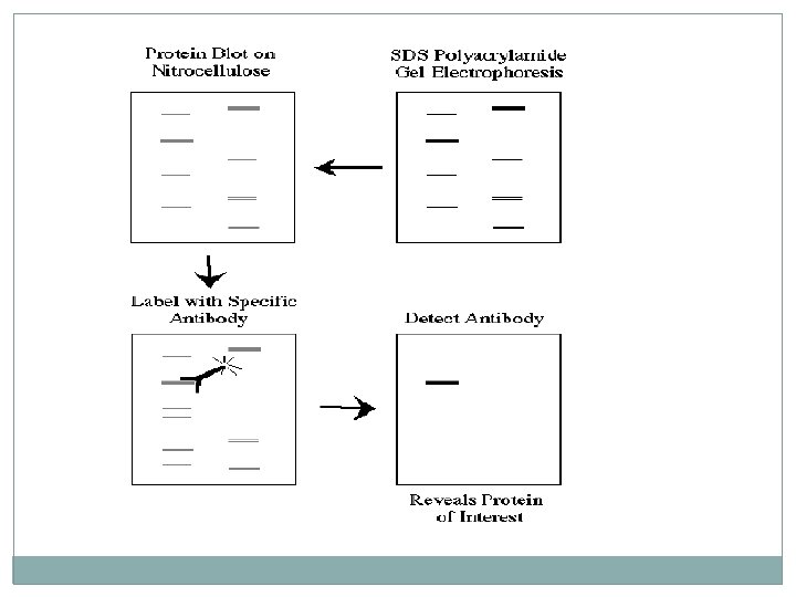
Flow chart of Western blotting Electrophoresing the protein sample Assembling the Western blot sandwich Transferring proteins from gel to nitrocellulose paper Staining of transferred proteins Blocking nonspecific antibody sites on the nitrocellulose paper Probing electroblotted proteins with primary antibody Washing away nonspecifically bound primary antibody Detecting bound antibody by horseradish peroxidase-anti-Ig conjugate and formation of a diaminobenzidine (DAB) precipitate Photographing the immunoblot
")
SDS polyacrylamide-gel electrophoresis (SDS-PAGE)
 Ø Ø Ø Stands for “SDS Polyacrylamide")
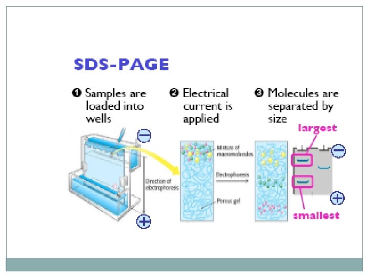
Sodium Dodecyl Sulfate- Polyacrylamide Gel Electrophoresis (SDS-PAGE) Ø Ø Ø Stands for “SDS Polyacrylamide Gel Electrophoresis 1) Thin gel separates molecules on the basis of size. 2) Negatively charged molecules are loaded into wells in a gel that is submerged in buffer. 3) Electrical current is applied to the gel, causing the molecules to move toward the positive electrode (the anode, usually marked red). 4) Smaller molecules move faster through the gel than larger molecules Thus, smaller molecules will be found at the bottom of the gel and larger molecules will be found at the top of the gel.
 SDS-PAGE is usually used to")
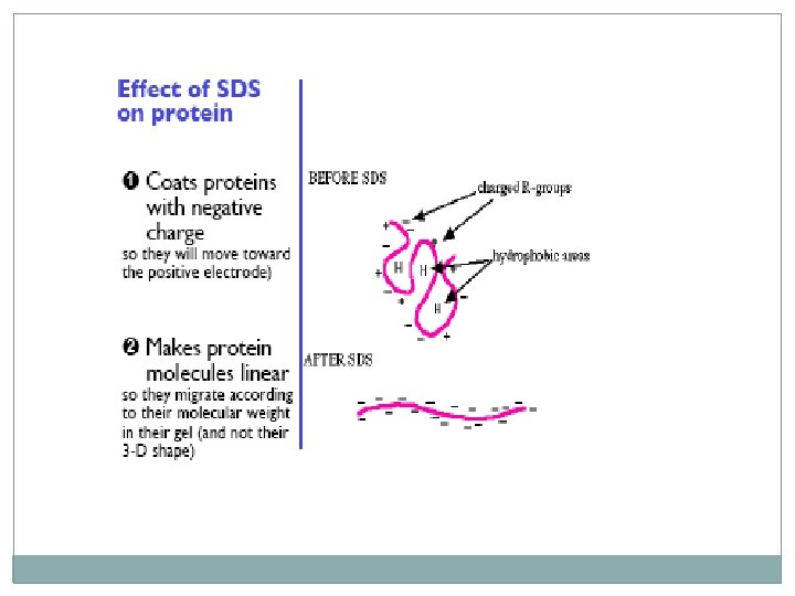
SDS-PAGE �Some differences from agarose gel electrophoresis: Ø 1) SDS-PAGE is usually used to separate protein molecules; Ø Agarose gels are usually used to separate DNA molecules. Ø 2) Proteins must be coated with SDS before being loaded in the gel. SDS is a negatively charged detergent that gives the proteins a negative charge and denatures them (makes them linear instead of 3 -D).

Components for SDS-PAGE Ø Tris buffer to provide appropriate p. H Ø SDS (sodium dodecyl sulfate) detergent to denature proteins and give them a negative charge. Ø Glycerol to make samples sink into wells Ø Blue dye to visualize samples as gel is run.

Western blot step-by-step � 1 The first step in a Western blotting procedure is to separate the macromolecules using gel electrophoresis. � 2 Size separation of the proteins in the mixture by Polyacrylamide Gel Elecrophoresis (PAGE). � 3 The separated molecules are transferred or blotted onto a second matrix, generally a nitrocellulose or polyvinylidene fluoride (PVDF) membrane. � 4 The membrane is blocked to prevent any nonspecific binding of antibodies to the surface of the membrane. � 5 The transferred protein is complexed with an enzyme-labeled antibody as a probe.

� 6 An appropriate substrate is then added to the enzyme and together they produce a detectable product such as a chromogenic or fluorogenic precipitate on the membrane for colorimetric or fluorimetric detection. Precaution � Blocking is a very important step in the immunodetection phase of Western blotting because it prevents non-specific binding of antibody to the blotting membrane � The most commonly used blocking solutions contain 3 -5% BSA or non-fat dried milk in a solution of PBS (phosphate buffered saline) or TBS (tris buffered saline) � Often, a small amount of Tween 20 detergent is added to blocking and washing solutions to reduce background staining, and the buffer is known as PBST or TBST

Immuno cyto chemistry
 To see if a specific protein of")
Why do a Western blot? � 1) To see if a specific protein of interest is present in a sample � 2) To compare the amounts of a protein of interest among different samples.

Analysis of protein samples by SDS polyacrylamide-gel electrophoresis and Western blotting

Applications � 1 - Western blot has been widely used in analyzing and identifying target proteins. � 2 - Western blot has also been used in clinical laboratories for assisting identification of certain antigen proteins (pathogen or biomarker).

Western blotting �Detects proteins and estimates their molecular weight. �Used to detect changes in protein expression.

Advantages � While ELISA being a not 100% specific test, Western blotting is a more specific test for detection of HIV. � It can detect one protein in a mixture of proteins while giving information about the size of the protein and so is more specific. � Western blot test is referred to as the 'Gold Standard’

Western Blot in Clinical Medicine � The confirmatory HIV test employs a Western blot to detect anti-HIV antibody in a human serum sample. Proteins from known HIV infected � cells are separated and blotted on a membrane then, the � serum to be tested is applied in the primary antibody incubation step; � free antibody is washed away, and a secondary anti-human antibody � linked to an enzyme signal is added. The stained bands then indicate � the proteins to which the patient's serum contains antibody.
- Slides: 20