
WELCOME TO THE TRAUMATOLOGY CHAIR HEAD OF DEPARTMENT



- Slides: 57
WELCOME TO THE TRAUMATOLOGY CHAIR !!!
HEAD OF DEPARTMENT PROFESSOR Fishchenko Vladimir Alexandrovich
PROFESSOR’S ASSISTANT BASHINSKIY GENNADIY PETROVICH
Traumatology: • acute injuries, • polytrauma. Orthopedics : • post-traumatic conditions, • axial deformities, • congenital and inherited systemic bone diseases, • limb developmental defciencies, • infammatory diseases of bones and joints, • musculoskeletal tumors.
EXAMINATION OF THE ORTHOPAEDIC PATIENT
Ø QUESTIONING Ø GENERAL APPEARANCE Ø LOCAL EXAMINATION OF THE AFFECTED AREA
QUESTIONING � Patient’s complaints (pain, limitation of function, deformity) � History (type of injury, cause, time, first aid) � Life history (occupation, habits, family history, social history, genetic history, previous injury
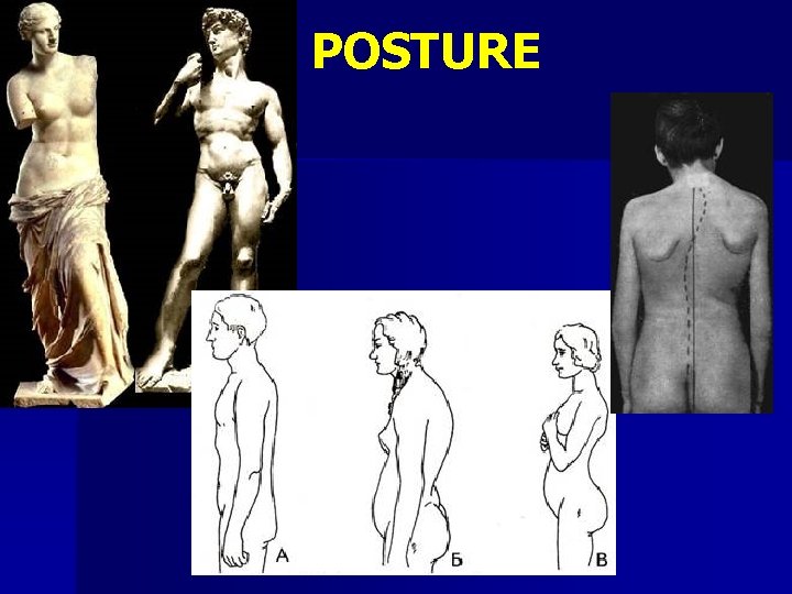
GENERAL APPEARANCE Ø Weight Ø Constitution Ø Position Ø Body build Ø Posture (attitude) Ø Gait
NOTA BENE! For the degree of abnormality aсessment, simultaneous examination of the opposite limb required
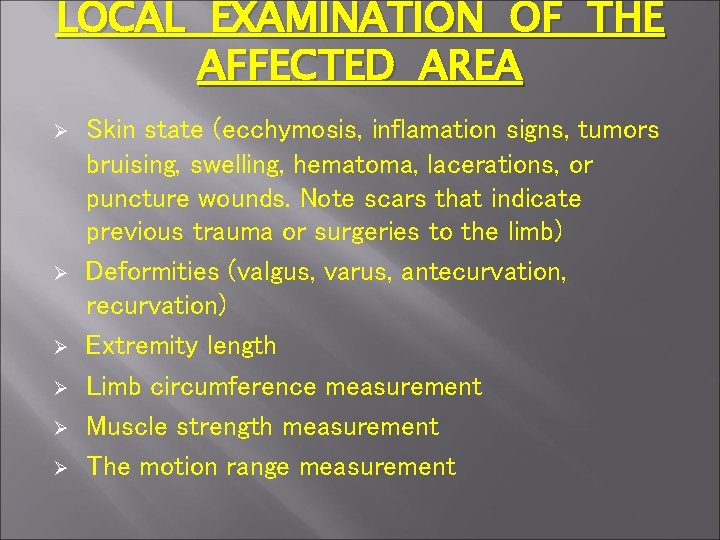
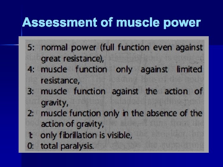
LOCAL EXAMINATION OF THE AFFECTED AREA Ø Ø Ø Skin state (ecchymosis, inflamation signs, tumors bruising, swelling, hematoma, lacerations, or puncture wounds. Note scars that indicate previous trauma or surgeries to the limb) Deformities (valgus, varus, antecurvation, recurvation) Extremity length Limb circumference measurement Muscle strength measurement The motion range measurement
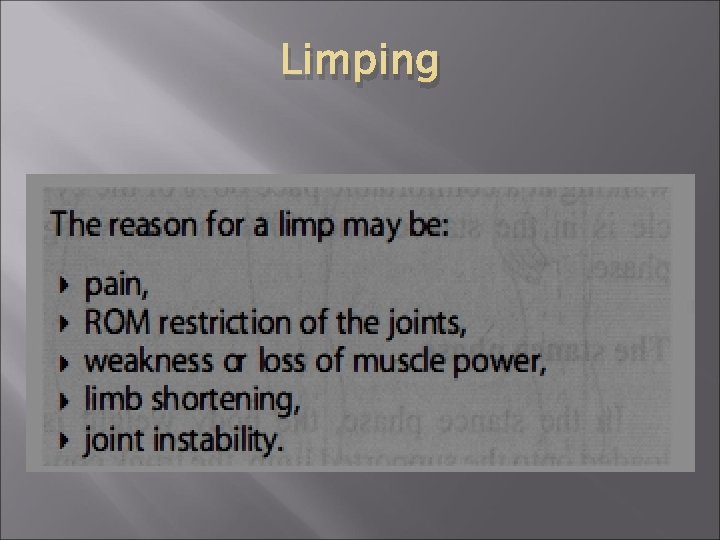
Limping
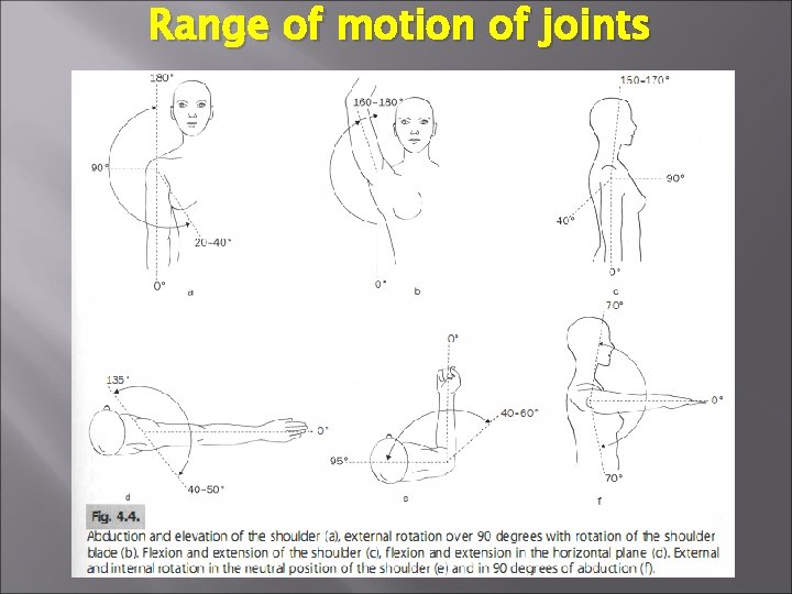
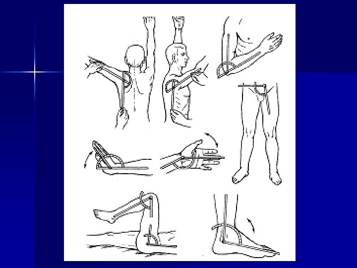
Range of motion of joints
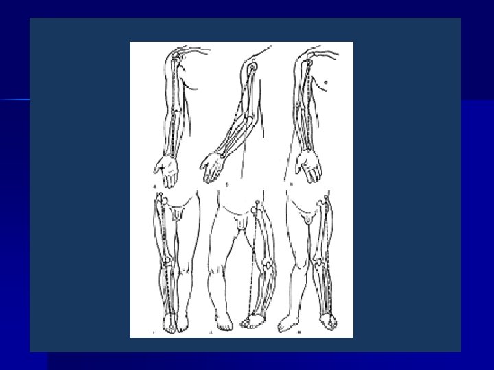
Axis of extremity n Axis of upper extremity – conditionally passes through the middle of shoulder, radial and ulnar heads.
Axis of lower extremity n Axis of lower extremity – from the front upper iliac horn through the middle of patella to the gap between 1 st and 2 nd fingers on feet.
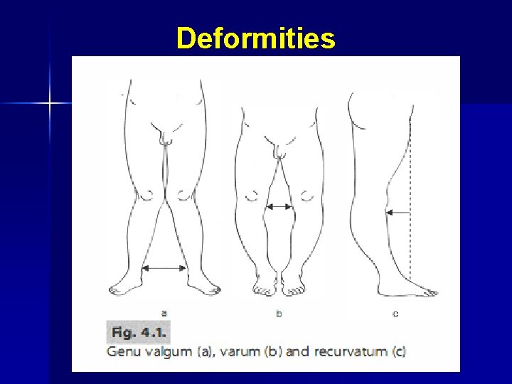
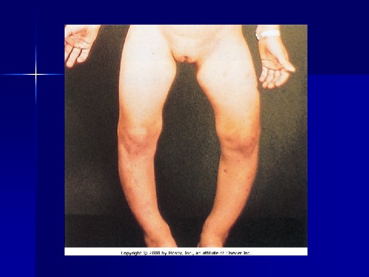
DEFORMATION OF EXTREMITY – Impairments of the axis are: outside – valgus
–Impairments of the axis are: inside – varus
n Impairments of the axis are: when the angle is open forward – recurvatum
n Impairments of the axis are: when the angle is open backwards – antecurvatum.
Deformities
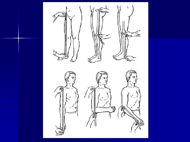
Measuring of length of limbs n Common lent of upper extremity measure from akromial process of scapula to styloid process of ulna
The common length of lower extremity is measured from the front upper iliac horn to the top of the inner ankle
n. The shoulder length is measured from the akromion to olecranon;
n The length of forearm is measured from olecranon to styloid process of ulna
n The length of the hip is measured from greater trohanter to the head of fibula
the length of the crus is measured from the head of fibula to the top of the lateral ankle n
There are following types of shortening 1) Anatomic shortening. n 2) Relative shortening n 3)projecting shortening n 4) Functional shortening n
n n The patient or limb position (attitude) – active, passive, involuntary (forced). - Active position indicates on absence of severe functional disorders in case of trauma, compensatory adjustments (adaptation) in orthopedic patients. - Passive position indicates on the severity of trauma, shock. It may be caused by fractured bones or paralysis. - Forced extremity or trunk attitude may be result of dislocation, inflammation, etc. After reposition of dislocation, reduction of the inflammatory process the forced position disappears.
n n n n n Contracture is a restriction of passive movements in the joint. 1. Antalgic (фprotective, analgetic) 2. Miogenic (due to one or group of muscles shortening) 3. Arthrogenic (due to posttraumatic, inflammatory or degenerative-dystrophic changes in the joints). 4. Desmogenic (scarring of fasciae, ligaments after trauma and operations) 5. Dermatogenic (after extensive burns) 6. Neurogenic (in cases of flaccid, spastic paralyses) 7. Tenogenic (reducing of a tendon or its fusion with its vagina) Contracture according to the limit of movements are divided into: flexional, extensional, abductory,
Joint stiffness is a state insignificant (that does not exceed 3 -5°) oscillatory motions are saved in a joint. n Ankylosis is a complete absence of movements. Ankyloses may be fibrous and bony (true), intra-articular and extra -articular, concordant (in functionally advantageous position) and discordant (in functionally n
Assessment of muscle power
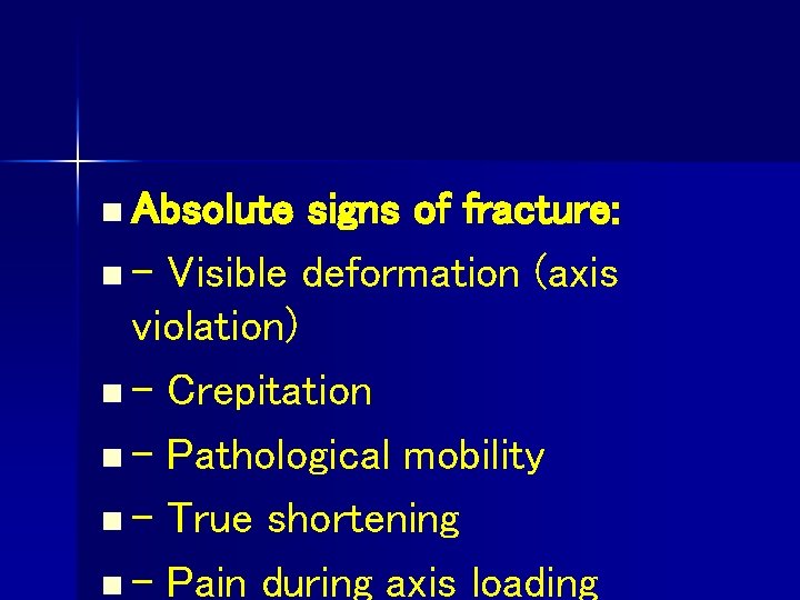
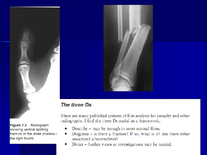
n Absolute signs of fracture: n - Visible deformation (axis violation) n - Crepitation n - Pathological mobility n - True shortening n - Pain during axis loading
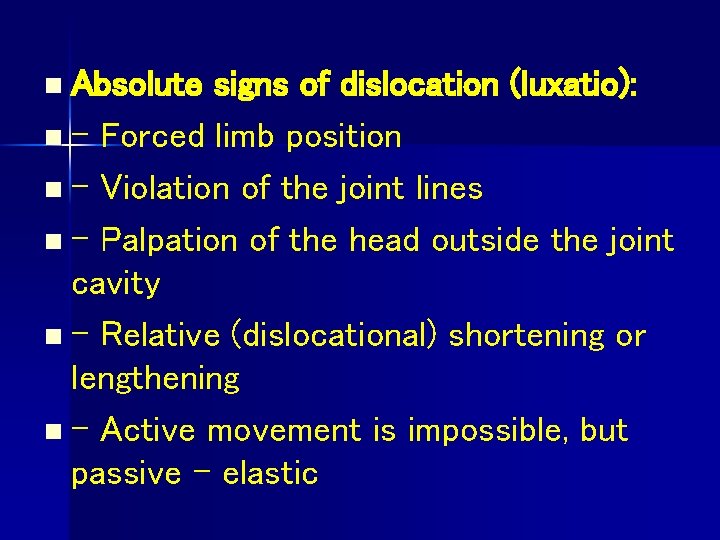
n Absolute signs of dislocation (luxatio): n - Forced limb position n - Violation of the joint lines n - Palpation of the head outside the joint cavity n - Relative (dislocational) shortening or lengthening n - Active movement is impossible, but passive - elastic
Rozer-Nelaton’s n Rozer-Nelaton’s line connects tuber of iscium and the front upper iliac horn. It is used to determine pathological states in the hip joint. Normally, hip is bent at an angle of 130°, the greater trochanter is palpated on this
Shemaker’s line n Shemaker’s line connects the tip of the greater trochanter and the front upper iliac horn. Conditional continue of the line usually passes above the navel.
Brian’s triangle n Brian’s triangle is formed by a line drawn along the axis of the aligned hip up to the crossing with the perpendicular, which starts from the front upper iliac horn. It connects it with a greater
Marx’s line n Marx’s line connects both epicondyles of the shoulder bone and normally is perpendicular to the longitudinal axis of the bone.
Huter’s triangle n Huter’s triangle is formed during forearm flexion at 90° by three bone shelves: both of epicondyles and the tip of olecranon.
Huter’s line n Huter’s line in the unbent position connects two epicondyles and the tip of olecranon.
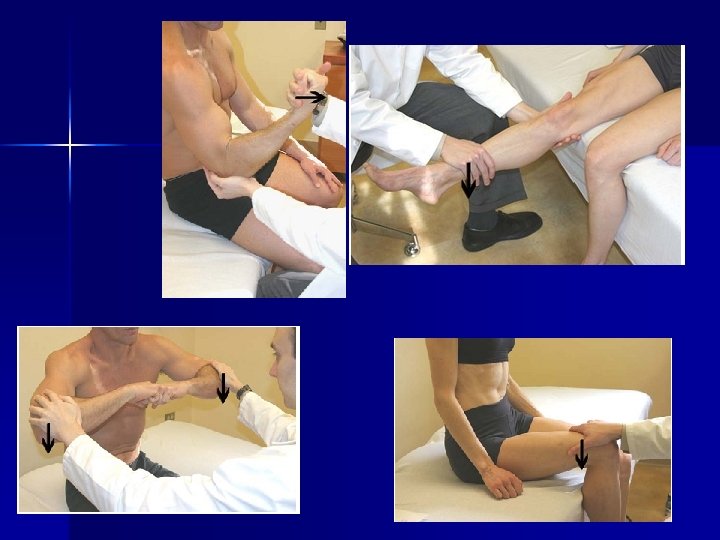
Local examination Look Ø Swelling Ø Bruising Ø Deformity Ø Overlying skin Ø Adjacent joint Ø Limb shortening
Local examination Feel Ø Temperature Ø Tenderness Ø Swelling Ø Peripheral sensation Ø Peripheral pulses
Local examination Move No attempt should be made to elicit abnormal mobility or crepitus in a fractured bone. Joint movements should only be tested if patients can perform them actively without
POSTURE
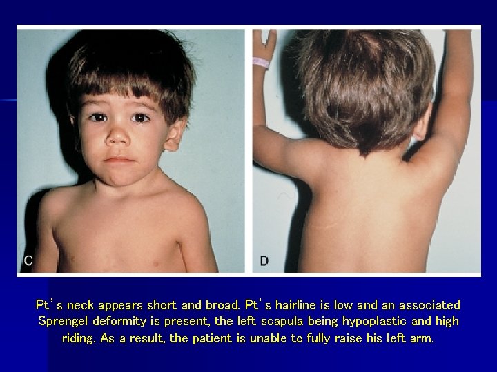
Pt’s neck appears short and broad. Pt’s hairline is low and an associated Sprengel deformity is present, the left scapula being hypoplastic and high riding. As a result, the patient is unable to fully raise his left arm.
CT
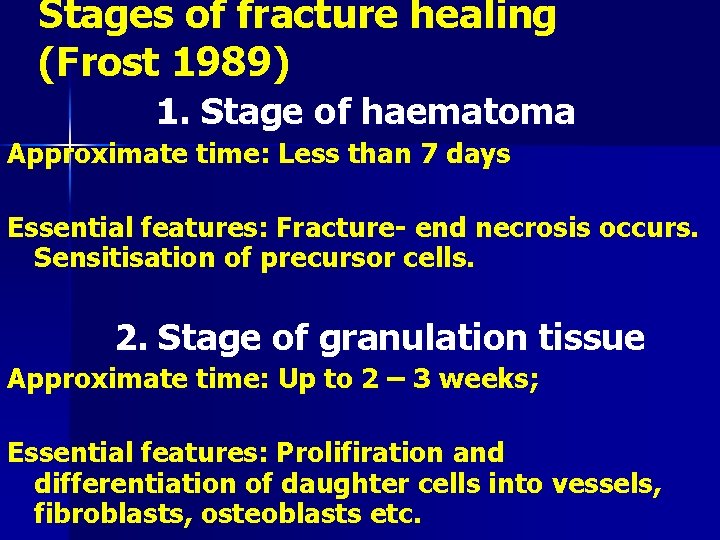
Stages of fracture healing (Frost 1989) 1. Stage of haematoma Approximate time: Less than 7 days Essential features: Fracture- end necrosis occurs. Sensitisation of precursor cells. 2. Stage of granulation tissue Approximate time: Up to 2 – 3 weeks; Essential features: Prolifiration and differentiation of daughter cells into vessels, fibroblasts, osteoblasts etc.
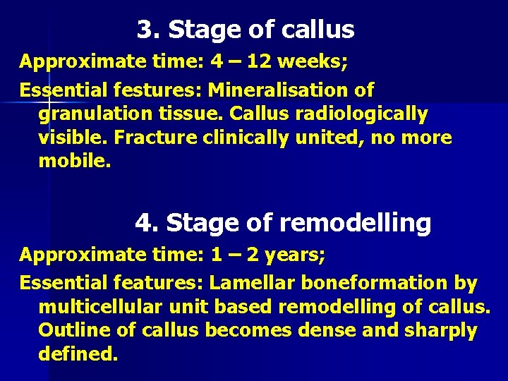
3. Stage of callus Approximate time: 4 – 12 weeks; Essential festures: Mineralisation of granulation tissue. Callus radiologically visible. Fracture clinically united, no more mobile. 4. Stage of remodelling Approximate time: 1 – 2 years; Essential features: Lamellar boneformation by multicellular unit based remodelling of callus. Outline of callus becomes dense and sharply defined.
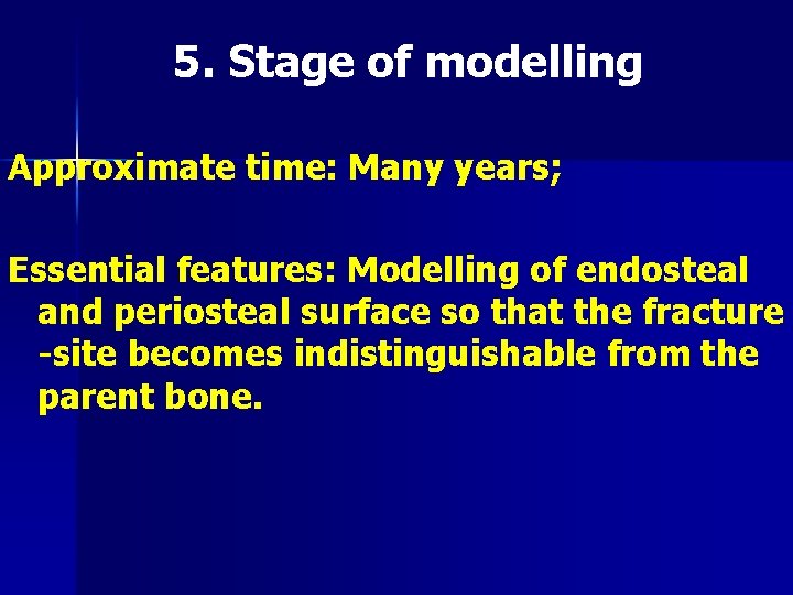
5. Stage of modelling Approximate time: Many years; Essential features: Modelling of endosteal and periosteal surface so that the fracture -site becomes indistinguishable from the parent bone.
Consilidation of bone tissue may be due to next bone callus: Endosteal n Periosteal n Paraosseous n Intermediary n
Consolidation of bone tissue devides to primary consolidation and secondary consolidation n Primary consolidation as rool due to intermedial callus. It’s much more complete; n And secondary as rool due to periostal callus.