WELCOME Macro Anatomy of the Liver Georgia Barker

WELCOME!!!

Macro Anatomy of the Liver Georgia Barker and Louise Young

Contents Location and Lobe Description Important Relations Peritoneal Ligaments of the Liver Blood Supply Calot’s Triangle Lymph Drainage Nerve Supply Bile Ducts

Regions of the Abdomen

What regions does the liver lie in? The liver lies in the right hypochondrium and epigastric region, extending into the left hypochondrium.

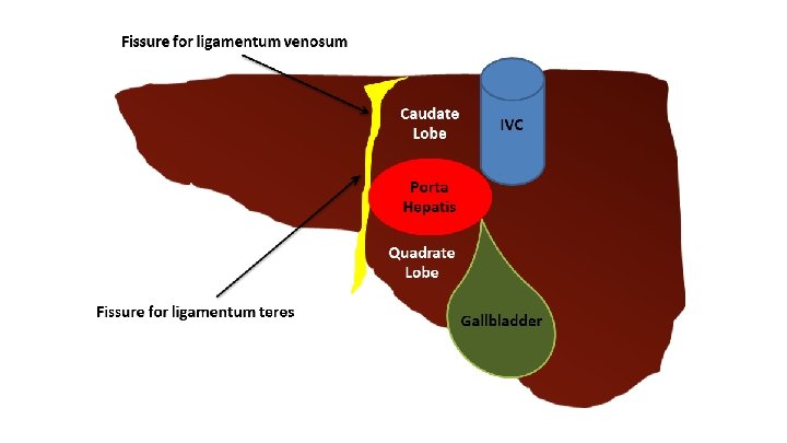
Lobes Divided into right and left lobes by the falciform ligament. Right lobe is larger and from it arise: ➢ Caudate lobe- bounded on the left by a groove for the ligamentum venosum and on the right by the IVC. ➢ Quadrate lobe- bounded on the left by a groove for the ligamentum teres and on the right by the gallbladder.

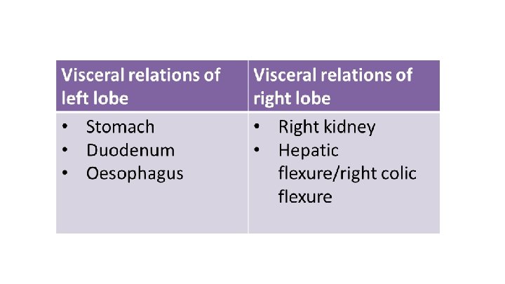
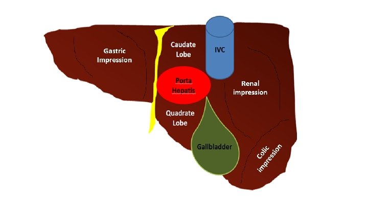
Important Relations of the Liver ● Anteriorly: diaphragm, costal margins, pleura, lower margins of both lungs, xiphoid process and anterior abdo wall. ● Posteriorly: diaphragm, right kidney, hepatic flexure of the colon, duodenum, gallbladder, IVC and oesophagus and fundus of the stomach.

Porta Hepatis ● Found on the posteroinferior surface, lies between the caudate and quadrate lobes. ● Contains the portal triad: ➢Hepatic portal vein ➢Hepatic artery proper ➢Common hepatic duct (Can remember this as VAD from posterior to anterior) ● Also has sympathetic and parasympathetic nerve fibres. As well as a few hepatic lymph nodes which drain into the celiac lymph nodes.

Calot’s Triangle

Peritoneal Ligaments ● Falciform ligament ● Coronary ligament- has upper and lower layers ● Right triangular ligament ● Left triangular ligament ● Ligaments making up the lesser omentum: ➢ Hepatogastric ligament ➢ Hepatoduodenal ligament

Blood Supply Afferent: • 25% from hepatic arteries- left and right hepatic arteries are from the hepatic artery proper, a branch of the common hepatic artery from the celiac trunk. • 75% from hepatic portal vein- supplies deoxygenated blood to the liver containing nutrients absorbed from the intestine. Efferent: • Hepatic veins which join the IVC

Portal System Deoxygenated blood, rich in absorbed nutrients Hepatic Portal Vein Splenic Vein Inferior Mesenteric Vein Superior Mesenteric Vein From Small Intestine From Large Intestine

Lymph ● The liver produces up to ½ of the bodies lymph. ● Efferent vessels pass through the porta hepatis and drain into the celiac nodes ● A few vessels from the bare area pass through the diaphragm to the posterior mediastinal lymph nodes.

Innervation ● Sympathetic and parasympathetic nerves from the celiac plexus. ● Also the anterior vagal trunk gives rise to a large hepatic branch, which passes directly to the liver.

Bile Ducts • Opens into the Ampulla of Vater • Which opens in the duodenum via the major duodenal papilla • Which is controlled by a small circular muscle known as the sphincter of Oddi • Secreted by liver cells at a rate of 40 m. L/hr
- Slides: 18