WELCOME ANATOMY OF FEMORAL TRIANGLE BY Dr Manjula

WELCOME

ANATOMY OF FEMORAL TRIANGLE BY: Dr. Manjula Vastrad Asst Prof Dept of Rachana Shareera SMVVS RKM AMC VIJAYAPURA

INTRODUCTION Is a triangular depression on the front of upper one third of the thigh immediately below the inguinal ligament

BOUNDARIES Laterally - medial border of sartorius Medially - medial border of adductor longus Base - inguinal ligament Apex - directed downwards, is formed by the point where medial & lateral borders meet.

ROOF Skin Superficial fascia containing superficial inguinal lymph nodes Femoral branch of genitofemoral nerve Branches of ilioinguinal nerve Superficial branches of femoral vessels Upper part of great saphenous vein Deep fascia with saphenous opening & cribriform fascia

FLOOR Medially-adductor longus & pectineus Laterally-psoas major & iliacus

CONTENTS 1. Femoral artery & its branches 2. Femoral vein & its tributaries. 3. Nerves Femoral nerve Nerve to pectineus Femoral branch of the gentiofemoral nerve Lateral cutaneous nerve of thigh 4. Femoral sheath 5. Femoral canal 6. Deep inguinal lymph nodes

CONTENTS

FEMORAL ARTERY ORIGIN Is the continuation of external iliac artery It traverses the triangle from its base at the mid inguinal point to the apex EXTENT AND COURSE Passes downwards and medially, first in femoral triangle and then in adductor canal Then passes through an opening in adductor Magnus to become continuous with popliteal artery

RELATIONS IN FEMORAL TRIANGLE Anterior - skin, superficial fascia, deep fascia and anterior wall of femoral sheath Posterior - rests on psoas major, pectineus and adductor longus Medially - Femoral vein Laterally - Femoral nerve and its branches, nerve to pectineus, femoral branch of genito femoral nerve, lateral cutaneous nerve of thigh.

RELATIONS

BRANCHES Gives 3 superficial and 3 deep branches in femoral triangle SUPERFICIAL BRANCHES Superficial external pudendal - supplies skin and external genital organs Superficial epigastric for skin and fasciae of lower part of anterior abdominal wall Superficial circumflex iliac for skin along iliac crest

BRANCHES

DEEP BRANCHES Profunda femoris - largest branch Arises from lateral side of femoral artery 4 cm below inguinal ligament, supplies all 3 compartments of thigh Branches Medial circumflex femoral artery Supplies adductor muscles and head of femur Lateral circumflex femoral artery Supplies Sartorius, rectus femoris, vastis lateralis.

Four perforating arteries arise in front of thigh. Second perforating artery gives rise to nutrient artery to femur 1 st at upper border of adductor brevis 2 nd in front of adductor brevis 3 rd immediately below adductor brevis 4 th is the termination of profunda femoris artery

Deep external pudendal artery Pass deep to spermatic cord / round ligament of uterus supplies scrotum or labium major Muscular branches Arise from femoral and profunda femoris artery or its branches to supply muscles of thigh

FEMORAL VEIN ORIGIN Begins as an upward continuation of the popleteal vein at the lower end of adductor canal. TERMINATION Ends by becoming continuous with the external iliac vein behind the inguinal ligament COURSE Accompanies the femoral artery, the vein is medial to artery in the base of the triangle ends at lower end of adductor canal.

TRIBUTARIES It receives great sephenous vein Veins accompanying three deep branches of femoral artery in femoral triangle, deep external pudendal and muscular. Lateral and medial circumflex femoral veins The descending genicular and muscular veins in adductor canal.

NERVES Femoral nerve Lies lateral to femoral artery, outside the femoral sheath, in the groove between ilicus and psoas major muscles. Branches. Muscular- ant supplies- sartorius & post supplies all the vasti and rectus femoris Cutaneous- ant div gives 2 cut i. e intermediate & medial cut nerve of thigh and post div gives 1 i. e saphenous nerve Articular branches- hip jt & knee jt Vascular branches to femoral artery & its branches

Nerve to pectineus Arises from femoral nerve just above the inguinal ligament. Passes behind femoral sheath to reach the surface of pectineus. Femoral branch of genito femoral nerve. Occupies the lateral compartment of femoral sheath along with femoral artery. It supplies most of skin over femoral triangle. Lateral cutaneous nerve of thigh Crosses lateral angle of triangle. Runs on lateral side of thigh dividing into anterior and posterior branches. Supplies anterolateral aspect of thigh and lateral aspect of gluteal region respectively

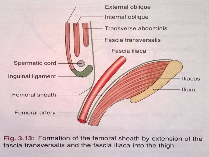
FEMORAL SHEATH Is a funnel shaped sleeve of fascia enclosing upper 3 -4 cm of femoral vessels Sheath is formed by downward extension of 2 layers of fascia of abdomen Anterior wall of sheath is formed by fascia transversalis which lies in anterior abdominal wall deep to transversalis abdominis. Posterior wall is formed by fascia iliaca, which covers the iliacus muscle. Inferiorly sheath merges with connective tissue around femoral vessels

Femoral sheath is asymmetrical Lateral wall is vertical Medial wall is oblique being directed downwards and laterally Sheath is divided into following 3 compartments by septa Lateral or arterial - contains femoral artery and femoral branch of genito femoral nerve Intermediate or venous contains femoral vein Medial or lymphatic - is smallest of all and is known as femoral canal

FEMORAL CANAL This is medial compartment of femoral sheath, conical in shape Being wide above or at base and narrow below Is 1. 5 cm long and 1. 5 cm wide at its base. Base or upper end of femoral canal is called femoral ring

FEMORAL RING Boundaries of ring Anteriorly inguinal ligament Posteriorly pectineus and its covering fascia Medially concave margin of lacunar ligament Laterally a septum separating it from femoral vein Femoral canal contains lymph node of colquet or of Rosen Muller, lymphatics and small amount of areolar tissue, lymph node drains glans penis in males and clitoris in females.

DEEP INGUINAL LYMPH NODES Lie deep to deep fascia Lie medial to upper part of femoral vein and receives lymph from superficial inguinal lymph nodes from glans penis or clitoris and deep lymphatics of lower limb

APPLIED ANATOMY

FEMORAL HERNIA Femoral canal is an area of potential weakness in the abdominal wall through which abdominal contents may bulge out forming a femoral hernia. Is more common in females because the femoral canal is wider in them than in males. This is associated with wider pelvis, and the small size of femoral vessels in the females. In cases of strangulation of the femoral hernia, the surgeon has to enlarge the femoral ring.

ABNORMAL OBTURATOR ARTERY Normal obturator artery is the branch of internal iliac, occasionally this appears to be branch of inferior epigastric. Usually the abnormal artery passes laterally to the femoral canal in contact with the femoral vein and is safe in an operation to enlarge femoral ring i. e along with the free margin of lacunar ligament. Such an artery is likely to cut if an attempt is made to enlarge the femoral ring cutting lacunar ligament.

NERVE INJURIES Injury to femoral nerve by wounds in the groin, though rare, causes paralysis of the quadriceps femoris and sensory deficit on the anterior and medial sides of thigh and medial side of leg. Lateral cutaneous nerve of thigh May get enlarged in the inguinal ligament. This leads to pain in on lateral side of thigh. It is called Meralgia parasthetica.

COMPRESSION OF FEMORAL ARTERY At the mid inguinal point against the head of femur or against the superior ramus of the pubis to control bleeding from the distal part of the limb in the thigh or leg. The femoral artery is exposed in the adductor canal for various surgical procedures.

PULSATION OF FEMORAL ARTERY Can be felt at mid inguinal point, against the head of femur and the tendon of psoas major. A bilateral absence or feebleness of the femoral pulse may result from narrowing of aorta or thrombosis, i. e the clotting of blood within the aorta.

LIGATION OR CATHETERISATION Since the femoral artery is quite superficial in femoral triangle, it can be easily exposed for ligation i. e tying or for passing a cannula or a thick needle. Catheters are passed upwards till the heart for certain minor operations.

STAB WOUNDS At the apex of femoral triangle may cut all the large vessels of lower limb because the femoral artery and vein, and the profunda femoris artery and vein are arranged in one line from before backwards at this site. Injury to femoral vessels results in fatal haemorrhage.

INTRAVENOUS INFUSIONS Femoral vein is commonly used for intravenous infusions in infants and in patients with peripheral circulatory failure.

THANK YOU
- Slides: 36