WarmUp 1 List and describe the 5 elements

Warm-Up 1. List and describe the 5 elements of a reflex arc. 2. List an example of a reflex. 3. What is the difference between a reflex and a voluntary reaction?

Human Brain

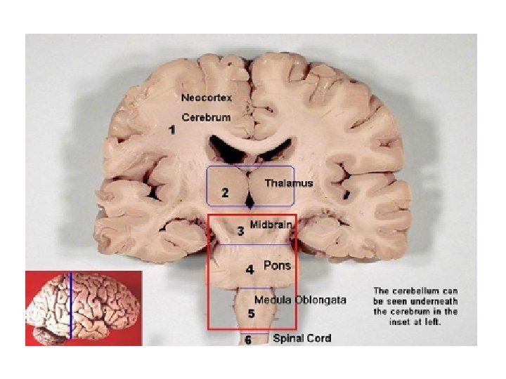
4 Major Regions Cerebral Hemispheres 2. Diencephalon 3. Brain stem 4. Cerebellum 1.
 L & R hemispheres Corpus callosum: large fiber tract; connects")
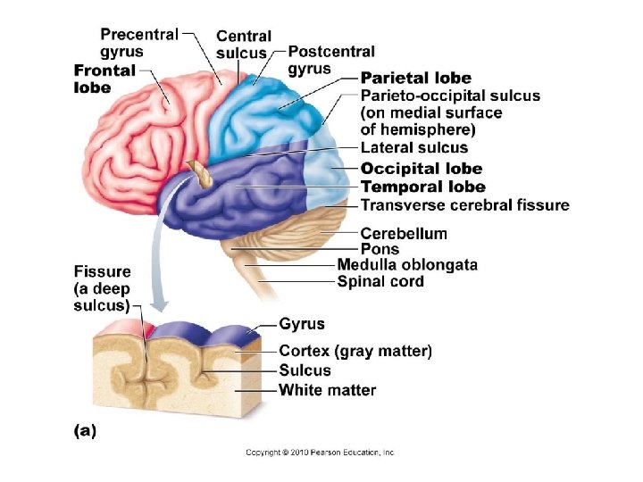
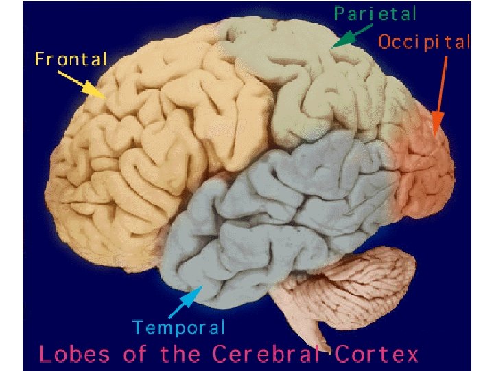
1. Cerebral Hemispheres (Cerebrum) L & R hemispheres Corpus callosum: large fiber tract; connects 2 hemispheres (internally) Lobes: major regions (named for cranial bones) Parietal, Parietal frontal, frontal occipital, occipital temporal Gyri (gyrus) = elevated ridges of tissue Sulci (sulcus) = shallow grooves Fissures = deeper grooves, separate large regions of brain

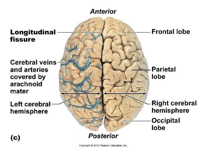
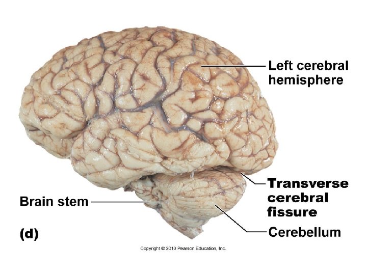
Cerebral Hemispheres Surface markings Central sulcus ▪ Separates the precentral gyrus of the frontal lobe and the postcentral gyrus of the parietal lobe Longitudinal fissure ▪ Separates the two hemispheres Transverse cerebral fissure ▪ Separates the cerebrum and the cerebellum

Precentral gyrus Frontal lobe Central sulcus Postcentral gyrus Parietal lobe Parieto-occipital sulcus (on medial surface of hemisphere) Lateral sulcus Occipital lobe Temporal lobe Transverse cerebral fissure Cerebellum Pons Medulla oblongata Spinal cord Fissure (a deep sulcus) Gyrus Cortex (gray matter) Sulcus White matter (a) Figure 12. 6 a

Cerebral Cortex Grey matter “Executive suite” conscious mind

Functions of the Major Lobes
 superficial layer of gray matter • 40%")
Cerebral Cortex • Thin (2– 4 mm) superficial layer of gray matter • 40% of the mass of the brain • Site of conscious mind: awareness, sensory perception, voluntary motor initiation, communication, memory storage, understanding Copyright © 2010 Pearson Education, Inc.

Functional Areas of the Cerebral Cortex • The three types of functional areas are: • Motor areas—control voluntary movement • Sensory areas—conscious awareness of sensation • Association areas—integrate diverse information • Conscious behavior involves the entire cortex Copyright © 2010 Pearson Education, Inc.

Cerebral Cortex: Frontal Lobe q. Movement of the body - Primary Motor Cortex, Premotor Cortex, Frontal Eye Field q. Meaning of words, Production of speech – Broca’s Speech Area Copyright © 2010 Pearson Education, Inc.

Posterior Motor map in precentral gyrus Anterior Toes Jaw Tongue Swallowing Copyright © 2010 Pearson Education, Inc. Primary motor cortex (precentral gyrus) Figure 12. 9

Prefrontal Cortex - Anterior Association Area • Most complicated cortical region • Involved with intellect, cognition, recall, and personality, concentration, planning, problem solving • Contains working memory needed for judgment, reasoning, persistence, and conscience • Development depends on feedback from social environment Copyright © 2010 Pearson Education, Inc.

Limbic Association Area • Located on the medial side of the frontal lobe. • Provides emotional impact that helps establish memories • Forms memories and translates that to motor responses • Processes emotions and guides emotional responses. Olfactory (Smell) Association • Important for social interactions and expressions of the personality. Copyright © 2010 Pearson Education, Inc.

Cerebral Cortex: Parietal Lobe q. Body Awareness - Primary Somatosensory Cortex q. Touch and Pressure q. Somatosensory Association Area Copyright © 2010 Pearson Education, Inc.
")
Posterior Sensory Anterior Sensory map in postcentral gyrus Genitals Primary somatosensory cortex (postcentral gyrus) Copyright © 2010 Pearson Education, Inc. Intraabdominal Figure 12. 9

Cerebral Cortex: Temporal Lobe q. Hearing – Auditory Cortex q. Smell – Olfactory Cortex Olfaction is the only sensory system that is not routed through the thalamus. q. Recognizing patterns and faces – Posterior Association Area q. Sound out words- Wernicke’s Speech Area q. Long Term Memory Insula (Deep to the Temporal Lobe) q. Taste – Gustatory Cortex Copyright © 2010 Pearson Education, Inc.

Cerebral Cortex: Occipital Lobe q. Primary Visual Cortex q. Visual Association Area Copyright © 2010 Pearson Education, Inc.

Motor areas Central sulcus Primary motor cortex Premotor cortex Frontal eye field Broca’s area (outlined by dashes) Prefrontal cortex Working memory for spatial tasks Executive area for task management Working memory for object-recall tasks Solving complex, multitask problems (a) Lateral view, left cerebral hemisphere Sensory areas and related association areas Primary somatosensory cortex Somatic Somatosensory sensation association cortex Gustatory cortex (in insula) Taste Wernicke’s area (outlined by dashes) Primary visual cortex Visual association area Auditory association area Primary auditory cortex Vision Hearing Motor association cortex Primary sensory cortex Primary motor cortex Sensory association cortex Multimodal association cortex Copyright © 2010 Pearson Education, Inc. Figure 12. 8 a

Copyright © 2010 Pearson Education, Inc.
 3 main structures: 1. Thalamus: relay station for incoming info 2.")
2. Diencephalon (interbrain) 3 main structures: 1. Thalamus: relay station for incoming info 2. Hypothalamus: A. Autonomic control center (heart rate, BP, digestion) B. Emotional response (limbic system) C. Control endocrine system pituitary gland at base 3. Epithalamus: pineal gland (sleep-wake cycle)

Diencephalon

Thalamic Function • 80% of diencephalon • Gateway to the cerebral cortex • Sorts, edits, and relays information • Mediates sensation, motor activities, cortical arousal, learning, and memory Copyright © 2010 Pearson Education, Inc.

Paraventricular nucleus Anterior commissure Preoptic nucleus Anterior hypothalamic nucleus Supraoptic nucleus Suprachiasmatic nucleus Fornix Arcuate nucleus Pituitary gland Optic chiasma Infundibulum (stalk of the pituitary gland) (b) The main hypothalamic nuclei. Copyright © 2010 Pearson Education, Inc. Dorsomedial nucleus Posterior hypothalamic nucleus Lateral hypothalamic area Ventromedial nucleus Mammillary body Figure 12. 13 b

Hypothalamic Function • Autonomic control center for many visceral functions (e. g. , blood pressure, rate and force of heartbeat, digestive tract motility) • Regulates body temperature, food intake, water balance, and thirst • Regulates sleep and the sleep cycle Copyright © 2010 Pearson Education, Inc.

Hypothalamic Function • Controls release of hormones by the anterior pituitary • Produces posterior pituitary hormones • Infundibulum—stalk that connects to the pituitary gland Copyright © 2010 Pearson Education, Inc.

Hypothalamic Function – Limbic System • The limbic system is where the subcortical structures meet the cerebral cortex. • Composed of a complex set of structures that lie on both sides of the thalamus. Includes the hypothalamus, hippocampus, amygdala, and several other nearby areas. Copyright © 2010 Pearson Education, Inc.

The Limbic System • Center for emotional response: Involved in perception of pleasure, fear, and rage and in biological rhythms and drives • Involved in motivation, emotion, learning, and memory. Copyright © 2010 Pearson Education, Inc.

Epithalamus • Pineal gland—extends from the posterior border and secretes melatonin • Melatonin—helps regulate sleep-wake cycles Copyright © 2010 Pearson Education, Inc.

3. Brain Stem Programmed, automatic behaviors for survival 3 regions: 1. Midbrain: vision, hearing, reflex 2. Pons: breathing 3. Medulla oblongata: heart rate, BP, breathing, and other autonomic responses

Brain Stem
 Optic chiasma Optic nerve")
Frontal lobe Olfactory bulb (synapse point of cranial nerve I) Optic chiasma Optic nerve (II) Optic tract Mammillary body Midbrain Pons Temporal lobe Medulla oblongata Cerebellum Spinal cord Copyright © 2010 Pearson Education, Inc. Figure 12. 14
 Crus cerebri of cerebral peduncles (midbrain) Diencephalon • Thalamus")
Optic chiasma Optic nerve (II) Crus cerebri of cerebral peduncles (midbrain) Diencephalon • Thalamus • Hypothalamus Mammillary body View (a) Thalamus Hypothalamus Diencephalon Midbrain Oculomotor nerve (III) Trochlear nerve (IV) Pons Brainstem Medulla oblongata Trigeminal nerve (V) Pons Middle cerebellar peduncle Abducens nerve (VI) Facial nerve (VII) Vestibulocochlear nerve (VIII) Pyramid Glossopharyngeal nerve (IX) Hypoglossal nerve (XII) Vagus nerve (X) Ventral root of first cervical nerve Decussation of pyramids Accessory nerve (XI) Spinal cord (a) Ventral view Copyright © 2010 Pearson Education, Inc. Figure 12. 15 a
 Thalamus View (b) Infundibulum Pituitary gland Superior colliculus")
Crus cerebri of cerebral peduncles (midbrain) Thalamus View (b) Infundibulum Pituitary gland Superior colliculus Inferior colliculus Trochlear nerve (IV) Trigeminal nerve (V) Pons Superior cerebellar peduncle Middle cerebellar peduncle Facial nerve (VII) Abducens nerve (VI) Glossopharyngeal nerve (IX) Hypoglossal nerve (XII) Inferior cerebellar peduncle Vestibulocochlear nerve (VIII) Olive Thalamus Vagus nerve (X) Hypothalamus Diencephalon Midbrain Accessory nerve (XI) Pons Brainstem Medulla oblongata (b) Left lateral view Copyright © 2010 Pearson Education, Inc. Figure 12. 15 b
 that control cranial nerves III")
Midbrain Nuclei • Nuclei (small areas of gray matter) that control cranial nerves III (oculomotor) and IV (trochlear) • Corpora Quadirgemina - Visual and Auditory reflex centers Copyright © 2010 Pearson Education, Inc.

Pons • Made mostly of fiber tracts - Connect higher brain centers and the spinal cord • Relay impulses between the motor cortex (frontal lobe) and the cerebellum • Nuclei that help maintain normal rhythm of breathing Copyright © 2010 Pearson Education, Inc.

Medulla Oblongata • Joins spinal cord at foramen magnum • Relay sensory information from muscles and joints to cerebellum • Mediates responses that maintain equilibrium Copyright © 2010 Pearson Education, Inc.

Medulla Oblongata • Autonomic reflex centers • Cardiovascular center • Cardiac center adjusts force and rate of heart contraction • Vasomotor center adjusts blood vessel diameter for blood pressure regulation • Respiratory centers • Generate respiratory rhythm • Control rate and depth of breathing Copyright © 2010 Pearson Education, Inc.

Medulla Oblongata • Additional centers regulate • Vomiting • Hiccuping • Swallowing • Coughing • Sneezing Copyright © 2010 Pearson Education, Inc.

Brain Stem – Reticular Formation • A network of gray matter that extends the entire length of the brain stem • Involved in motor control of visceral organs • A special group of neurons forms the Reticular Activating System RAS • Plays a role in consciousness and sleep/wake cycle • Filter for the flood of sensory inputs – weak or repetitive impulses get ignored Copyright © 2010 Pearson Education, Inc.

4. Cerebellum Balance, equilibrium, timing of skeletal muscle activity

The Cerebellum • 11% of brain mass • Dorsal to the pons and medulla • precise timing and appropriate patterns of skeletal muscle contraction • Sports, dancing, etc. Copyright © 2010 Pearson Education, Inc.

Cognitive Function of the Cerebellum • sequences of events during complex movements • nonmotor functions such as word association and puzzle solving • Fibers from the semi-circular canals in the inner ear and from proprioceptors in the skeletal muscles and tendons reach the cerebellum – controls balance and equilibrium • Motor memory Copyright © 2010 Pearson Education, Inc.

12 Cranial Nerve Mnemonic: On old Olympus' towering top a Fin and German viewed some hops • O: olfactory nerve (CN I) • O: optic nerve (CN II) • O: oculomotor nerve (CN III) • T: trochlear nerve (CN IV) • T: trigeminal nerve(CN V) • A: abducens nerve (CN VI) • F: facial nerve (CN VII) • A: auditory (or vestibulocochlear) nerve (CN VIII) • G: glossopharyngeal nerve (CN IX) • V: vagus nerve (CN X) • S: spinal accessory nerve (CN XI) • H: hypoglossal nerve (CN XII) Copyright © 2010 Pearson Education, Inc.
, arachnoid")
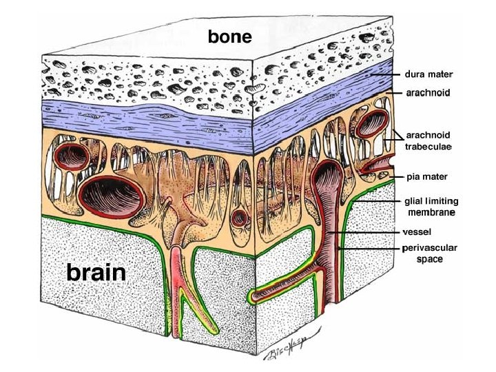
Protection of CNS Meninges: connective tissue covering CNS structures Dura mater (leathery outer), arachnoid mater (web -like middle), pia mater (surface of brain) Meningitis: inflammation of meninges; bacterial or viral infection Cerebrospinal fluid (CSF): watery cushion to protect NS from trauma and provides oxygen, nutrients and vital chemicals to the brain and spinal cord Lumbar (spinal) tap – test for infection, tumors, multiple sclerosis

Meningitis

Symptoms of Meningitis

Treatment for Meningitis Bacterial antibiotics Herpes meningitis antiviral meds IV fluids Prevention: vaccines for bacterial infections (Hi. B)

Blood-Brain Barrier: endothelial cells in capillaries prevent substances from crossing into brain NO: YES: Urea Toxins Proteins White blood cells Bacteria Most drugs Water Glucose Amino acids Gases Fat-soluble substances Some drugs: anesthetics, alcohol, nicotine
- Slides: 55