VULVAR DERMATOLOGIC LESIONS LINDSAY CHURCHLEY MD FRCSC Northern


 • Risk factors: – – – Abx use Steroid")

 • Treatment: – Vulvar tx:")


 BID for 2 -4 weeks")
 •")


- Slides: 45
VULVAR DERMATOLOGIC LESIONS LINDSAY CHURCHLEY MD, FRCSC Northern Ontario Women’s Health Conference November 22, 2019
Disclosure • I have no relationship with for-profit or not-for -profit organizations • This session has not received financial or inkind support
Objectives • Key aspects of history and physical • Indications for vulvar biopsy • Common lesions: – Candidiasis – Vulvar Dermatitis – Linchen simplex chronicus – Lichen planus – Lichen sclerosis – Psoriasis – HPV – Squamous intraepthelial lesions
Key aspects of history & physical HISTORY PHYSICAL Duration, location Pain Pruritus Discharge bleeding Potential provocative events • Medical history • Sexual history • inspection: • Symmetry, erythema, edema, focal lesion • Distorted anatomy • Swabs • Biopsy • Oral lesions • • •
Indications for vulvar biopsy • Uncertain of diagnosis • Suspicion for malignancy • Failure of Initial treatment
How to do a vulvar biopsy • Clean with antiseptic solution • Local anesthetic – 1% or 2% lidocaine – Can also use liposomal lidocaine or EMLA, but need to apply 30 -60 min before bx • Keyes punch biopsy (usually 3 -5 mm) • Sample down to dermis • Hemostasis – Monsel’s solution – silver nitrate sticks – simple interrupted suture
How to do a vulvar biopsy
Where to do a vulvar biopsy • • • Ulcerative lesion: Sample at the edge Hyperpigmented lesion: darkest area Thickened lesion: thickest area Larger lesion: May need to take a few samples Homogeneous lesion: sample at the centre Heterogenous lesion: biopsy each area
COMMON VULVAR LESIONS
Candidiasis • Candida albicans (90%) • Risk factors: – – – Abx use Steroid use Imunocompromised Diabetes HIV • Vulvar itching, irritation, burning • dysparunia
Vulvar Candidiasis • Physical exam: – Erythema – Vulvar edema – Fissures, dryness or cracks in vulvar skin – Vaginal discharge – white, clumpy, thick, curdy
Candidiasis • Swabs, KOH whiff test (if discharge present) • Treatment: – Vulvar tx: miconazole OR clotrimazole topical cream BID 10 -14 day – Vaginal tx: miconazole OR clotrimazole vaginal insert 3, 6, or 7 nights OR - Floconazole 150 mg tab PO x 1
Keys to candidiasis • If on Antibiotics – treat x 6 days • Avoid grapefruit juice if on Fluconazole • Treatment failure: – Can try boric acid 600 mg PV OD x 14 d (vaginal symptoms) – May be non-albicans strain (Candida glabrata)
Vulvar Dermatitis • Atopic dermatitis – Endogenous • Contact Dermatitis – Exogenous – Allergic (20%) • Onset: 2 weeks after first exposure • 1 week after repeat exposure • Very ITCHY – Irritant (80%) • Onset: rapid (hours to a day) after exposure • More stingy and burning than itchy
Common irritants • • • Soap, bubble bath, shampoo Shaving gel/cream Pads (menstrual or incontinence) Synthetic fabric underwear Sweat Urine Perfumes Alcohol Condoms spermicides
Common allergens • • Latex Lanolin Tea tree oil Propylene glycol Perfumes Chlorhexidine benzocaine
Contact dermatitis Symmetrical Raised Bright red Intense itching Extends over the area of contact • No loss of architecture • • •
Treatment • Remove irritant • Reduce inflammation: – Clobetasol 0. 05% BID x 1 -3 weeks • Restore skin barrier and hydrate: – sitz baths – Emollient – white petrolatum, olive oil • Break the itch-scratch cycle – Cool packs – Plain cold yogurt on sanitary napkin for 5 -10 min • If no improvement in 3 weeks – Consider allergy testing, biopsy
itch-scratch-itch-scratch
Lichen Simplex Chronicus • End stage itch/scratch cycle • Most common vulvar dermatosis (30 -50%) • Lichenification • Edema • Excoriations • Fissures • Broken/spare pubic hair • Maintain architecture
Lichen Simplex Chronicus
LSC treatment • Remove irritant • Search for/treat infection • Reduce inflammation – Longer course of corticosteroid – Clobetasol 0. 05% ointment BID x 4 -6 wk, then taper • • Hydrate (sitz baths) Bland emollients (petrolatum, oil) Consider antihistamine or TCA Xylocaine/lidocaine 5% ointment
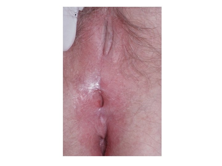
LICHEN SCLEROSIS
Lichen Sclerosis • • 1: 300 -1: 1000 women Hormonal and autoimmune Bimodal – postmenopausal and premenarchal Symptoms: – Itchy, sore, irritated – Dysuria – Dysparuneia – Decreased sexual sensation – Asymptomatic
Lichen Sclerosis • Anogenital area – 85% perianal involvment • Figure of 8, hour glass • Loss of architecture – – Labia minora regression Clitoral concealment Urethral obstruction Introital stenosis • Skin – think and crinkled • Porcelain white plaques
Lichen sclerosis
Lichen sclerosis - Treatment • There is no cure • Treatment goal – reduce symptoms, prevent anatomical distortion • Clobetasol 0. 5% ointment • Initial: daily x 2 -3 months • Maintenance: every other day for 6 months • Can reduce to 1/week if chronic and stable • Rarely – surgical treatment required • Follow up – q 6 -12 months
Lichen sclerosis – treatment failure • • • Undertreatment Superinfection Treat atrophy with PV estrogen Change steroid class Biopsy Note: 4 -6% of patient with stable disease progress to SCC - Biopsy any new, changing, or persistently symptomatic patients
LICHEN PLANUS • • Found at introtius +/- inside vagina T-cell autoimmune disease Age 30 -60 Three variants: – Erosive (most common) – Papulosquamous – Hypertrophic • can be drug induced: NSAIDs, B-blocker, methyldopa • Look for lesions elsewhere (mouth)
Lichen planus – signs/symptoms • • Intense Pruritis Chronic discharge Burning, soreness Dyspareunia Post-coidal bleeding Abnormal pap Vulva and vagina bleed easily with spec exam Adhesions/synechiae
Lichen planus - diagnosis • Glazed erythema • White border • Architectural destruction • Vaginal involvment – 70%: denuded, adhesion, sero-purlent exudate, bleeding • Biopsy • LOOK AT MOUTH
Lichen planus - Treatment • d/c any trigger meds • Vulvar tx: clobetasol 0. 05% BID upto 3 months, with slow taper • Alternative tx: – Preparation containing: Clobetasol 0. 05%, 3% oxytetracycline, 100, 000 U/g nystatin – Sometimes need systemic steroids • Vaginal tx: hydrocortisone acetate 25 mg suppositories qhs in vagina • Vaginal dilators • Watch for candidiasis
Psoriasis • 1 -2% of population have psoriasis • Occasionally, can be found on labia, mons pubis • Thick, red plaques • Covered with silvery scales • Triggers: stress, menses, cold, some drugs • Remission: pregnancy, summer months
Psoriasis: treatment • High potency corticosteroid (clobetasol 0. 05%) BID for 2 -4 weeks too affected areas, then taper to weekly • long term use: Decreased response, skin atrophy • Refer to dermatology – Vitamin D anologs – Phototherapy – Biologics (infliximab)
Human papillomavirus - HPV • Low risk strains – 6, 11 (genital warts) • High risk strains – 16, 18, 31, 33, 45 • Seen in 70 -80% of pre-invasive vulvar lesions (HSIL) • Seen in 20 -50% of vulvar CA • Risk stronger with: – Smoking – Co-infection Herpes simplex virus
Squamous Intraepithelial Lesions **USED TO BE CALL VIN** International Society for the Study of Vulvovaginal Disease
Vulvar SIL • LSIL – genital warts - not pre-cancer – Treatment only for aesthetics or discomfort – Tx: excision, liquid nitrogen, laser, imiquimod • HSIL - pre-cancerous – Younger, smokers, immunosuppressed – Requires treatment: excision, laser, imiquimod • d. VIN – pre-cancerous – Older, lichen sclerosis, lichen planus – Higher chance of progressing to cancer – Requires treatment (excision)
Vulvar SIL - symptoms • • Pruritis, pain, burning Bleeding Discharge Dysuria Persistent ulcer Abnormal skin colour or texture Change in exsisting nevus Lump or wart-like growth
LSIL
HSIL
d. VIN
Summary • Take a thorough history and physical • How/when to do a vulvar biopsy • Review of common lesions – Candidiasis – Vulvar Dermatitis – Lichen Simplex Chronicus – Lichen sclerosis – Lichen planus – Psoriasis – HPV – Vulvar Squamous intraepithelial lesions
References • Williams Gynecology • Vulvar Squamous intraepithelial Lesions – International Society for the Study of Vulvovaginal Disease • Management of Vulvar Intraepithelial Neoplasia – ACOG guideline • Dr. Helene Gagne – Vulvar Gynecologist • Genital Dermatology – Dr. Michael Policar
Thank you!!