
von Willebrands Disease Robert Gosselin MT ASCP CLS
von Willebrands Disease Robert Gosselin MT (ASCP), CLS
von Willebrand disease (VWD): evidence-based diagnosis and management guidelines, the National Heart, Lung, and Blood Institute (NHLBI) Expert Panel report (USA) (Hemophilia 2008; 14: 171 -232) von Willebrand Factor Assays: Guideline – Second Edition (H 51 -2) (in progress)
1924 Erik von. Willebrand evaluated a 5 year old girl with bleeding diathesis and ultimately 66 family members in Finland noted: – Mucocutaneous bleeding – Autosomal inheritance – Prolonged bleeding times – Normal clotting times – Not corrected with blood transfusions
In 1950 plasma protein defect causing prolonged bleeding time Deemed “von Willebrand factor” As cryoprecipitate and F 8 concentrates were produced, it was noted that they complexed together
VWF produced in two areas endothelium – synthesized and stored in Weibel-Palade bodies megakaryocytes – stored in alpha granules Assembled from identical subunits to form linear strings multimers formed in the Golgi complex
In circulation, VWF travels as a complex with F 8 Upon injury, VWF undergoes conformational change and adheres to the subendothelium under high shear conditions Platelets adhere via GPIb-IX Half-life is ~12 hours
VWF multimers – Degradation by ADAMTS 13 [A Disintegrin-like And Metalloprotease domain with Thrombo. Spondin type 1 motif, member 13] Deficiency of ADAMTS 13 have been associated with TTP
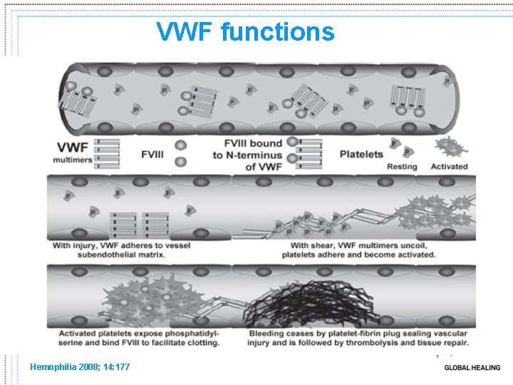
VWF functions Hemophilia 2008; 14: 177
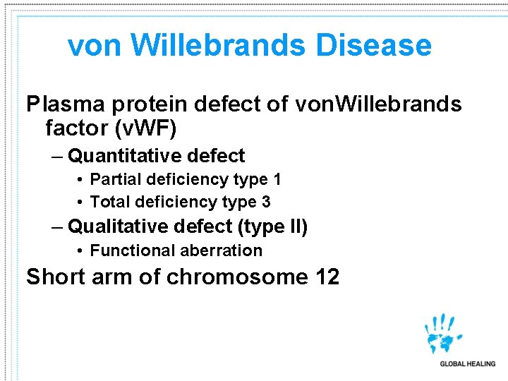
von Willebrands Disease Plasma protein defect of von. Willebrands factor (v. WF) – Quantitative defect • Partial deficiency type 1 • Total deficiency type 3 – Qualitative defect (type II) • Functional aberration Short arm of chromosome 12
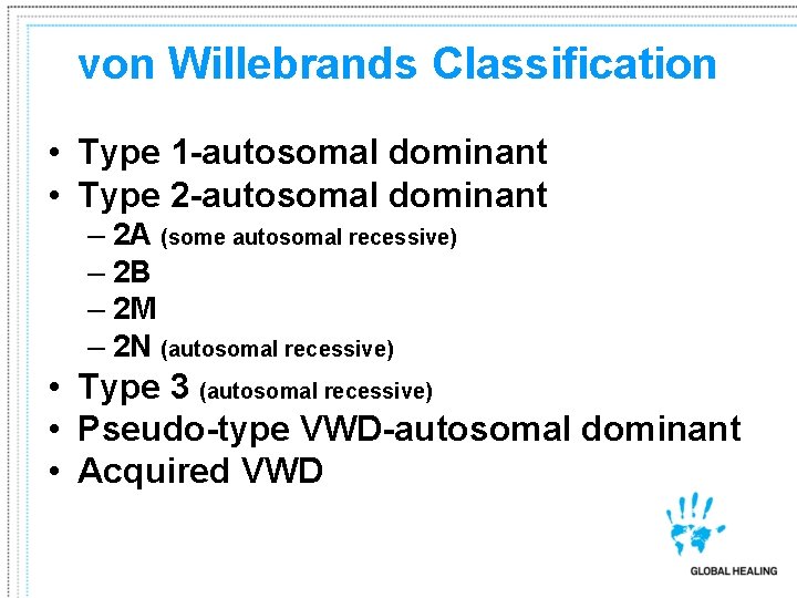
von Willebrands Classification • Type 1 -autosomal dominant • Type 2 -autosomal dominant – 2 A (some autosomal recessive) – 2 B – 2 M – 2 N (autosomal recessive) • Type 3 (autosomal recessive) • Pseudo-type VWD-autosomal dominant • Acquired VWD
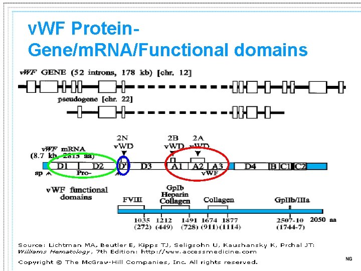
v. WF Protein. Gene/m. RNA/Functional domains

von Willebrands Disease 1% of population with v. WF – 125/1, 000 with significant bleeding – In UK, 6294 registered v. WD patients, but only 10% received treatment in calendar year 2001 Type I accounts for ~75% of all v. WD Dx Mild to severe – Hx • • Easy bruising Nose bleeds Oral cavity bleeding Menorrhagia Bleeding variability – Mild to severe
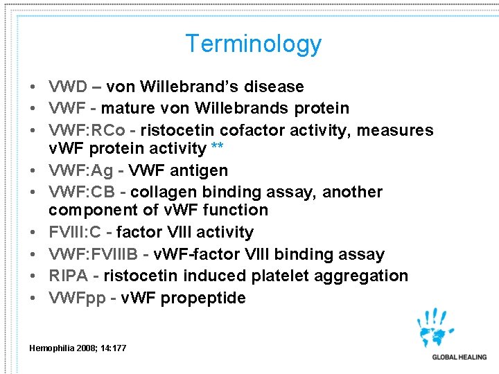
Terminology • VWD – von Willebrand’s disease • VWF - mature von Willebrands protein • VWF: RCo - ristocetin cofactor activity, measures v. WF protein activity ** • VWF: Ag - VWF antigen • VWF: CB - collagen binding assay, another component of v. WF function • FVIII: C - factor VIII activity • VWF: FVIIIB - v. WF-factor VIII binding assay • RIPA - ristocetin induced platelet aggregation • VWFpp - v. WF propeptide Hemophilia 2008; 14: 177
Laboratory Testing for v. WD and PLT defects
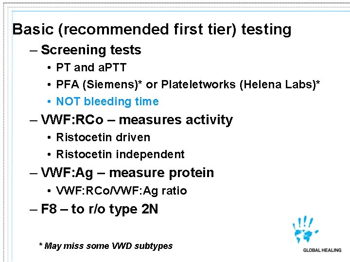
Basic (recommended first tier) testing – Screening tests • PT and a. PTT • PFA (Siemens)* or Plateletworks (Helena Labs)* • NOT bleeding time – VWF: RCo – measures activity • Ristocetin driven • Ristocetin independent – VWF: Ag – measure protein • VWF: RCo/VWF: Ag ratio – F 8 – to r/o type 2 N * May miss some VWD subtypes
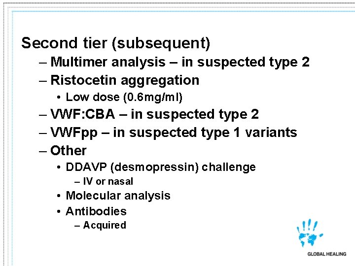
Second tier (subsequent) – Multimer analysis – in suspected type 2 – Ristocetin aggregation • Low dose (0. 6 mg/ml) – VWF: CBA – in suspected type 2 – VWFpp – in suspected type 1 variants – Other • DDAVP (desmopressin) challenge – IV or nasal • Molecular analysis • Antibodies – Acquired
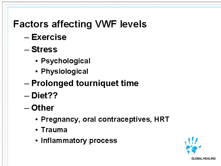
Factors affecting VWF levels – Exercise – Stress • Psychological • Physiological – Prolonged tourniquet time – Diet? ? – Other • Pregnancy, oral contraceptives, HRT • Trauma • Inflammatory process
Other factors influencing VWF levels – Sample type • 3. 2% sodium citrate preferred – ABO blood group VWF: Ag mean (range)** • • Type O: 75 (36 -157) Type A: 106 (48 -234) Type B: 117 (57 -241) Type AB: 123 (64 -238) – Using filters to achieve PPP ** Hemophilia 2008; 14: 177
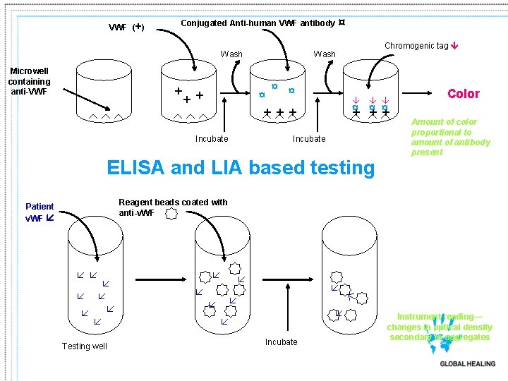
VWF (+) Conjugated Anti-human VWF antibody Wash Microwell containing anti-VWF + ¤ ¤ ++ + Incubate Chromogenic tag Wash ¤ + ¤ + Incubate ELISA and LIA based testing Color Amount of color proportional to amount of antibody present Reagent beads coated with anti-v. WF Patient v. WF Testing well Incubate Instrument reading— changes in optical density secondary to aggregates
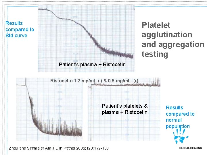
Results compared to Std curve Platelet agglutination and aggregation testing Patient’s plasma + Ristocetin 1. 2 mg/m. L (l) & 0. 6 mg/m. L (r) Patient’s platelets & plasma + Ristocetin Zhou and Schmaier Am J Clin Pathol 2005; 123: 172 -183 Results compared to normal population
VWF: RCo • Platelet poor plasma • Stabilized platelets – Commercially prepared – May be combined with ristocetin • Ristocetin – Initially used as antibiotic – Peptide from Amycolatopsis lurida – Thought to mimic shear effect of shear stress
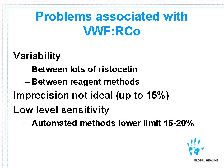
Problems associated with VWF: RCo Variability – Between lots of ristocetin – Between reagent methods Imprecision not ideal (up to 15%) Low level sensitivity – Automated methods lower limit 15 -20%
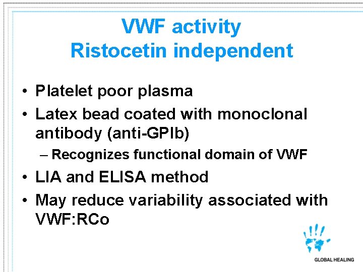
VWF activity Ristocetin independent • Platelet poor plasma • Latex bead coated with monoclonal antibody (anti-GPIb) – Recognizes functional domain of VWF • LIA and ELISA method • May reduce variability associated with VWF: RCo
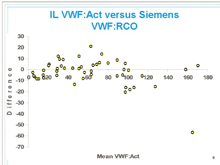
IL VWF: Act versus Siemens VWF: RCO
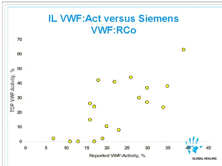
IL VWF: Act versus Siemens VWF: RCo
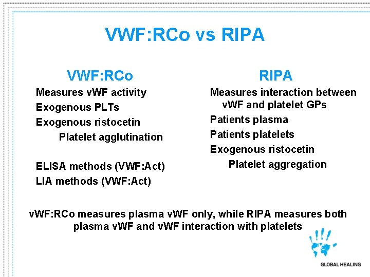
VWF: RCo vs RIPA VWF: RCo Measures v. WF activity Exogenous PLTs Exogenous ristocetin Platelet agglutination ELISA methods (VWF: Act) LIA methods (VWF: Act) RIPA Measures interaction between v. WF and platelet GPs Patients plasma Patients platelets Exogenous ristocetin Platelet aggregation v. WF: RCo measures plasma v. WF only, while RIPA measures both plasma v. WF and v. WF interaction with platelets
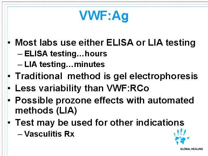
VWF: Ag • Most labs use either ELISA or LIA testing – ELISA testing…hours – LIA testing…minutes • Traditional method is gel electrophoresis • Less variability than VWF: RCo • Possible prozone effects with automated methods (LIA) • Test may be used for other indications – Vasculitis Rx
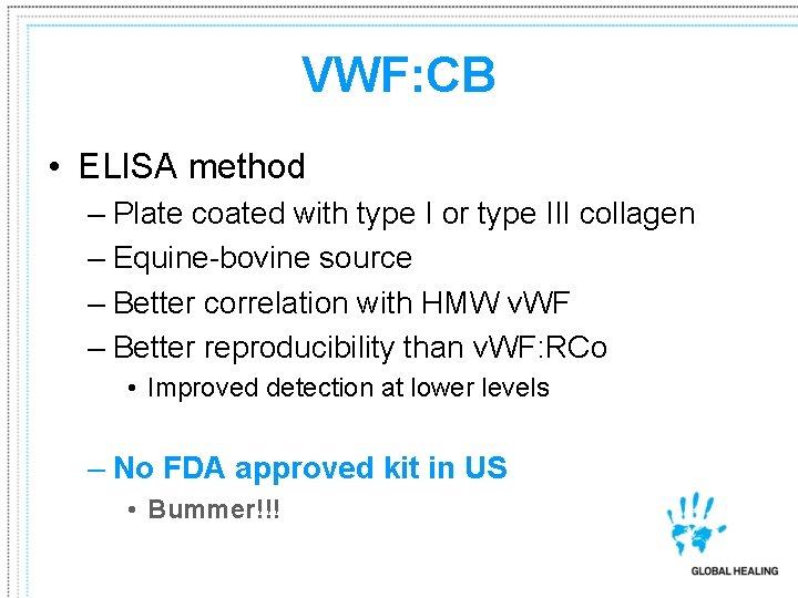
VWF: CB • ELISA method – Plate coated with type I or type III collagen – Equine-bovine source – Better correlation with HMW v. WF – Better reproducibility than v. WF: RCo • Improved detection at lower levels – No FDA approved kit in US • Bummer!!!
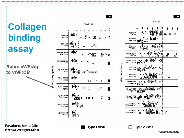
Collagen binding assay Ratio: v. WF: Ag to v. WF: CB Favaloro, Am J Clin Pathol 2000; 608 -618 Type 1 VWD Type 2 VWD
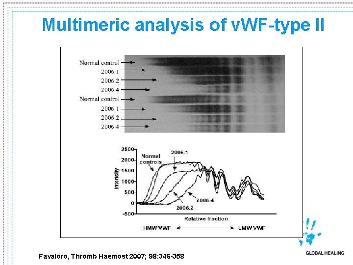
Multimeric analysis of v. WF-type II Favaloro, Thromb Haemost 2007; 98: 346 -358
v. WF propeptide (v. WF: pp) • Stored in α granules or Weibel-Palade bodies • After secretion into plasma, t½: 2 -3 hrs – vs mature v. WF t½: ~10 hrs • Plasma concentrations ~1μg/m. L – vs mature v. WF: 10 μg/m. L • Studies suggest: – Increased v. WF: pp to v. WF: Ag ratio with reduced v. WF: Ag true genetic defect with v. WF survival – v. WF survival would also suggest alternative Rx strategies
Pseudo-VWD • Similar to type 2 – Decreased PLT count – “Gain in function” • Aggregation with low dose ristocetin – Must perform VWF: PB • Increased with VWD: 2 B • Normal with psuedo-VWD – Abnormal GPIb receptor
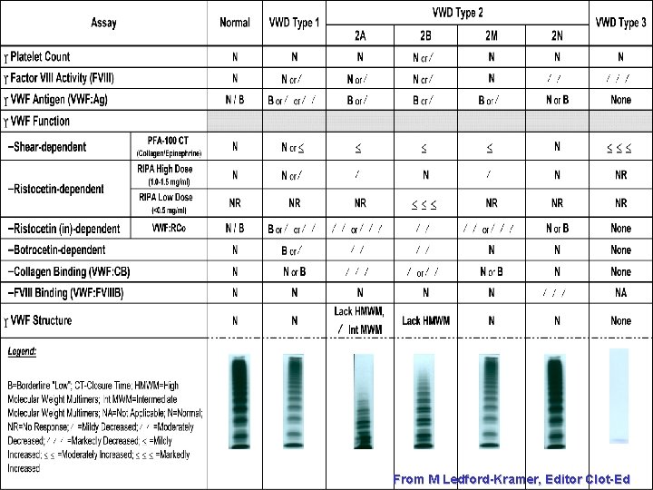
From M Ledford-Kramer, Editor Clot-Ed
CLSI Guideline H 51 -2 • Recommendations for – Sample type, processing – Testing – Calibration and results reporting • • Same calibrator for VWF: Act and VWF: Ag Different calibrator for therapeutics Units Ratios – Implementation/validation proposals? ? • Implementing new method • Lot-to-lot
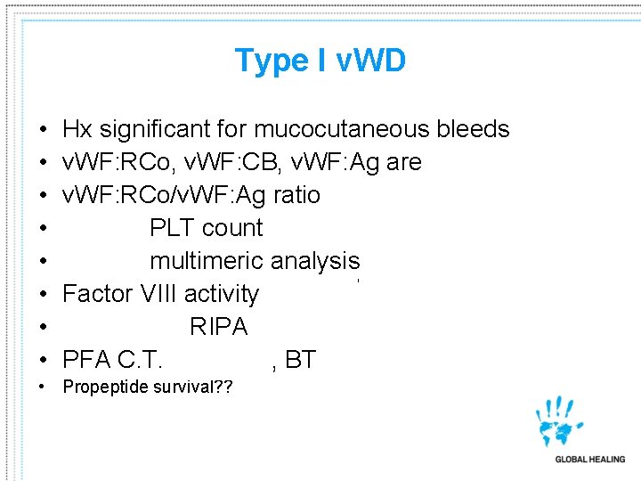
Type I v. WD • • Hx significant for mucocutaneous bleeds v. WF: RCo, v. WF: CB, v. WF: Ag are <50 IU/ml v. WF: RCo/v. WF: Ag ratio >0. 7 Normal PLT count Normal multimeric analysis Factor VIII activity normal/ Decreased RIPA PFA C. T. usually , BT slightly better than coin • Propeptide survival? ?
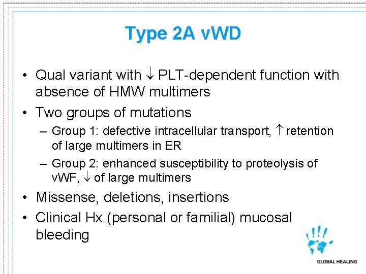
Type 2 A v. WD • Qual variant with PLT-dependent function with absence of HMW multimers • Two groups of mutations – Group 1: defective intracellular transport, retention of large multimers in ER – Group 2: enhanced susceptibility to proteolysis of v. WF, of large multimers • Missense, deletions, insertions • Clinical Hx (personal or familial) mucosal bleeding
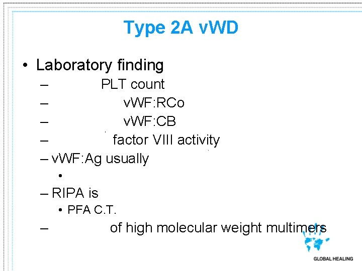
Type 2 A v. WD • Laboratory finding – Normal PLT count – Decreased v. WF: RCo – Decreased v. WF: CB – Normal/ factor VIII activity – v. WF: Ag usually normal/ • Ratio of v. WF: RCo or v. WF: CBA to v. WF: Ag is <0. 7 – RIPA is reduced • PFA C. T. usually – Absence of high molecular weight multimers

Type 2 B v. WD • Qual variant with “gain of function” • Increased affinity for platelet Gp. Ib • 4 mutations (nucleotide substitutions) within the v. WFA 1 domain account for ~90% of type IIb • Similar laboratory parameters as pseudotype v. WD • Clinical Hx (personal or familial) mucosal bleeding
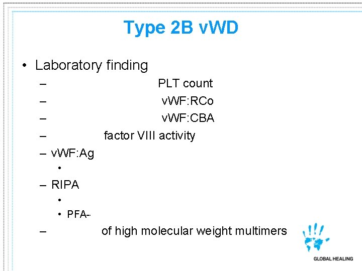
Type 2 B v. WD • Laboratory finding – – – Normal to slightly PLT count Decreased (usually) v. WF: RCo Decreased (usually) v. WF: CBA Normal/ factor VIII activity v. WF: Ag usually normal/ • Ratio of v. WF: RCo or v. WF: CBA to v. WF: Ag is <0. 7 – RIPA is • low dose of ristocetin (0. 75 mg/m. L) • PFA---normal to – Absence of high molecular weight multimers
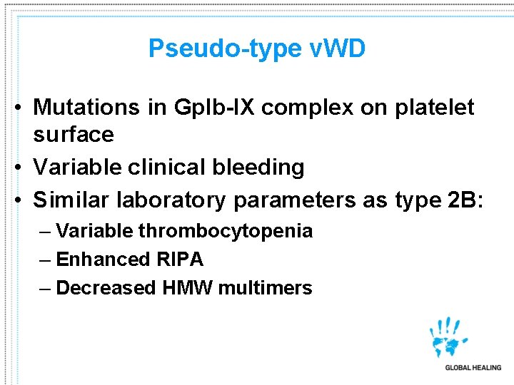
Pseudo-type v. WD • Mutations in Gp. Ib-IX complex on platelet surface • Variable clinical bleeding • Similar laboratory parameters as type 2 B: – Variable thrombocytopenia – Enhanced RIPA – Decreased HMW multimers
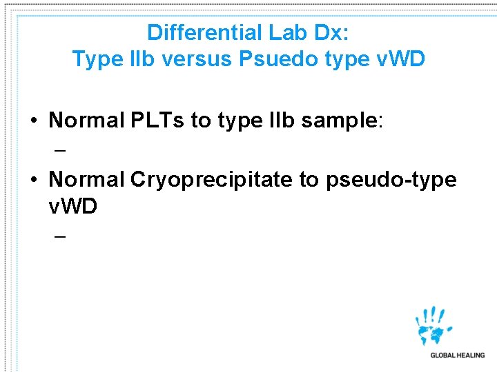
Differential Lab Dx: Type IIb versus Psuedo type v. WD • Normal PLTs to type IIb sample: – Enhanced RIPA • Normal Cryoprecipitate to pseudo-type v. WD – Spontaneous aggregation VWF: PB assay preferred/definitive
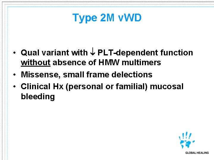
Type 2 M v. WD • Qual variant with PLT-dependent function without absence of HMW multimers • Missense, small frame delections • Clinical Hx (personal or familial) mucosal bleeding
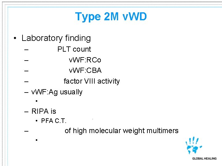
Type 2 M v. WD • Laboratory finding – – – Normal PLT count Decreased v. WF: RCo Decreased v. WF: CBA Normal/ factor VIII activity v. WF: Ag usually normal/ • Ratio of v. WF: RCo or v. WF: CBA to v. WF: Ag is <0. 7 – RIPA is reduced • PFA C. T. usually – Presence of high molecular weight multimers • increased amounts may be seen
Type 2 N v. WD • A qualitative defect resulting in decreased v. WF: FVIII binding • Autosomal recessive • Heterozygotes with normal factor VIII • Bleeding Hx related to surgery or trauma
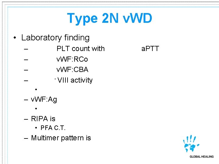
Type 2 N v. WD • Laboratory finding – – Normal PLT count with prolonged a. PTT Normal v. WF: RCo Normal v. WF: CBA factor VIII activity (usually between 5 -30 IU/ml) • Decreased v. WF: FVIIIC binding – v. WF: Ag normal • Ratio of v. WF: RCo or v. WF: CBA to v. WF: Ag is >0. 8 – RIPA is normal • PFA C. T. normal – Multimer pattern is normal
Type III v. WD • A virtual absence of v. WF protein • Presentation early in life • Multiple mutations, including frame shifts and nonsense mutations, as well as deletions
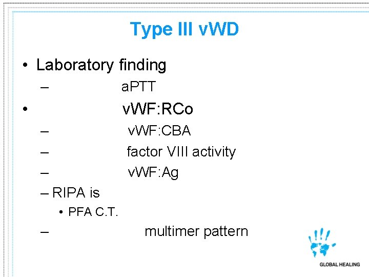
Type III v. WD • Laboratory finding – Prolonged a. PTT • Absence of v. WF: RCo – Absence of v. WF: CBA – Markedly factor VIII activity – Absence of v. WF: Ag – RIPA is markedly abnormal • PFA C. T. markedly abnormal – No detectable multimer pattern
- Slides: 47