Vitamin D Vitamin D Vitamin D is not


 Vit. D 3(Cholecalciferol )")


 per day. • Sign")
- Slides: 28
Vitamin D
Vitamin D • Vitamin D is not strictly a vitamin, since it can be synthesized in the skin, and synthesis into a vitamin. • it is only when sunlight exposure is inadequate that a dietary source is required. Its main function is in the regulation of calcium absorption and homeostasis; most of its actions are mediated by nuclear receptors that regulate gene expression. • Deficiency, leading to rickets in children and osteomalacia in adults, continues to be a problem in countries where sunlight exposure is poor.
Sources – Liver, beef, veal, eggs yolk , dairy products , some saltwater fish (salmon, tuna and sardine). – Fortified food (milk, yogurt, cheese, margarine, orange juice, breads and cereals). – It is stable in cooking and storage. – Synthesis in skin. � 2009 Cengage-Wadsworth
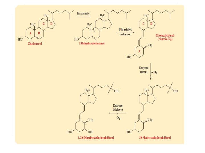
Vitamin D • The normal dietary form of vitamin D is cholecalciferol (also known as calciol). • This is also the compound that is formed in the skin by ultraviolet (UV) irradiation of 7 dehydrocholesterol. • Some foods are enriched or fortified with (synthetic) ergocalciferol, which undergoes the same metabolism as cholecalciferol and has the same biological activity.
International unit • Like vitamin A, vitamin D was originally measured in international units of biological activity before the pure compound was isolated: • 1 IU = 25 ng of cholecalciferol • 1 μg of cholecalciferol = 40 IU.
Vitamin D Chemical nature and properties � types:Vit. D 2(Ergocalciferol) Vit. D 3(Cholecalciferol ) � pro-Vit. D 2:Ergosterol Pro-Vit. D 3: 7 -hydro-cholesterol Ergosterol→Vit. D 2 cholesterol→ 7 -hydro cholesterol→Vit. D 3 � active form of Vit. D 3: 1, 25 - (OH)2 -Vit. D 3 transportation: DBP
OH OH
Absorption and metabolism • Vitamin D is absorbed in lipid micelles and incorporated into chylomicrons; therefore, people on a low fat diet will absorb little of such dietary vitamin D as is available. • Indeed, it is noteworthy that at the time that rickets was a major public health problem in Scotland, herrings (a rich source) were a significant part of the diet: it can only be assumed that the diet was so low in fat the absorption of the vitamin was impaired.
Synthesis of vitamin D in the skin • The steroid 7 -dehydrocholesterol (an intermediate in the synthesis of cholesterol that accumulates in the skin but not other tissues) undergoes a non-enzymic reaction on exposure to UV light, yielding previtamin D, which undergoes a further reaction over a period of hours to form cholecalciferol, which is absorbed into the bloodstream.
Synthesis of vitamin D in the skin • Structurally, Vitamin D is derived from a steroid. • It contains 3 intact rings (A, C and D) with a break between carbon 9 and 10 in the B ring. • The steroid 7 - dehydrocholestrol is synthesized and secreted on the skin surface. • The conjugated double bonds (57) B allow the absorption of UV light. • In the skin with UV cause ring B opining and forming previtamin D 3, followed by rearranged to D 3 (cholcalciferol).
Synthesis of vitamin D in the skin • In temperate climates there is a marked seasonal variation in the plasma concentration of vitamin D; it is highest at the end of summer and lowest at the end of winter. • • The synthesis of cholecalciferol is low in winter. By contrast, in summer, when the sun is more or less overhead, there is a considerable amount of UV light even on a moderately cloudy day, and enough can penetrate thin clothes to result in significant formation of vitamin D. • In northerly climates, and especially in polluted industrial cities with little sunlight, people may well not be exposed to enough UV light to meet their vitamin D needs, and they will be reliant on the few dietary sources of the vitamin.
Regulation of vitamin D metabolism • The main function of vitamin D is in the control of calcium homeostasis and, in turn, vitamin D metabolism in the kidney is regulated, at the level of 1 or 24 -hydroxylation, by factors that respond to plasma concentrations of calcium and phosphate. • In tissues other than the kidney that hydroxylate calcidiol to calcitriol, the enzyme is not regulated in response to plasma calcium.
Regulation of vitamin D metabolism ● Calcitriol acts to reduce its own synthesis and increase formation of 24 -hydroxycalcidiol, by regulating the expression of the genes for the two hydroxylases. ● Parathyroid hormone is secreted in response to a fall in plasma calcium. In the kidney it acts to increase the activity of calcidiol 1 -hydroxylase and decrease that of 24 -hydroxylase. In turn, both calcitriol and high concentrations of calcium repress the synthesis of parathyroid hormone; calcium also inhibits the secretion of the hormone from the parathyroid gland.
Regulation of vitamin D metabolism • Calcium exerts its main effect on the synthesis and secretion of parathyroid hormone. However, calcium ions also have a direct effect on the kidney, reducing the activity of calcidiol 1 hydroxylase. ● Phosphate also affects calcidiol metabolism; throughout the day there is an inverse fluctuation of plasma phosphate and calcitriol, and feeding • people on a low-phosphate diet results in increased circulating concentrations of calcitriol.
Absorption, transport & storage • Cholecalciferol diffuses from the skin into the blood. • It is transported in the blood by vitamin D binding protein (DBP) (transcalciferin) that is synthesized in the liver. • Dietary vitamin D 3 (cholecalceferol) is absorbed from a micelle, in association with fat and with the aid of bile salts, in the intestinal cell. • About 50% of dietary vitamin D 3 is absorbed. • The rate if absorption is the most rapid in the duodenum, but the largest amount is absorbed in the distal small intestine.
Absorption, transport & storage • In the intestinal cell: vitamin D is incorporated primarily into chylomicrons, which then enter the lymphatic system with subsequent entry into the blood. • In the blood: Chylomicrons transport around 40% of the cholecalciferol. • Some vitamin D may be transferred from the chylomicron to DBP for delivery to extrahepatic tissues.
Absorption, transport & storage • Cholecalciferol, which slowly diffuse from the skin into the blood, is picked up for transport by DBP. • About 60% of plasma cholecalciferol is bond to DBP for transport. • The vitamin D-DBP complex travels primarily to the liver but may be picked up by other tissues, especially muscle and adipose tissue, before hepatic uptake. • Cholecalciferol reaching the liver either by chylomicron remnant or by DBP typically is metabolized by a couple of different hydrolases to generate active form of the vitamin.
Absorption, transport & storage • In the liver: 25 - hydroxylase functions in the mitochondria to hydroxylate cholecalciferol at carbon 25 to form 25 -OH (vitamin) D 3 (25 -OH cholecalciferol). • The efficiency of the liver 25 -hydroxylase appears to be related to vitamin D concentrations and its metabolites. • Liver expresses most of 25 -hydroxylase, but the enzyme is found also in other organs (lungs, intestine & kidneys).
Synthesis of active form of vitamin D ACTIVE FORM 1, 25 -dihdroxy cholecalciferol or calcitriol
1 - α- hydroxylase
Metabolic functions of vitamin D • The principal function of vitamin D is to maintain the plasma concentration of calcium; calcitriol achieves this in three ways: ● increased intestinal absorption of calcium ● reduced excretion of calcium by stimulating resorption • in the distal renal tubules (due to increased vit. D synthesis)
Metabolic functions of vitamin D ● mobilization of bone mineral. synthesis and secretion of insulin, parathyroid, and thyroid hormones; ● inhibition of production of interleukin by activated T -lymphocytes and of immunoglobulin by activated • B-lymphocytes; differentiation of monocyte precursor cells; ● modulation of cell differentiation, proliferation and • apoptosis.
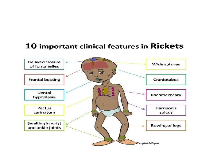
Vitamin D deficiency: rickets and osteomalacia Historically, rickets is a disease of toddlers, especially in northern industrial cities. Their bones are undermineralized as a result of poor absorption of calcium in the absence of adequate amounts of calcitriol. When the child begins to walk, the long bones of the legs are deformed, leading to bow-legs or knock knees. More seriously, rickets can also lead to collapse of the ribcage and deformities of the bones of the pelvis. Similar problems may also occur in adolescents who are deficient in vitamin D during the adolescent growth spurt, when there is again a high demand for calcium for new bone formation.
Vitamin D deficiency: rickets and osteomalacia • Osteomalacia is the adult equivalent of rickets. It results from the demineralization of bone, rather than the failure to mineralize it in the first place, as is the case with rickets. • Women who have little exposure to sunlight are especially at risk from osteomalacia after several pregnancies, because of the strain that pregnancy places on their marginal reserve of calcium. • Osteomalacia also occurs in the older people. The problem may be inadequate exposure to sunlight, but there is also evidence that the capacity to form 7 -dehydrocholesterol in the skin decreases with advancing age, so that older people are more reliant on the few dietary sources of vitamin D.
Interactions with drugs and other nutrients • Iron deficiency decreases in vitamin D absorption. • The interaction of calcitriol and vitamin K dependent protein. � 2009 Cengage-Wadsworth
Toxicity • Tolerable upper intake level 50µg (2. 000 IU) per day. • Sign and symptoms: • Over absorption of calcium (hypercalcemia), increase calcium execration. • Calcium deposits in kidneys, heart and blood vessels. • In infants: anorexia, nausea, vomiting hypertension. � 2009 Cengage-Wadsworth