
Vitamin D Effects on Microbial Flora Justin Beiriger

Vitamin D Effects on Microbial Flora Justin Beiriger Central Catholic High School Grade 10 2 nd Year in PJAS
Vitamin D • A group of fat-soluble secosteroids. • Nicknamed “the sunshine vitamin”. – Because the body can synthesize it with adequate sun exposure. • Effects of supplementation are uncertain. • Needed for bone growth. – Can cause a build-up of calcium.
Vitamin D Toxicity • • Also called hypervitaminosis D. Results from excess vitamin D supplements. Can cause liver or kidney conditions. Main consequence is a build-up of calcium in the bloodstream. – Called hypercalcemia
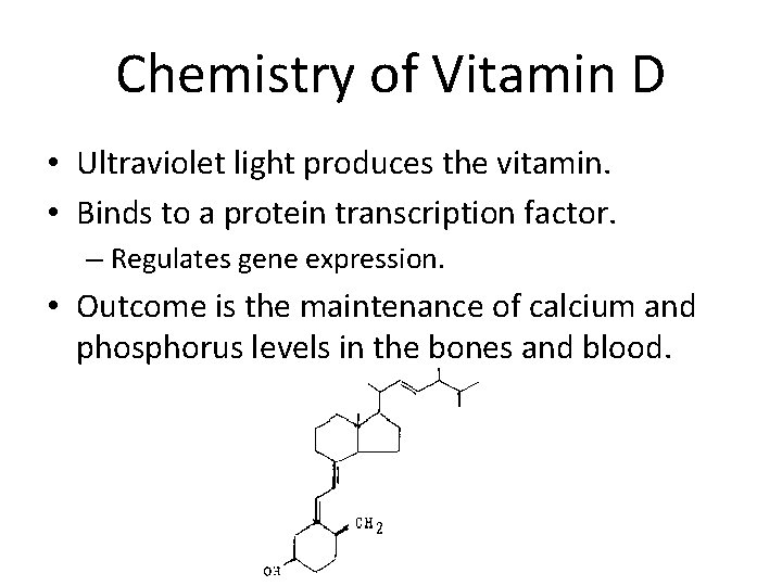
Chemistry of Vitamin D • Ultraviolet light produces the vitamin. • Binds to a protein transcription factor. – Regulates gene expression. • Outcome is the maintenance of calcium and phosphorus levels in the bones and blood.
Previous Studies • Edward Mellanby first discovered vitamin D. – 1918 -1920 – He wrote extensively on vitamin deficiency. • Previous studies of vitamin D supplementation have produced inconsistent results, with some trials showing a decrease in inflammatory markers and others showing no effect.
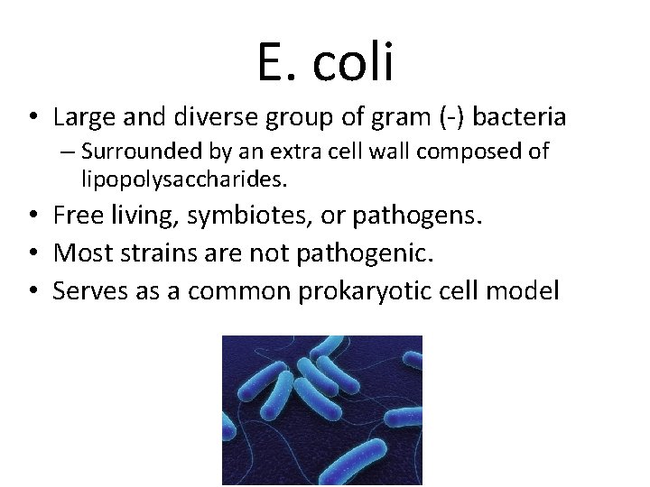
E. coli • Large and diverse group of gram (-) bacteria – Surrounded by an extra cell wall composed of lipopolysaccharides. • Free living, symbiotes, or pathogens. • Most strains are not pathogenic. • Serves as a common prokaryotic cell model
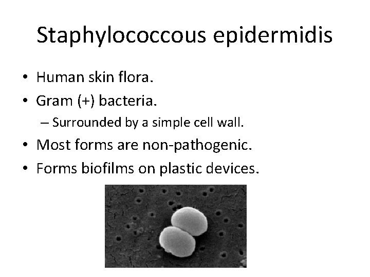
Staphylococcous epidermidis • Human skin flora. • Gram (+) bacteria. – Surrounded by a simple cell wall. • Most forms are non-pathogenic. • Forms biofilms on plastic devices.
Purpose To determine what effect vitamin D has on E. coli and Staph survivorship.
Hypotheses • Null hypothesis: Vitamin D will not significantly affect the survivorship of E. coli or Staph. • Alternative hypothesis: Vitamin D will significantly affect the survivorship of E. coli and/or Staph.
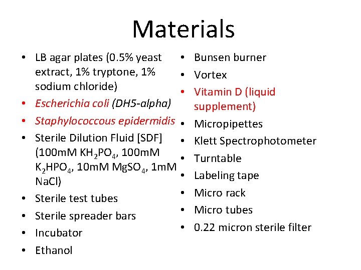
Materials • • LB agar plates (0. 5% yeast extract, 1% tryptone, 1% • sodium chloride) • • Escherichia coli (DH 5 -alpha) • Staphylococcous epidermidis • • Sterile Dilution Fluid [SDF] • (100 m. M KH 2 PO 4, 100 m. M • K 2 HPO 4, 10 m. M Mg. SO 4, 1 m. M • Na. Cl) • • Sterile test tubes • • Sterile spreader bars • • Incubator • Ethanol Bunsen burner Vortex Vitamin D (liquid supplement) Micropipettes Klett Spectrophotometer Turntable Labeling tape Micro rack Micro tubes 0. 22 micron sterile filter
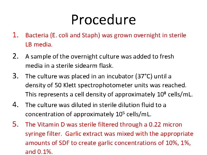
Procedure 1. Bacteria (E. coli and Staph) was grown overnight in sterile LB media. 2. A sample of the overnight culture was added to fresh media in a sterile sidearm flask. 3. The culture was placed in an incubator (37°C) until a density of 50 Klett spectrophotometer units was reached. This represents a cell density of approximately 108 cells/m. L. 4. The culture was diluted in sterile dilution fluid to a concentration of approximately 105 cells/m. L. 5. The Vitamin D was sterile filtered through a 0. 22 micron syringe filter. Garlic extract was mixed with the appropriate amounts of SDF to create garlic concentrations of 10%, 1%, and 0. 1%.
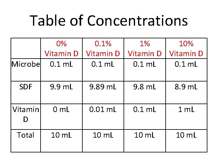
Table of Concentrations 0% 0. 1% 1% 10% Vitamin D Microbe 0. 1 m. L SDF 9. 9 m. L 9. 8 m. L 8. 9 m. L Vitamin D 0 m. L 0. 01 m. L 0. 1 m. L Total 10 m. L
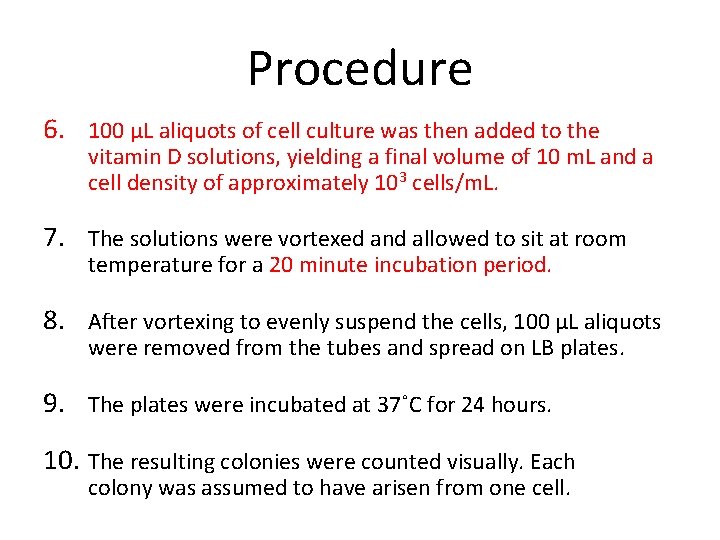
Procedure 6. 100 µL aliquots of cell culture was then added to the vitamin D solutions, yielding a final volume of 10 m. L and a cell density of approximately 103 cells/m. L. 7. The solutions were vortexed and allowed to sit at room temperature for a 20 minute incubation period. 8. After vortexing to evenly suspend the cells, 100 µL aliquots were removed from the tubes and spread on LB plates. 9. The plates were incubated at 37˚C for 24 hours. 10. The resulting colonies were counted visually. Each colony was assumed to have arisen from one cell.
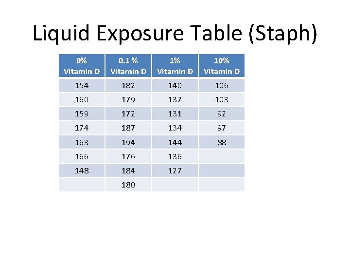
Liquid Exposure Table (Staph) 0% Vitamin D 0. 1 % Vitamin D 10% Vitamin D 154 182 140 106 160 179 137 103 159 172 131 92 174 187 134 97 163 194 144 88 166 176 136 148 184 127 180
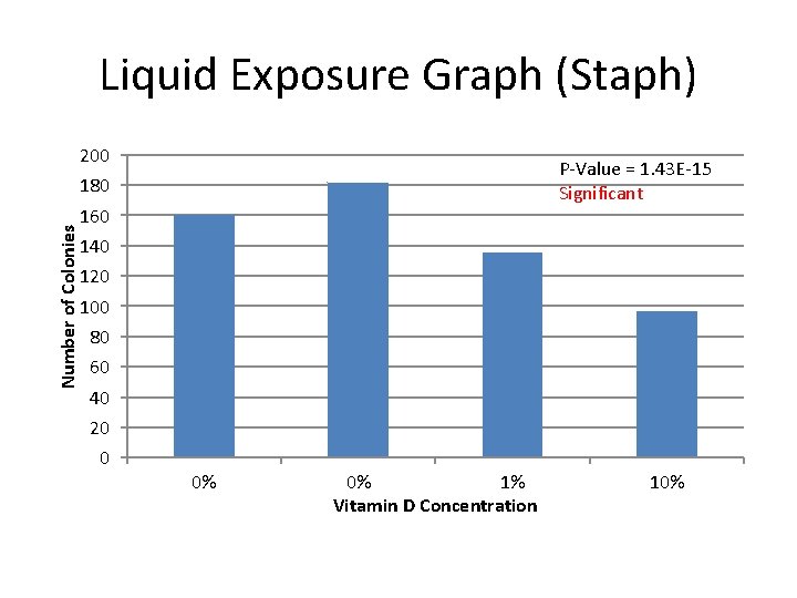
Liquid Exposure Graph (Staph) 200 P-Value = 1. 43 E-15 Significant Number of Colonies 180 160 140 120 100 80 60 40 20 0 0% 0% 1% Vitamin D Concentration 10%
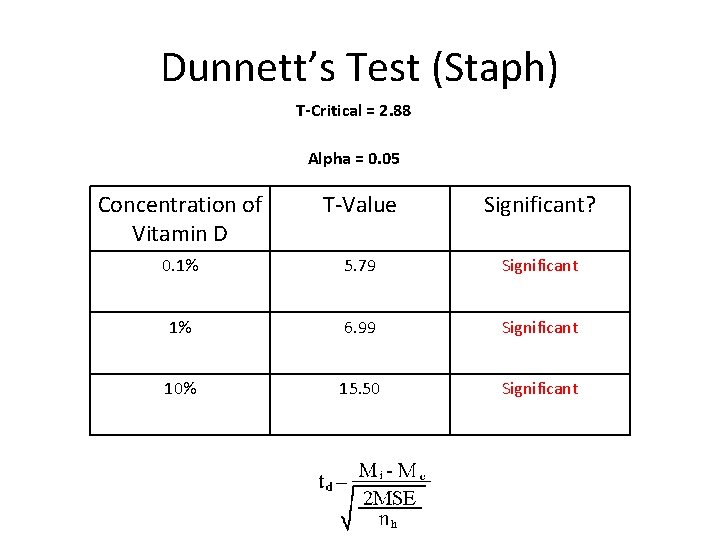
Dunnett’s Test (Staph) T-Critical = 2. 88 Alpha = 0. 05 Concentration of Vitamin D T-Value Significant? 0. 1% 5. 79 Significant 1% 6. 99 Significant 10% 15. 50 Significant
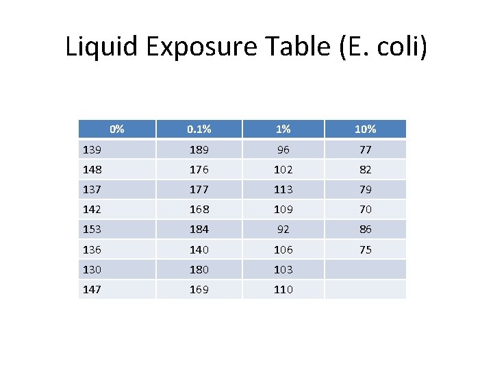
Liquid Exposure Table (E. coli) 0% 0. 1% 1% 10% 139 189 96 77 148 176 102 82 137 177 113 79 142 168 109 70 153 184 92 86 136 140 106 75 130 180 103 147 169 110
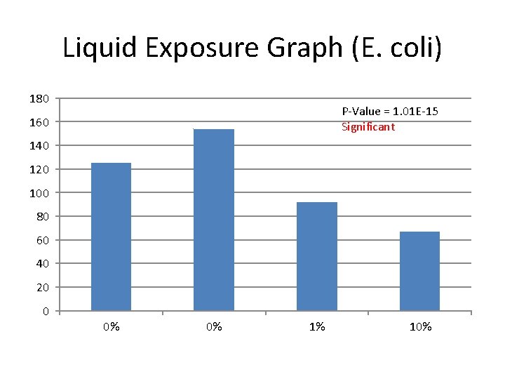
Liquid Exposure Graph (E. coli) 180 P-Value = 1. 01 E-15 Significant 160 140 120 100 80 60 40 20 0 0% 0% 1% 10%
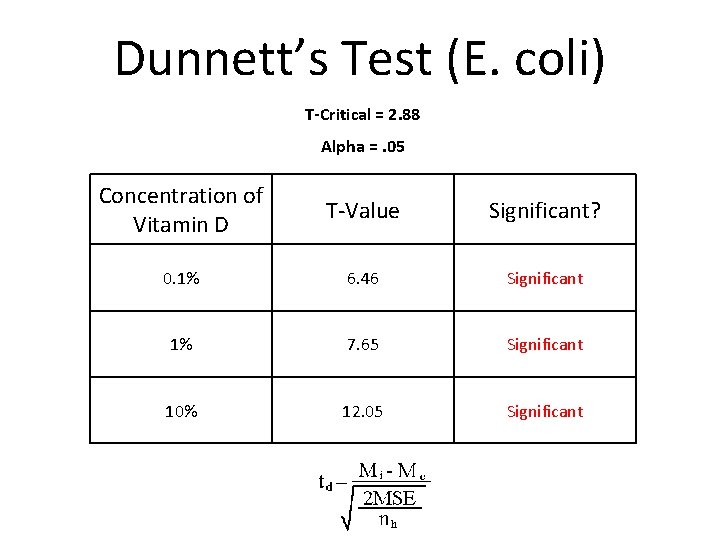
Dunnett’s Test (E. coli) T-Critical = 2. 88 Alpha =. 05 Concentration of Vitamin D T-Value Significant? 0. 1% 6. 46 Significant 1% 7. 65 Significant 10% 12. 05 Significant
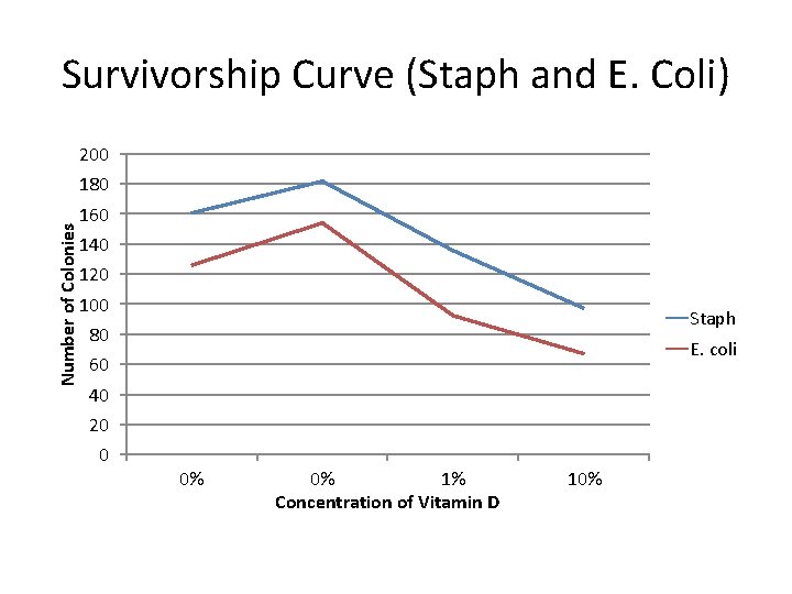
Survivorship Curve (Staph and E. Coli) 200 Number of Colonies 180 160 140 120 100 Staph 80 E. coli 60 40 20 0 0% 0% 1% Concentration of Vitamin D 10%
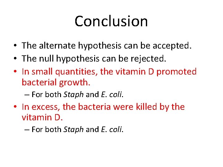
Conclusion • The alternate hypothesis can be accepted. • The null hypothesis can be rejected. • In small quantities, the vitamin D promoted bacterial growth. – For both Staph and E. coli. • In excess, the bacteria were killed by the vitamin D. – For both Staph and E. coli.
Limitations • The plating was not perfectly synchronized. • Some cells could have had longer or shorter exposure times. • The liquid vitamin D was removed from pill form, so it possibly contained excess pieces of the capsule.
Extensions • More trials to create a better basis for evaluating the results • Prolonged exposure test using vitamin D infused into the agar • Using an antibiotic resistant strain of bacteria for the test model
References • • • http: //ods. od. nih. gov/factsheets/vitamind/ http: //www. medicalnewstoday. com/articles/161618. php http: //www. webmd. com/vitamins-supplements/ingredientmono-929 VITAMIN%20 D. aspx? active. Ingredient. Id=929&active. Ingredient. Name=VITAMIN%20 D http: //www. mayoclinic. com/health/vitamin-d/NS_patient-vitamind http: //www. webmd. com/a-to-z-guides/e-coli-infection-topic-overview http: //www. medicalnewstoday. com/articles/68511. php http: //kidshealth. org/kid/stay_healthy/food/ecoli. html http: //www. about-ecoli. com/ http: //web. uconn. edu/mcbstaff/graf/Student%20 presentations/S%20 epidermidis/ sepidermidis. html http: //microbewiki. kenyon. edu/index. php/Staphylococcus_epidermidis http: //www. plospathogens. org/article/info%3 Adoi%2 F 10. 1371%2 Fjournal. ppat. 10 01133 http: //wiki. medpedia. com/Staphylococcus_epidermidis
- Slides: 24