Vitamin classification Lipidsoluble vitamins A D E and

 • hydrophobic compounds, absorbed efficiently")
Vitamin classification Lipid-soluble vitamins (A, D, E and K) • hydrophobic compounds, absorbed efficiently with lipids, • transport in the blood in lipoproteins or attached to specific binding proteins, • more likely to accumulate in the body, • more likely to lead to hypervitaminosis • single large doses may prevent deficiently

ANTIXEROPHTALMIC VITAMIN Vitamin A is the first vitamin discovered by scientists.
. NAD(P)+")
It is a family of natural compounds called Retinol, Retinal and Carotenoids (Beta-Carotenes). NAD(P)+ NAD(P)H+H+

Sources of vitamin A • • • cod liver oil meat egg milk dairy products β-carotene §carrot § broccoli § spinach § papaya § apricots • Vitamin A - 0. 75 – 2. 5 mg/day • β-carotene - 5 mg/day

Vit. A transport and metabolism β-carotene dioxygenase Retinal reductase

Functions of Vitamin A • Vision: Vitamin A is a component of the visual pigment rhodopsin. Retinal is bound to the protein opsin. • Growth: Vitamin A deficiency causes loss of appetite. Slow bone growth. Affects CNS. • Reproduction: Retinol and retinal are essential for normal reproduction • Maintenance of epithelial cells: Essential for normal differentiation of epithelial tissues and mucus secretion • Both retinoids and carotenoids have anticancer activity.
 Rhodopsin -opsin Nerve impulse to brain")
Role of vitamin A in vision (lipoprotein) Rhodopsin -opsin Nerve impulse to brain

Vitamin A – Retinal’s Role in Vision

Vitamin A – Retinal’s Role in Vision

Role of vitamin A in vision

Other biochemical functions vit A. • Vitamin A is necessary for the maintenance of normal epithelium and skin. Retinoic acid is found to have an important role in glycoprotein synthesis. Retinol and retinoic acid act like steroid hormones in the controlling the expression of certain genes and they are essential for normal spermatogenesis in the male and preventing fetal resorption in the female.

Mechanism of vitamin A action fat-soluble vitamin RBP c. RBP

The biochemical functions vit A. Vitamin A maintains healthy cells in the mucous membranes. Mucus Without vitamin A, the normal structure and function of the cells in the mucous membranes are impaired. Goblet cells

Vitamin A • Roles in the Body – Vitamin A in Reproduction and Growth • Sperm development in men • Normal fetal development in women • Growth in children • Remodeling of the bone involves osteclasts, osteoblasts, and lysosomes. – Osteoclasts are cells that destroy bone growth. – Osteoblasts are cells that build bones. – Lysosomes are sacs of degradative enzymes that destroy bones.

Other biochemical functions vit A. • Both retinoids and carotenoids have anticancer activity. Beta-carotene is an antioxidant and may play a role in trapping peroxyl and free radicals in tissues the same as major anti-oxidant vitamin E, but at low partial pressures of oxygen (arise in epithelial tissues). β-carotenes may be useful in preventing cancer and heart attacks.

Deficiency. 1. Night blindness or nyctalopia. Visual acuity is diminished in dim light. The patient cannot read or drive a car in poor light. The dark adaptation time is increased. 2. Xerophthalmia. The conjunctiva gets keratinised and loses its normal transparency. Dryness spreads to cornea. It becomes glazy and lusterless due to keratinisation of corneal epithelium. Infections may supersede. 3. Keratomalacia. The softening of the cornea. There is degeneration of corneal epithelium which may get vascularised. Bacterial infection leads to corneal ulceration perforation of cornea and total blindness.

Night blindness –is an early symptom of A deficiency Visual acuity is diminished in dim light. The patient cannot read or drive a car in poor light. The dark adaptation time is increased.

Reproductive system Vitamin A deficiency impairs fertility in both sexes. Fetuses may be aborted or reabsorbed, or born with hydrocephalus, as shown in the lower pup on this image.

FOLLICULAR HYPERKERATOSIS – Follicular hyperkeratosis resuts from hyperkeratinisation of the epithelium lining the follicles. The skin becomes rough (plus bacterial infection can lead to acne). Keratinising metaplasia of the epithelium of the lungs, gastrointestinal, and genitourinary tracts, coupled with reduction in mucous secretion. Epithelium is atrophied.

Too Much Can Be Toxic!! • Hypervitaminosis A leads to toxic symptoms: – – – – Dry, itchy skin Headaches and fatigue Hair loss Liver damage Blurred vision Loss of appetite Skin coloration
")
Polar Bear Liver One ounce of polar bear liver contains enough vitamin A (retinol) to kill a person!

Vitamin A Toxicity During Pregnancy Cleft Lip Examples of birth defects were : • hydrocephaly (enlargement of the fluidfilled spaces in the brain); microcephaly (small head); • mental retardation; • ear and eye abnormalities, • cleft lip and palate, and • other facial abnormalities and • heart defects

Vitamin. D 3 Vitamin. D 2

Ergocalciferol-D 2 found in plants, cholecalciferol-D 3 found in animal tissues. Daily norm 5 -10 mcg for children.

• Vitamin D, as either D 3 or D 2, does not have significant biological activity. Rather, it must be metabolized within the body to the hormonally-active form. This transformation occurs in two steps. • Cholecalciferol (or Ergocalciferol) are absorbed from the intestine and transported to the liver bound to a specific vitamin D-binding protein. • Within the liver, cholecalciferol is hydroxylated to 25 hydroxycholecalciferol by the enzyme 25 -hydroxylase 1. Within the kidney, 25 -vitamin D serves as a substrate for 1 -alphahydroxylase, yielding 1, 25 dihydroxycholecalciferol, the biologically active form of vitamin D

• In conjunction of the parathyroid gland. It maintains the proper levels of serum calcium, and phosphorus, which promotes the formation, calcification and repair of bones. • Stimulates renal tubular transport of calcium and phosphorus(Deluca). • Maintain blood levels of calcium and phosphate for bone formation, mineralization, growth, and repair • Improves muscle strength and immune function • Reduces inflammation

Functions: It is to maintain adequate plasma level of calcium. § On the intestine. Increases uptake of calcium (binds to a cytosolic receptors, moves to the nucleus where it selectively interacts with the cellular DNA, as a result - increases synthesis of a specific calcium-binding protein). § The absorption of calcium is increased. § In kidney. Calcitriol increases the reabsorption of calcium and phosphorus by renal tubules, therefore, both minerals are conserved. § Bone. Stimulating mineralisation of bone, when the ionic product of calcium and phosphorus increases due to increasing activity of alkaline phosphotase.

Mechanism of vitamin D action Vit D Ca++ binding protein R R

In adults is osteomalacia is characterized by demineralization of previously formed bone.

Vitamin D deficiency RICKETS

Vitamin D deficiency

Rickets

Vitamin D Deficiency – Osteomalacia • Affects adults • Soft, flexible, brittle, deformed bones • Progressive weakness • Pain in pelvis, lower back, and legs Osteoporosis • Loss of calcium from the bones due to inadequate synthesis of vitamin D • Results in a reduced bone density

Vitamin D Toxicity High-dose supplements may cause toxicity. – Toxicity symptoms • Elevated blood calcium • Calcification of soft tissues (blood vessels, kidneys, heart, lungs, and tissues around joints) • Frequent urination

Synthesis from intestinal bacteria

They are naphthoquinone derivatives, with a long isoprenoid side chain. K 1 Vitamin K is found in plants as phylloquinone (vitamin K 1) and in animals as menaquinone (vitamin K 2).

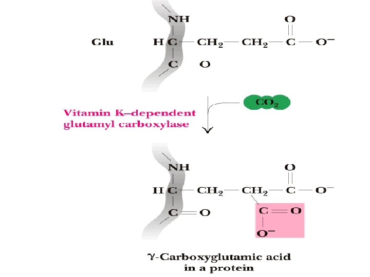
Vitamin K is necessary for hepatic synthesis of prothrombin and the blood clotting factors, and to maintain proper bone density.

Vitamin K cycle • Anticoagulants such as Warfarin and Dicoumarol block the reduction of vitamin K oxide to vitamin K, explaining their antagonistic effects on this cycle.

The role of vitamin K in blood clotting

Hemophilia

Vitamin K Deficiencies - A Common Link in Connective Tissue Disorders Other symptoms that associated with vitamin K deficiencies are osteoporosis and mitral valve prolapse, scoliosis and hypermobility. Heavy menstrual bleeding Gastrointestinal bleeding EHLER’S DANLOS SYNDROME

Hypervitaminosis K • Administration of large amount of menadione may result in toxicity. In hypervitaminosis K massive trombosis, hemolysis , hyperbilirubinemia, and brain damage may occur.

Is a mixture of tocopherols

Sources of vitamin E • • fortified cereals seeds and seed oils, like sunflower nuts and nut oils, like almonds and hazelnuts green leafy vegetables, broccoli cabbage celery http: //health. allrefer. com/health/nutrition. html

d-Alpha-Tocopherol Alfa-tocopherol is the most active. Tocopherol is absorbed and transported as chylomicrons. It export from liver in very low density lipoproteins. It is store in adipose tissue.

Antioxidant Vitamin E is necessary for fertility • Prevents loss of spermatogenesis in males • Muscle development. •

As a biological antioxidant Prevents lipid peroxidation of biological membrane Vitamin E Prevents peroxidation of vitamin A & PUFA Reproductive functions & prevents sterility

Vitamin E deficiency occurs only rarely in humans and virtually never as a result of dietary deficiencies. Vitamin E deficiency does occur as a result of genetic abnormalities in the tocopherol transfer protein (-TTP) and as a result of various at malabsorption syndromes. • Spino-cerebellar ataxia • Myopathies • Peripheral neuropathy • Ataxia • Skeletal myopathy • Retinopathy • Impairment of the immune response • Red blood cell destruction

Antivitamins Currently, the antivitamins are conventionally divided into two groups: • Antivitamins structurally related to the native vitamin and competitively antagonistic to it. • Antivitamins that modify the chemical nature of the native vitamin or inhibit its absorption and transport, with the ensuing diminution or loss of its biological activity. • These include, for example: • 1. thiaminases I and II causing the breaking down of the vitamin B 1 molecules. • 2. acsorbate oxidase, that catalyzes the degradation of vitamin C. • 3. the protein avidin that can bind biotin into a biologically inactive complex.

Most antivitamins are used as drugs of strictly selective action aimed at definite biocamical and physiological processes. Dicumarol, warfarin, and tromexane (antagonists of the fatsoluble vitamin K are often used as anticoagulants. Well studied thiamin antivitamins are also hydroxythiamine, pyrithiamine, and neopyrithiamine; riboflavin antivitamins aterbin, quinocrine ydrochloride, galactoflavin, isoriboflavin – all of them competitors for vitamin B 2 in the biosynthesis of coenzymes FAD and FMN; pyridoxine antivitamins deoxypyridoxine, cycloserine, isoniazid, an antibacterial for tuberculosis microbacteria. Antivitamins for folic acids are amino- and ametopterin; for vitamin B 12, derivatives of 2 -aminomethylpropanol-B 12; for nicotinic acid, isoniazid and 3 -acetylpyridine; for para-aminobenzoic acid, sulphanilamide preparations. All of this preparations have found application as antibacterial and antiblastic agents in the controlled cellular synthesis of proteins and nucleic acids.

- Slides: 53