
VITAMIN C E Dr JACOB INTRODUCTION Vitamins are

VITAMIN C & E Dr JACOB

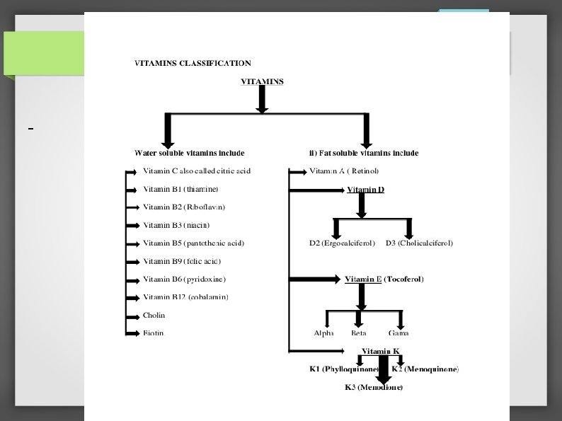
INTRODUCTION -Vitamins: are essential organic compounds that are required in very small amounts (micronutrients) - Involved in fundamental functions in the body, such as growth, maintenance of health, and metabolism. - Because our bodies cannot biosynthesize vitamins, vitamins must be supplied by the diet or as supplements. - Deficiencies are common in many developing countries and are often associated with global malnutrition.

VITAMIN C
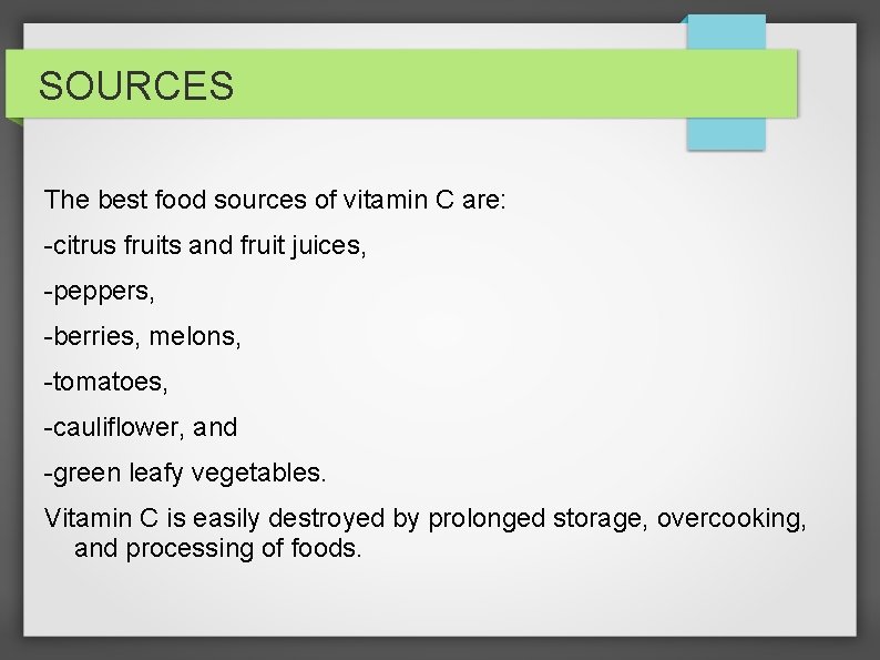
SOURCES The best food sources of vitamin C are: -citrus fruits and fruit juices, -peppers, -berries, melons, -tomatoes, -cauliflower, and -green leafy vegetables. Vitamin C is easily destroyed by prolonged storage, overcooking, and processing of foods.
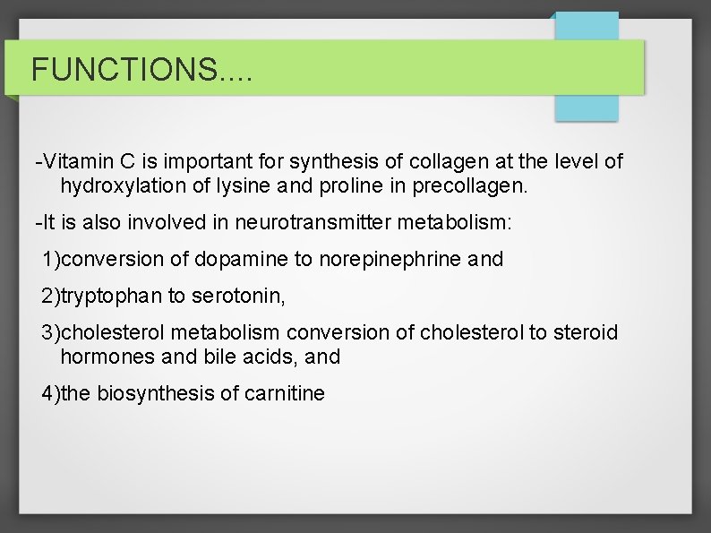
FUNCTIONS. . -Vitamin C is important for synthesis of collagen at the level of hydroxylation of lysine and proline in precollagen. -It is also involved in neurotransmitter metabolism: 1)conversion of dopamine to norepinephrine and 2)tryptophan to serotonin, 3)cholesterol metabolism conversion of cholesterol to steroid hormones and bile acids, and 4)the biosynthesis of carnitine
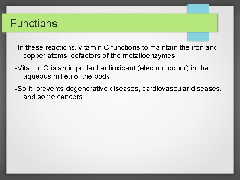
Functions -In these reactions, vitamin C functions to maintain the iron and copper atoms, cofactors of the metalloenzymes, -Vitamin C is an important antioxidant (electron donor) in the aqueous milieu of the body -So it prevents degenerative diseases, cardiovascular diseases, and some cancers. -
Functions Vitamin C enhances : 1)nonheme iron absorption, 2)the transfer of iron from transferrin to ferritin, 3)formation of tetrahydrofolic acid and thus can affect the cellular and immunologic functions of the hematopoietic system.
Dietary needs Humans depend on dietary sources for vitamin C. An adequate intake is : -40 mg for age 0 -6 mo and -50 mg for age 6 -12 mo. -For older children: The RDA is 15 mg for age 1 -3 yr, -25 mg for age 4 -8 yr, -45 mg for age 9 -13 yr, and -65 -75 mg for age 14 -18 yr.
Dietary needs. . -The requirement for vitamin C is increased during infectious and diarrheal diseases. -Children exposed to smoking or environmental tobacco smoke also require increased amounts of foods rich in vitamin C.
Absorption & storage -Absorption of vitamin C occurs in the upper small intestine. -It is by an active process or by simple diffusion when large amounts are ingested. -Vitamin C is not stored in the body but is taken up by all tissues. -The highest levels are found in the pituitary and adrenal glands. -The brain ascorbate content in the fetus and neonate is manyfold higher than the content in the adult brain, a finding probably related to its function in neurotransmitter synthesis.
RISK -Breast milk contains sufficient vitamin C to prevent deficiency throughout infancy. -Infants consuming pasteurized or boiled animal milk are at significant risk of developing deficiency if the other sources of vitamin C are also lacking in the diet. -Neonates whose feeding has been delayed because of clinical condition can also suffer from ascorbic acid deficiency.
-For patients on total parenteral nutrition (TPN); 1)a parenteral dose of 80 mg/day is recommended for full-term infants. 2)a parenteral dose of 25 mg/kg/day is recommended for preterm infants.
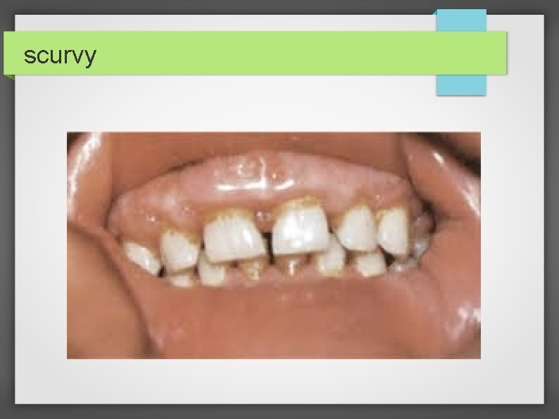
Deficiency A deficiency of vitamin C results in the clinical presentation of scurvy. Children fed 1)predominantly heat-treated (ultra-hightemperature or pasteurized) milk 2)unfortified formulas and not receiving fruits and fruit juices are at significant risk for symptomatic disease.
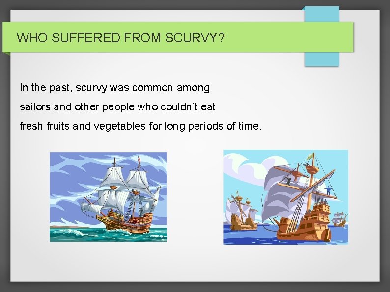
WHO SUFFERED FROM SCURVY? In the past, scurvy was common among sailors and other people who couldn’t eat fresh fruits and vegetables for long periods of time.
Deficiency. . In scurvy; -There is defective formation of connective tissues and collagen in skin, Cartilage, dentine, bone, and blood vessels, leading to their fragility. In the long bones: -Osteoid is not deposited by osteoblasts, -Cortex is thin, and -Trabeculae become brittle and fracture easily.
scurvy
Clinical manifestations The early manifestations are: 1) irritability, 2)loss of appetite, 3)low-grade fever, and 4)tenderness in the legs. These signs and symptoms are followed by leg swelling—most marked at the knees and the ankles— and pseudoparalysis.
Clinical manifestations. . -The infant might lie in the “pithed frog” position: with the hips and knees semiflexed and the feet rotated outward. -Subperiosteal hemorrhages in the lower limb bones. These sometimes acutely increase the swelling and pain, and the condition might mimic : 1)acute osteomyelitis or 2)arthritis.
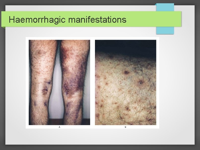
Haemorrhagic manifestations
Clinical manifestations. . -A “rosary” at the costochondral junctions and depression of the sternum are other typical features. -The angulation of scorbutic beads is usually sharper than the angulation of a rachitic rosary. -Gum changes are seen in older children after teeth have erupted and are manifested as: Bluish purple, spongy swellings of the mucous membrane, especially over the upper incisors.
Clinical features. . . -Anemia, a common finding in infants and young children with scurvy, is related to impaired iron absorption and coexistent hematopoietic nutrient deficiencies including iron, vitamin B 12, and folate. -Hemorrhagic manifestations of scurvy include : -petechiae, -purpura, and -ecchymoses at pressure points; -epistaxis; -gum bleeding; and the characteristic perifollicular hemorrhages
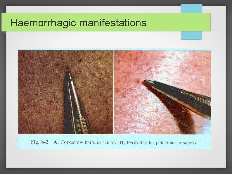
Haemorrhagic manifestations
Lab findings & diagnosis The diagnosis of vitamin C deficiency is usually based on the: 1)Characteristic clinical picture, 2)The radiographic appearance of the long bones, and 3)A history of poor vitamin C intake.
Radiological findings. . . -The shafts of the long bones have a ground-glass appearance due to trabecular atrophy. -The cortex is thin and dense, giving the appearance of pencil outlining of the diaphysis and epiphysis. -The white line of Fr? nkel, an irregular but thickened white line at the metaphysis, represents the zone of well-calcified cartilage. -The epiphyseal centers of ossification also have a ground-glass appearance and are surrounded by a sclerotic ring
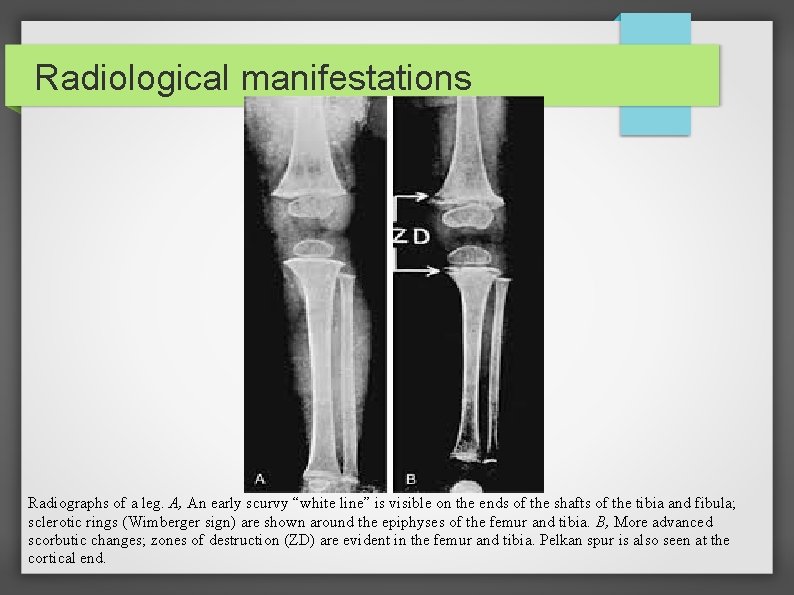
Radiological manifestations Radiographs of a leg. A, An early scurvy “white line” is visible on the ends of the shafts of the tibia and fibula; sclerotic rings (Wimberger sign) are shown around the epiphyses of the femur and tibia. B, More advanced scorbutic changes; zones of destruction (ZD) are evident in the femur and tibia. Pelkan spur is also seen at the cortical end.
Lab findings & diagnosis. . . -Biochemical tests are not very useful in the diagnosis of scurvy, because they do not reflect the tissue status. -A plasma ascorbate concentration of <0. 2 mg/d. L usually is considered deficient. -Leukocyte concentration of vitamin C is a better indicator of body stores, but this measurement is technically more difficult to perform.
Lab findings & diagnosis. . . -Saturation of the tissues with vitamin C can be estimated from the urinary excretion of the vitamin after a test dose of ascorbic acid. -In healthy children, 80% of the test dose appears in the urine within 3 -5 hr after parenteral administration. -Generalized nonspecific aminoaciduria is common in scurvy, whereas plasma amino acid levels remain normal.
Differential diagnosis -Arthritis, -Osteomyelitis, -Battered child syndrome, or acrodynia. -The early irritability and bone pain are sometimes attributed to nonspecific pains or other nutritional deficiencies. -Copper deficiency results in a radiographic picture very similar to that of scurvy. -Henoch-Schonlein purpura, -Thrombocytopenic purpura, or leukemia is sometimes suspected in children presenting with hemorrhagic manifestations.
TREATMENT -Vitamin C supplements of 100 -200 mg/day orally or parenterally ensure rapid and complete cure. -The clinical improvement is seen within a week in most cases, but the treatment should be continued for up to 3 months for complete recovery. -Dietary modifications
Prevention -Breast-feeding protects against vitamin C deficiency throughout infancy. -In children consuming milk formula, fortification with vitamin C must be ensured. -Children consuming heat-treated milk should consume adequate vitamin C–rich foods in infancy. -Dietary or medicinal supplements are required in severely malnourished children.
Toxicity -Daily intake of <2 g of vitamin C is generally without adverse effects in adults. -Larger doses can cause gastrointestinal problems, such as abdominal pain and osmotic diarrhea. -In general, megadoses of vitamin C should be avoided in patients with a history of urolithiasis or conditions related to excessive iron accumulation such as thalassemia and hemochromatosis.
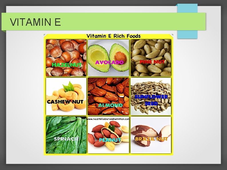
VITAMIN E
VITAMIN E -Vitamin E is a fat-soluble vitamin and functions as an antioxidant, but its precise biochemical functions are not known. -Vitamin E deficiency can cause hemolysis or neurologic manifestations and occurs in premature infants, in patients with malabsorption, and in an autosomal recessive disorder affecting vitamin E transport.
Pathogenesis -The majority of vitamin E is located within cell membranes, where it prevents lipid peroxidation and the formation of free radicals. -Other antioxidants, such as ascorbic acid, enhance the antioxidant activity of vitamin E. -Premature infants are particularly susceptible to vitamin E deficiency, because there is significant transfer of vitamin E during the last trimester of pregnancy.
Pathogenesis. . -Vitamin E deficiency in premature infants causes thrombocytosis, edema, and hemolysis potentially causing anemia. -The risk of symptomatic vitamin E deficiency was increased by the use of formulas for premature infants that had a high content of polyunsaturated fatty acids (PUFAs). -These formulas led to a high content of PUFAs in red blood cell membranes, making them more susceptible to oxidative stress, which could be ameliorated by vitamin E.
-Vitamin E deficiency does occur in children with fat malabsorption secondary to the need for bile acid for vitamin E absorption. -Although symptomatic disease is most common in children with cholestatic liver disease, it can occur in patients with : 1)cystic fibrosis, 2)celiac disease, 3)short-bowel syndrome, or Crohn disease. In ataxia with isolated vitamin E deficiency (AVED), a rare autosomal recessive disorder, there are mutations in the gene for αtocopherol transfer protein.
Clinical features -A severe, progressive neurologic disorder occurs in patients with prolonged vitamin E deficiency. -Clinical manifestations do not appear until after 1 yr of age, even in children with cholestasis since birth. -Patients may have cerebellar disease, posterior column dysfunction, and retinal disease.
Clinical features. . -Loss of deep tendon reflexes is usually the initial finding. Subsequent manifestations include: 1)Limb ataxia (intention tremor, dysdiadokokinesia), 2)Truncal ataxia (wide-based, unsteady gait), 3)Dysarthria, 4)ophthalmoplegia (limited upward gaze), 5)nystagmus, 6)decreased proprioception (positive Romberg test), 7)decreased vibratory sensation, 8)pigmentary retinopathy
Clinical features. . -Visual field constriction can progress to blindness. -Cognition and behavior can also be affected. -Myopathy and cardiac arrhythmias are less common findings. -In premature infants, hemolysis due to vitamin E deficiency typically develops during the 2 nd month of life. -Edema may also be present.
Lab findings -Vitamin E status is best determined by measuring the ratio of vitamin E to serum lipids; 1) A ratio <0. 8 mg/g is abnormal in older children and adults; 2) <0. 6 mg/g is abnormal in infants <1 yr. -Premature infants with hemolysis due to vitamin E deficiency also often have elevated platelet counts. -Neurologic involvement can cause abnormal somatosensory evoked potentials and nerve conduction studies.
DIAGNOSIS -Premature infants with unexplained hemolytic anemia after the 1 st month of life, especially if thrombocytosis is present, either should be empirically treated with vitamin E or should have serum vitamin E and lipid levels measured. -Children with neurologic findings and a disease that causes fat malabsorption should have their vitamin E status evaluated. -
DIAGNOSIS. . . -Because children with AVED do not have symptoms of malabsorption, a correct diagnosis requires a high index of suspicion. -Friedreich ataxia has been misdiagnosed in some patients. -Children with unexplained ataxia should be screened for vitamin E deficiency.
DIAGNOSIS. . . -Because children with AVED do not have symptoms of malabsorption, a correct diagnosis requires a high index of suspicion. -Friedreich ataxia has been misdiagnosed in some patients. -Children with unexplained ataxia should be screened for vitamin E deficiency.
TREATMENT -For correction of deficiency in neonates, the dose of vitamin E is 25 -50 units/day for 1 wk, followed by adequate dietary intake. -Children with deficiency due to malabsorption should receive 1 unit/kg/day, with the dose adjusted based on levels; ongoing treatment is necessary. -Children with AVED normalize their serum vitamin E levels with high doses of vitamin E.
PREVENTION -Premature infants should receive sufficient vitamin E via formula or breast milk fortifier and formula without a high content of PUFAs. -Children at risk for vitamin E deficiency due to malabsorption should be screened for deficiency and given adequate vitamin E supplementation.
● ● REFERENCES: NELSON textbook of paediatrics 19 th edition: chapter on nutrition ● IAP textbook of paediatrics ● IAP official website: latest recommendations ● SURAJ GUPTA: book on nutrition

THANK YOU
- Slides: 48